Keratoacanthoma (ICD-10: D23) 🚨
Keratoacanthoma
Keratoacanthoma is a rapidly growing, non-pigmented, and typically benign skin neoplasm that often resembles squamous cell carcinoma in both clinical and histological appearance. Despite its malignant-like features, keratoacanthoma frequently undergoes spontaneous regression within several months of its initial appearance. This tumor usually arises in adulthood, predominantly after the age of 35–40 years, and is more common in men than in women.
Predisposing Factors
While no definitive cause has been identified for keratoacanthoma, several contributing factors are believed to increase the risk of its development. These predisposing factors include:
- Excessive Sun Exposure: Chronic or intense exposure to ultraviolet radiation (natural or artificial) is a significant risk factor, particularly in fair-skinned individuals.
- Ionizing Radiation: Previous radiation therapy or environmental exposure may induce tumor formation.
- Chemical Irritants: Prolonged skin contact with carcinogenic or irritant substances can contribute to keratoacanthoma formation.
- Chronic Trauma: Persistent irritation, wounds, or burns in the same skin area may predispose to tumor development.
- Foreign Bodies: Embedded splinters, metal shavings, or other foreign material in the skin may trigger a reactive keratoacanthoma.
Diagnosis
Diagnosis of keratoacanthoma is based on thorough clinical evaluation, which includes physical inspection of the lesion and dermatoscopic examination. Due to its striking similarity to basal cell carcinoma and especially squamous cell carcinoma, a biopsy is typically performed to confirm the diagnosis and rule out malignancy. Histopathological analysis is essential to distinguish keratoacanthoma from more aggressive forms of skin cancer.
Clinical Presentation
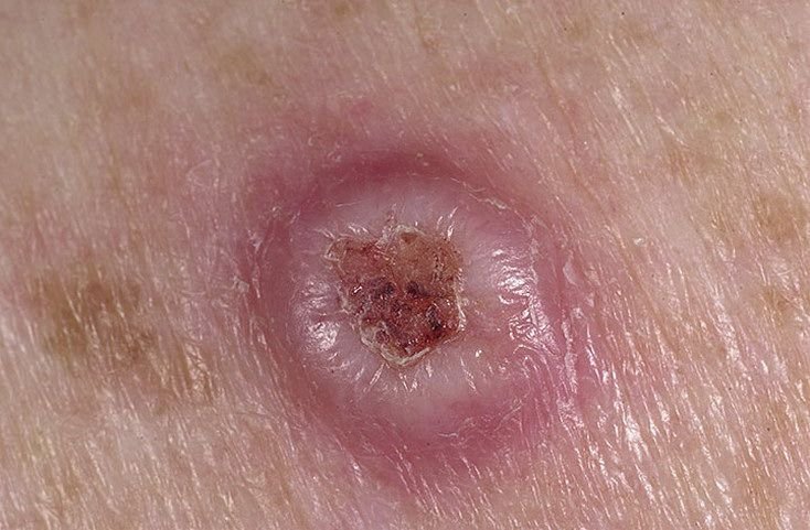
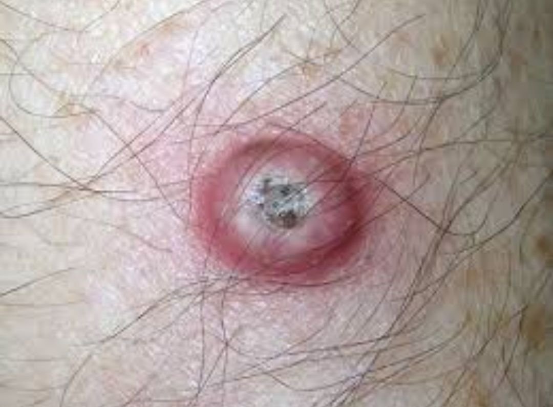
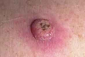
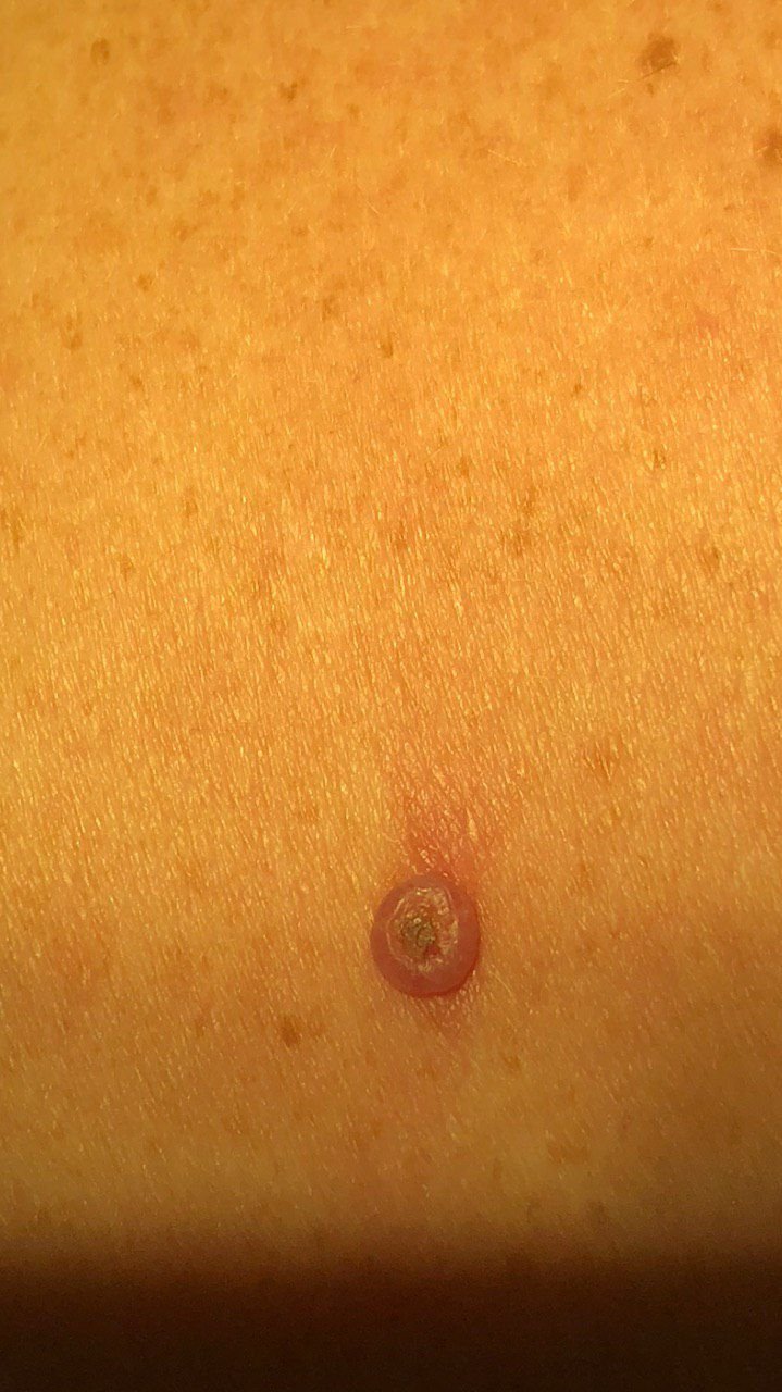
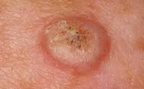
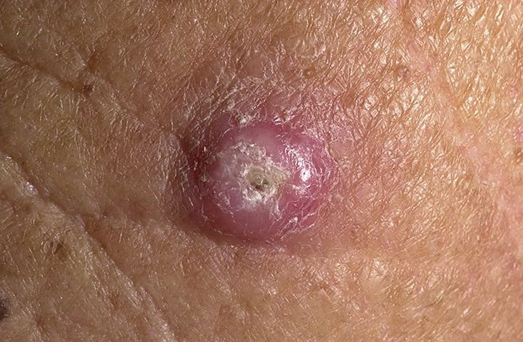
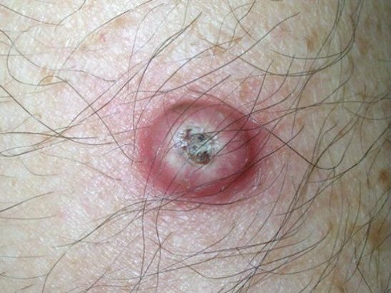
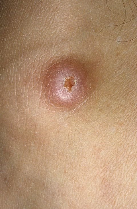
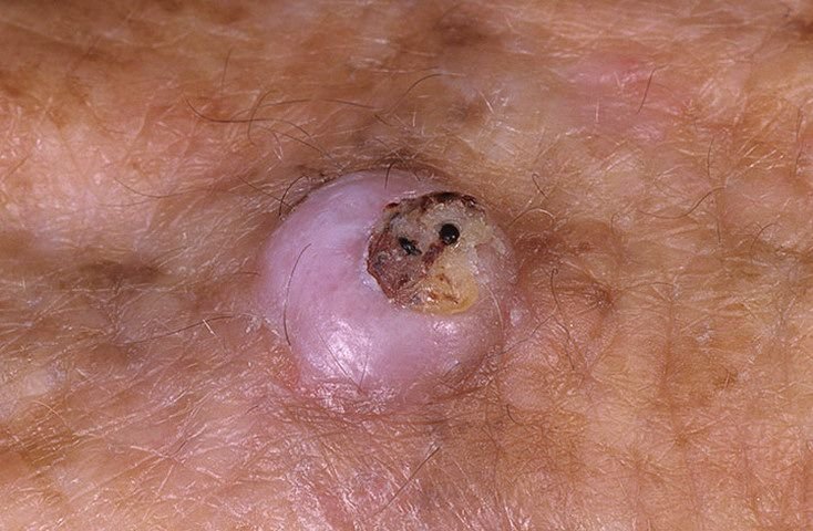
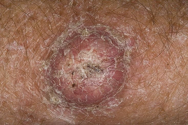
Keratoacanthoma often appears as a raised, dome-shaped lesion with a central crater or keratin-filled core. The peripheral surface is usually smooth and devoid of normal skin lines. In some cases, the center is ulcerated or crusted. The tumor may initially grow rapidly over weeks, reaching a size of 10–20 mm, after which growth typically slows. Lesions larger than 20 mm may develop bleeding or pain with minor trauma.
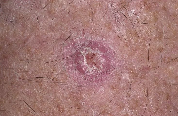
The borders of the lesion are usually symmetrical and regular, although some cases may show diffuse, ill-defined edges with surrounding erythema. The central area may appear grayish due to keratinization, while the periphery is often pink, red, or yellowish. Hair does not grow on the lesion’s surface. On palpation, the lesion feels firm yet mobile relative to deeper tissues. In smaller lesions, there are usually no subjective symptoms, but larger tumors may cause tenderness or discomfort.
Keratoacanthomas most frequently occur on sun-exposed areas of the body. Common sites include the forearms, hands, face, neck, back, and lower legs. Less commonly, lesions may arise on the chest, abdomen, or thighs.
Dermatoscopic Features
Dermatoscopic examination of keratoacanthoma may reveal the following features:
- Homogeneous Pink Peripheral Coloration: A uniform pink background in the periphery of the lesion.
- Whitish-White Annular Zone: Surrounding the central core, this ring-like zone reflects keratin-filled tissue.
- Central Keratin Plug: A hallmark of keratoacanthoma, seen as yellowish or grayish masses of keratin in the center.
- Blood Clots or Hemorrhagic Spots: Common in larger tumors or those subject to trauma.
- Peripheral Vascular Patterns: Linear, hairpin, or radially oriented vessels in the surrounding tissue.
Differential Diagnosis
Keratoacanthoma must be distinguished from several other dermatologic conditions, some of which are malignant. These include:
- Cutaneous horn
- Dermatofibroma
- Open comedone (blackhead)
- Seborrheic keratosis
- Bowen’s disease
- Squamous cell carcinoma
- Basal cell carcinoma
- Melanoma (especially amelanotic variants)
Risks and Prognosis
Although keratoacanthoma is generally benign and may undergo spontaneous regression, it is generally managed as a lesion that can be difficult to distinguish from squamous cell carcinoma. The risk of malignant transformation into squamous cell carcinoma is relatively low but increases in the presence of additional risk factors, such as chronic trauma, burns, or chemical exposure.
Furthermore, individuals with a history of keratoacanthoma may have an elevated risk of developing other skin cancers elsewhere on the body. This necessitates careful follow-up and regular skin examinations to ensure early detection and differentiation of new lesions.
Management and Clinical Strategy
Upon suspicion or confirmation of keratoacanthoma, referral to a dermatologist or oncologist is recommended. Due to its clinical resemblance to invasive carcinomas, histological confirmation is essential. Even with a confirmed benign diagnosis, surgical excision is generally recommended due to the potential for growth, discomfort, bleeding, and malignant transformation.
If surgery is declined, the patient must undergo active dynamic observation, including photographic documentation of the lesion to monitor for subtle changes. Monitoring is especially important for tumors exceeding 20 mm or those that exhibit new symptoms.
Routine skin examinations are advisable in the spring and autumn, particularly in individuals with a history of skin cancer or keratoacanthoma. Full-body skin mapping aids in long-term surveillance and allows for early detection of suspicious changes in skin morphology.
Treatment
The gold-standard treatment is surgical excision with a margin of healthy tissue, ensuring complete removal and minimizing recurrence risk. Excision should be full-thickness to capture the entire lesion.
Flat plane excision or superficial removal techniques are discouraged, as they increase the chance of recurrence. Similarly, methods such as laser ablation or cryodestruction are not recommended for keratoacanthoma due to poor histological control and a higher rate of local relapse.
Prevention
Preventive strategies aim to reduce the likelihood of developing keratoacanthoma and limit environmental and physical insults to the skin:
- Limit sun exposure and avoid artificial UV sources like tanning beds.
- Use broad-spectrum sunscreens and wear protective clothing during sun exposure.
- Prevent chronic trauma or mechanical irritation to the skin.
- Avoid occupational hazards involving ionizing radiation or toxic chemicals.
- Practice good skin hygiene and regularly inspect the skin for changes.
- Undergo periodic dermatological evaluations to detect potentially malignant lesions early.
Early identification and timely management are critical to minimizing complications and improving outcomes in patients with keratoacanthoma.