Dermatitis (ICD-10: L20) ⚠️
Contact Dermatitis: Inflammatory Skin Response to External Irritants and Allergens
Overview
Contact dermatitis is a term used to describe acute or chronic inflammation of the skin that arises at the site of direct interaction with physical, chemical, or biological agents. It is one of the most commonly diagnosed dermatologic conditions, accounting for up to 15–20% of all visits to dermatologists.
The term “dermatitis” derives from the Greek word for skin (“derma“) and the Latin suffix “-itis”, denoting inflammation. In modern clinical practice, the terms “dermatitis” and “eczema” are often used interchangeably. However, a general distinction is made: “dermatitis” is often used to describe acute skin reactions that resolve more quickly, while “eczema” may refer to chronic and relapsing inflammatory skin conditions.
Contact dermatitis is divided into several major types based on pathogenesis and duration:
- Acute contact dermatitis
- Simple (irritant)
- Allergic
- Chronic contact dermatitis
- Cumulative-toxic
- Allergic
Simple (Irritant) Contact Dermatitis
Simple or irritant contact dermatitis results from direct skin damage by an external irritant without involvement of the immune system. This is the most common form of contact dermatitis, responsible for approximately 80% of all cases.
Mechanism of Development
The severity of the reaction depends on the concentration and duration of contact with the irritant. It may result from a single exposure to a strong substance (e.g., acid or alkali), or from repeated exposure to milder irritants (e.g., water, soap, detergents, friction, cold air). The threshold for skin irritation varies individually and is often lower in those with atopic predisposition.
Common Irritants Include:
- Detergents and cleaning agents
- Alkalis and acids (industrial or domestic chemicals)
- Oils, solvents, resins
- Oxidizing and reducing agents
- Fiberglass, dust, wood particles
- Prolonged moisture exposure and wet-dry cycles (common on hands, diaper area, wounds)
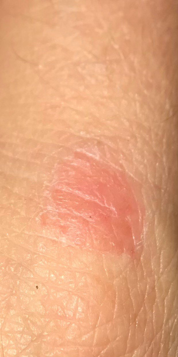
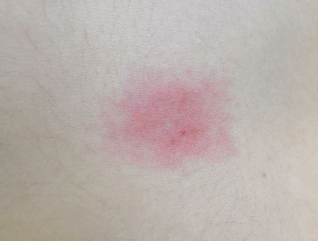
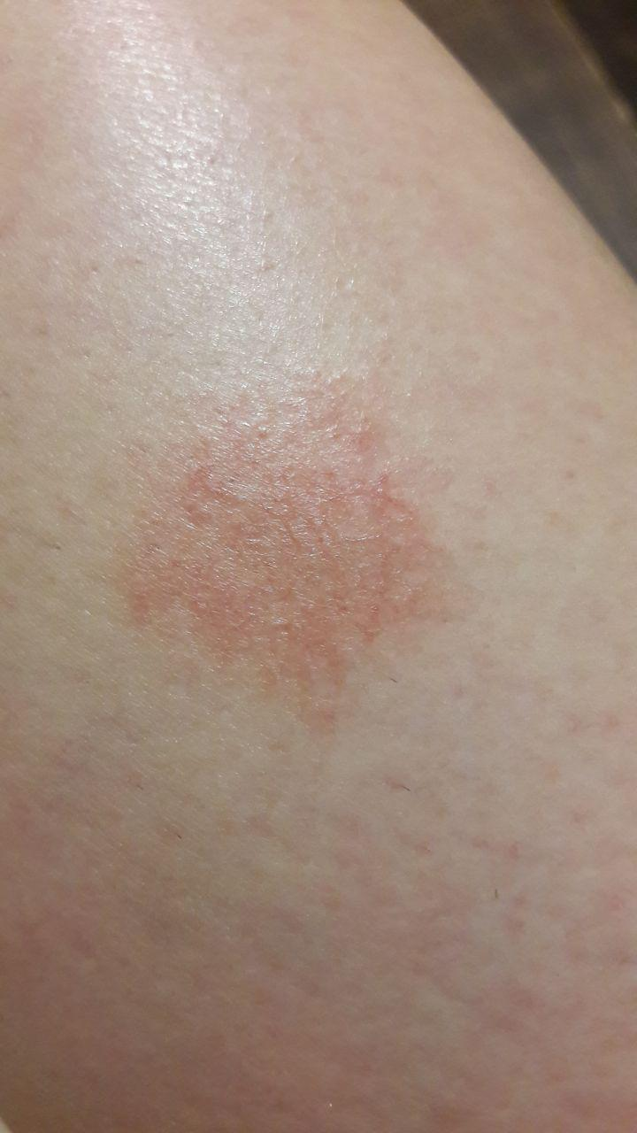
Symptoms
Clinical presentation varies based on exposure duration and irritant potency:
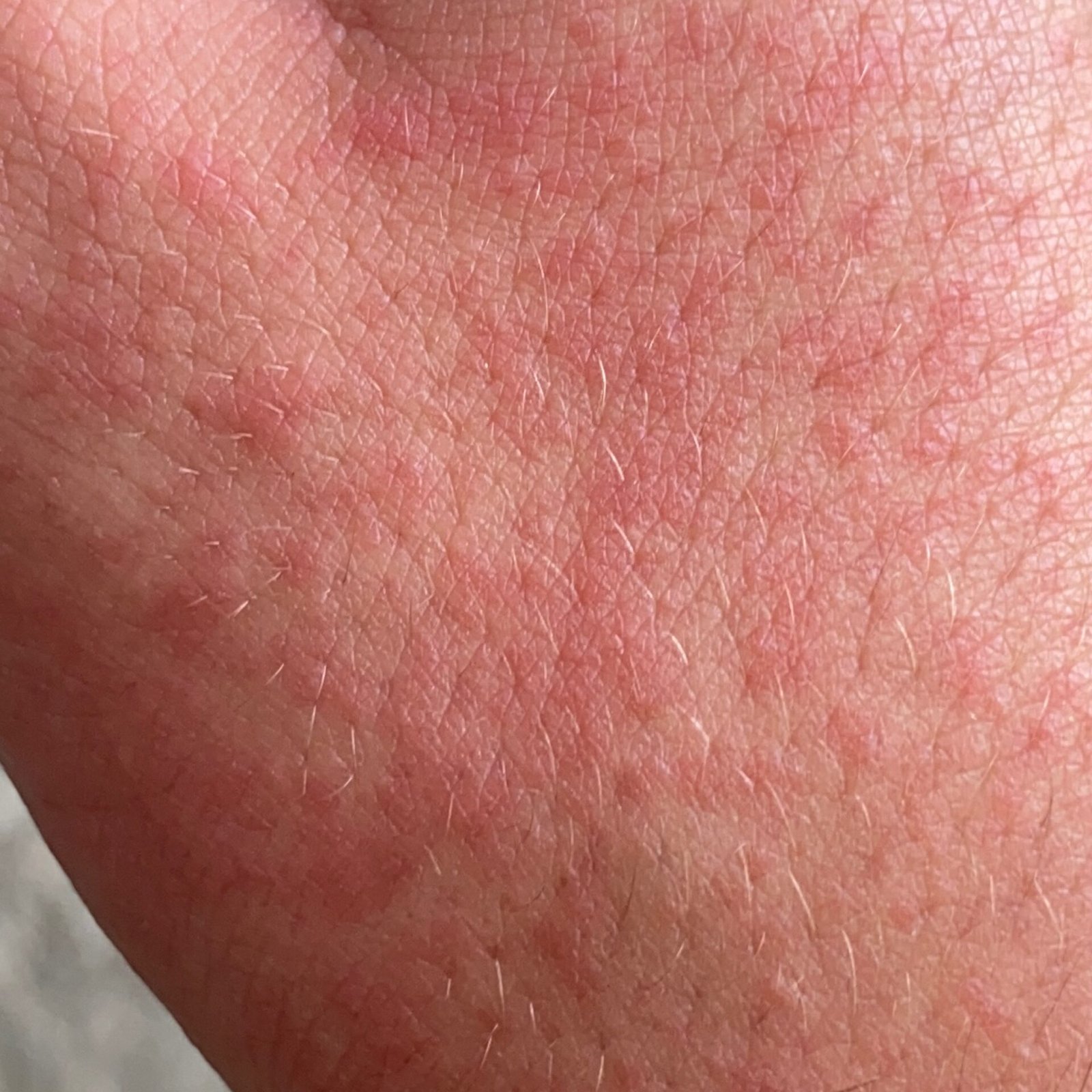
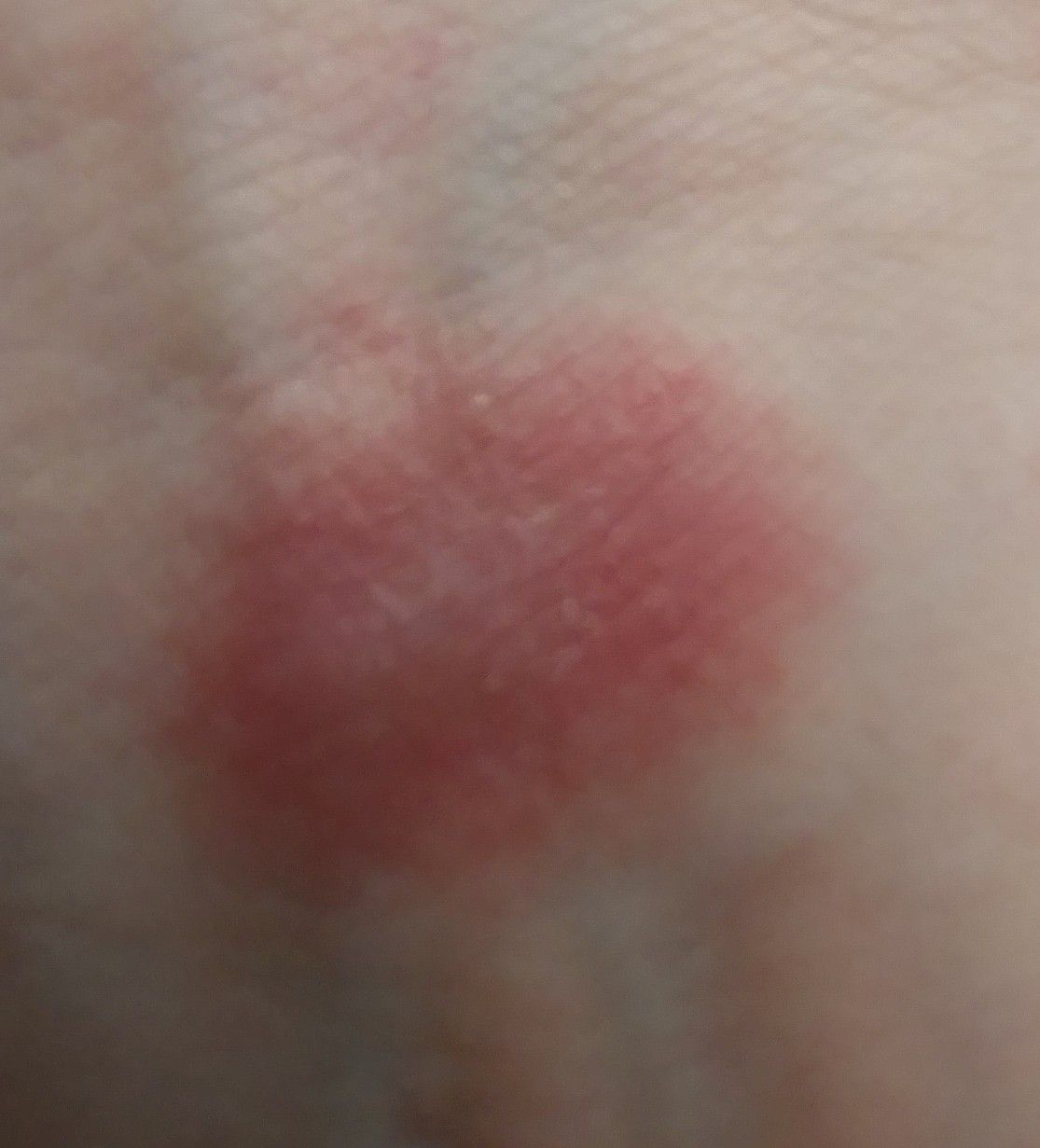
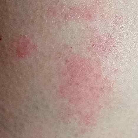
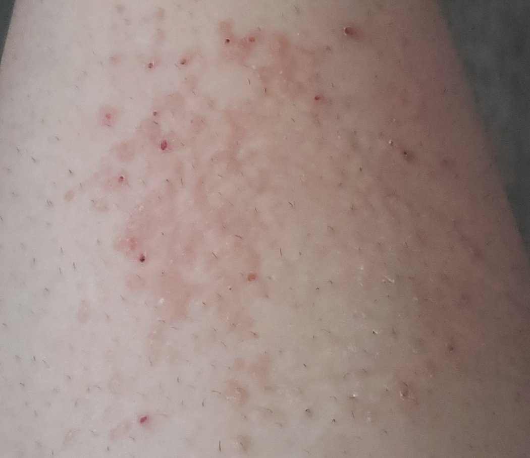
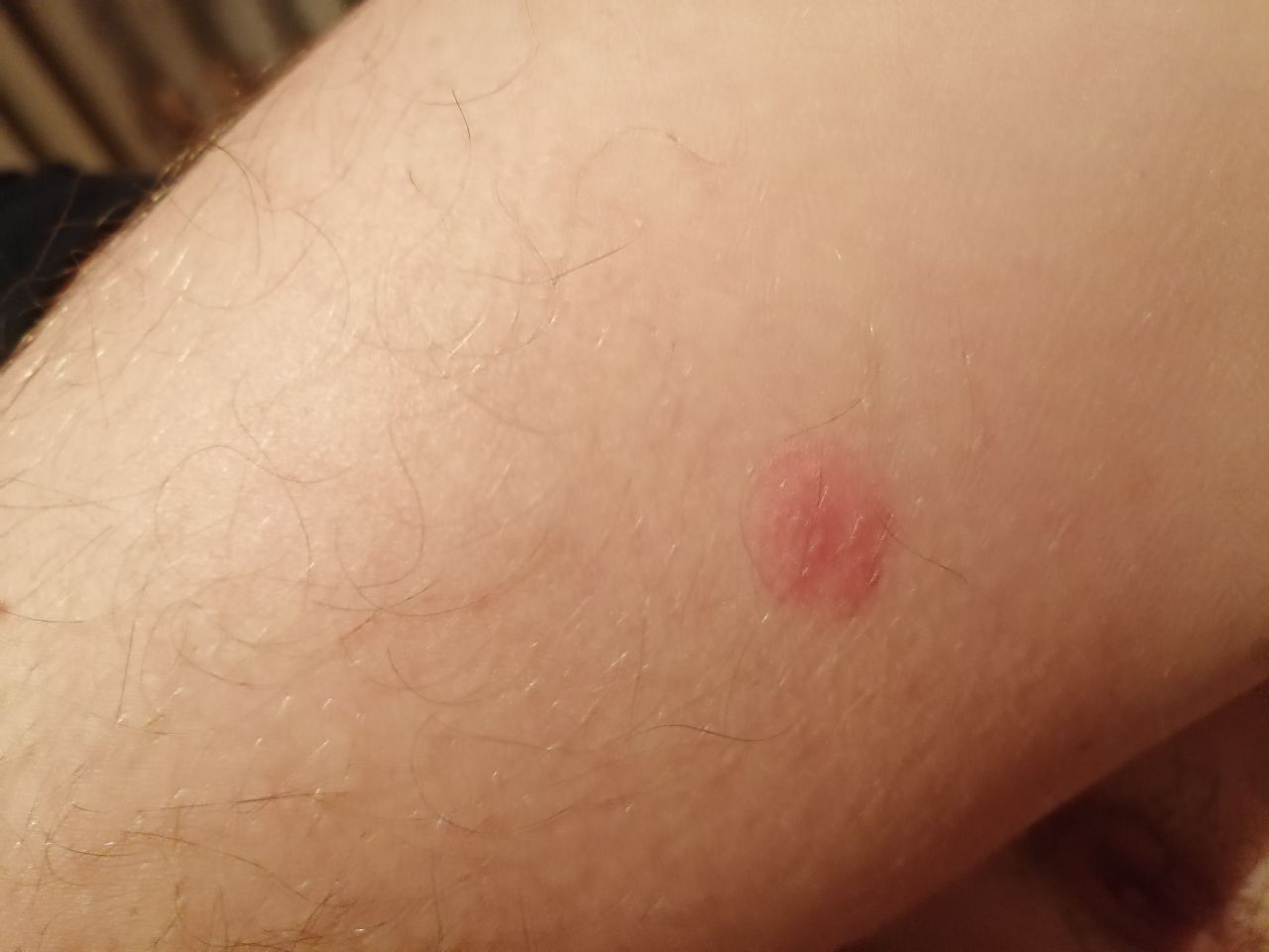
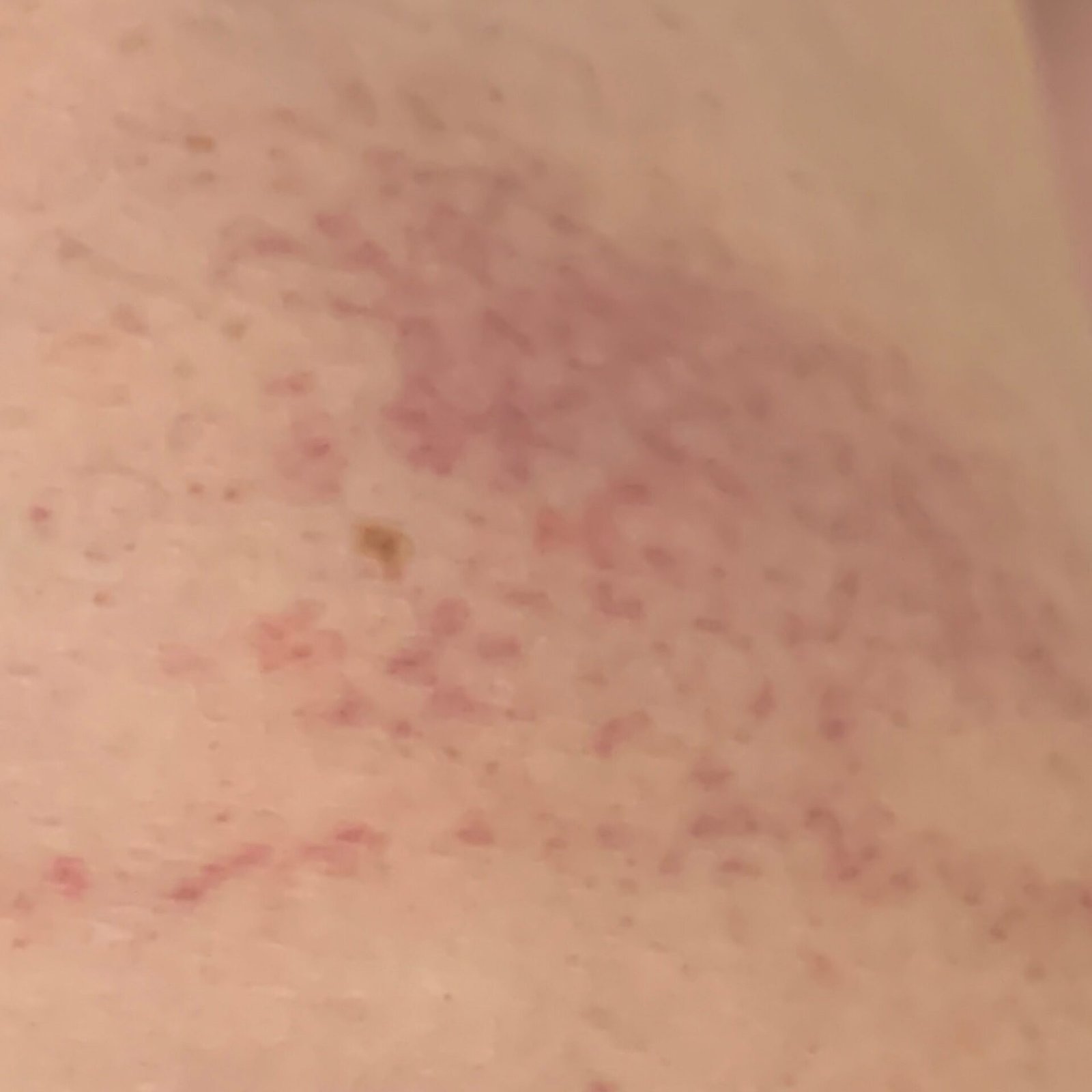
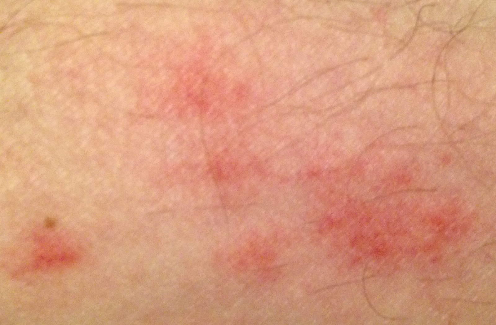
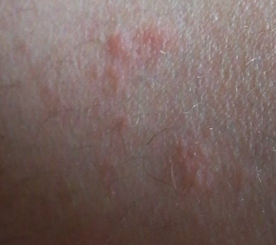
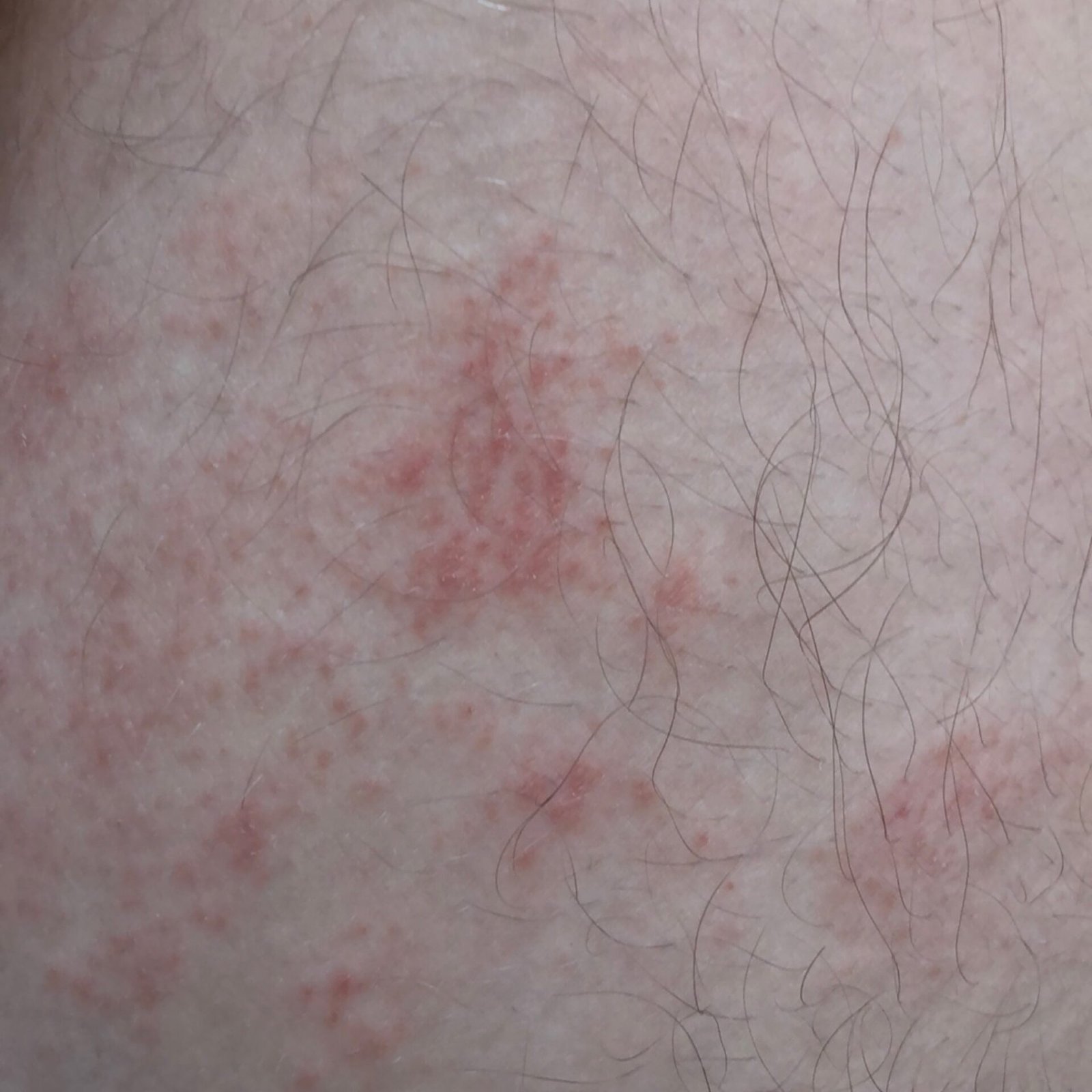
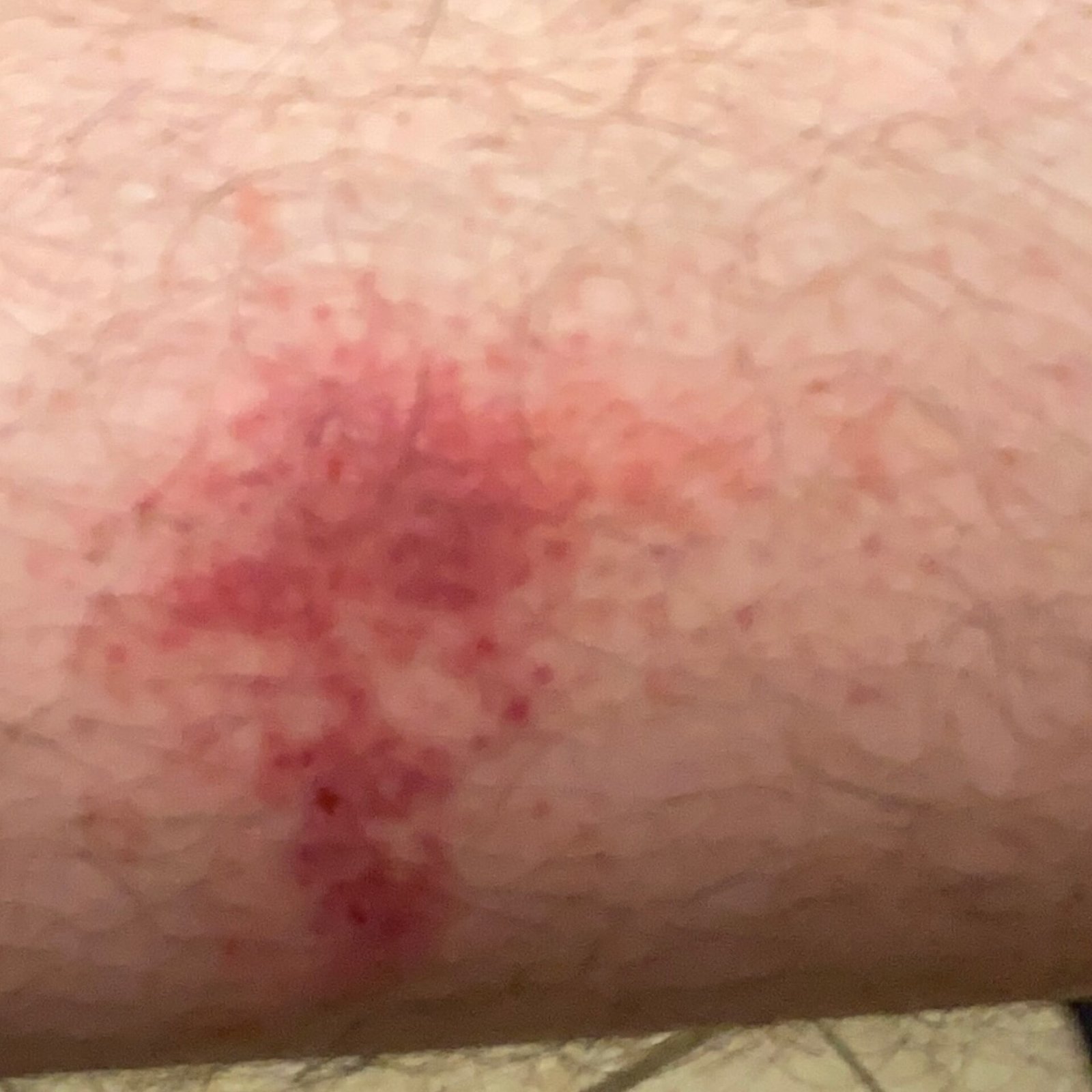
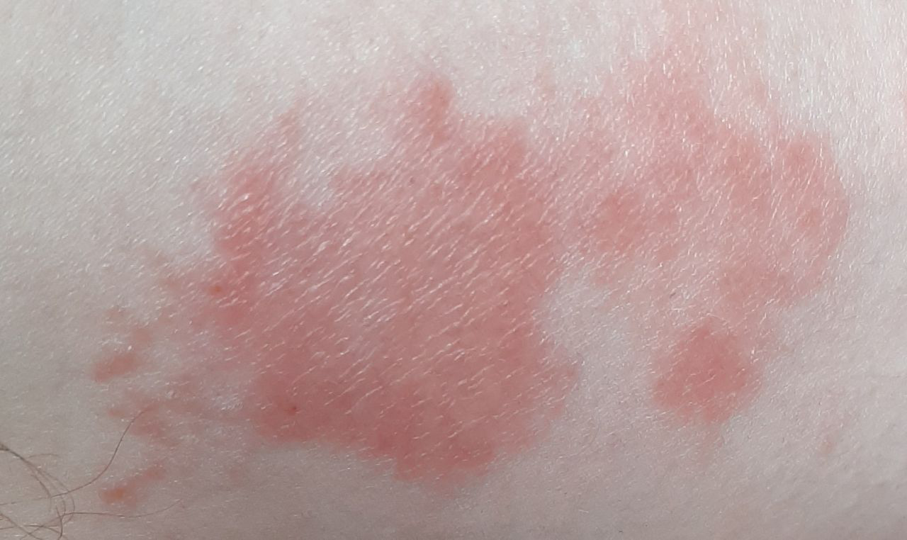
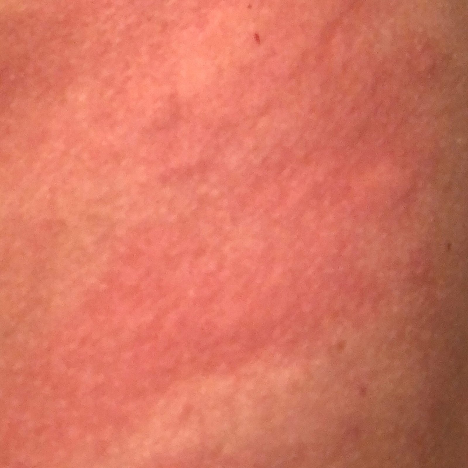
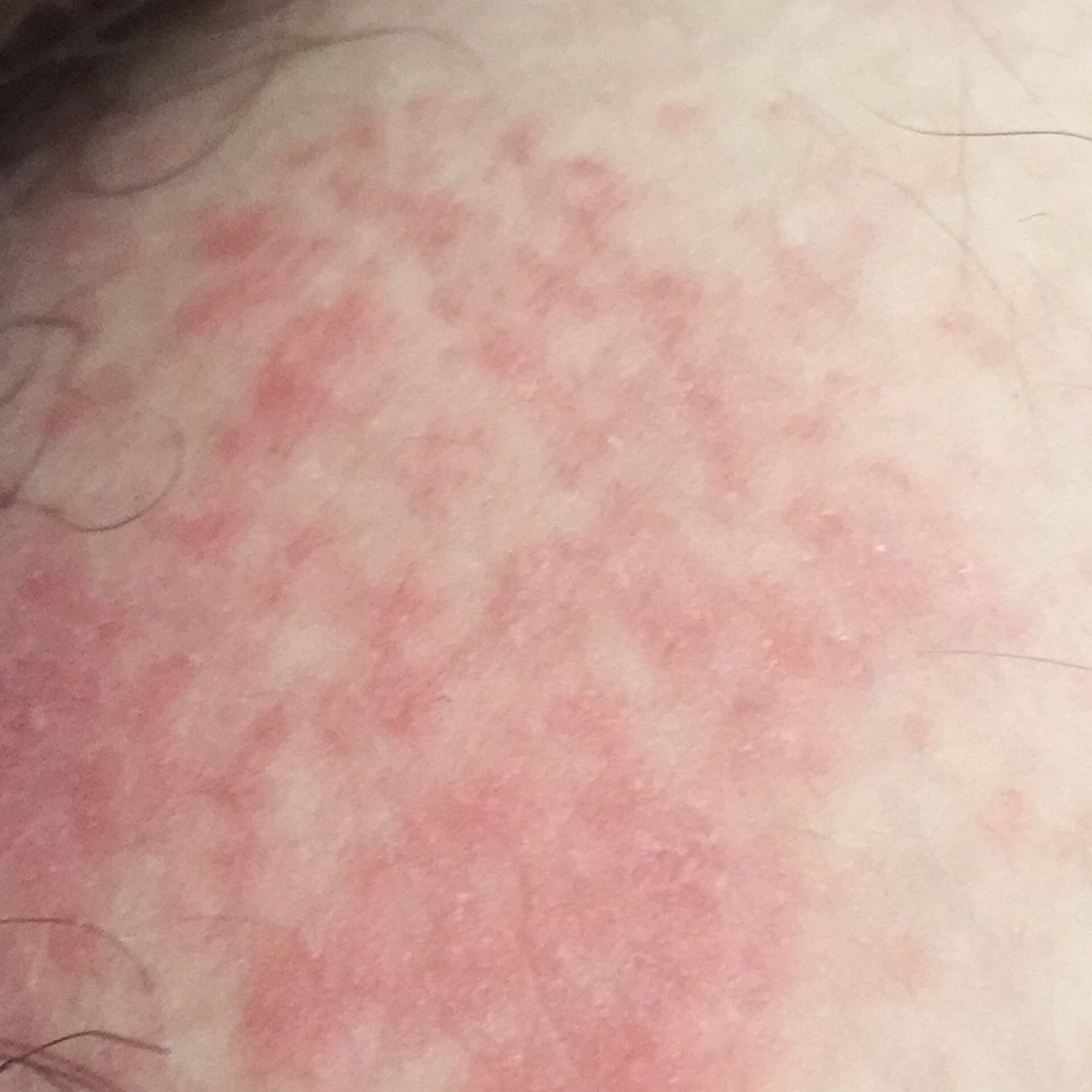
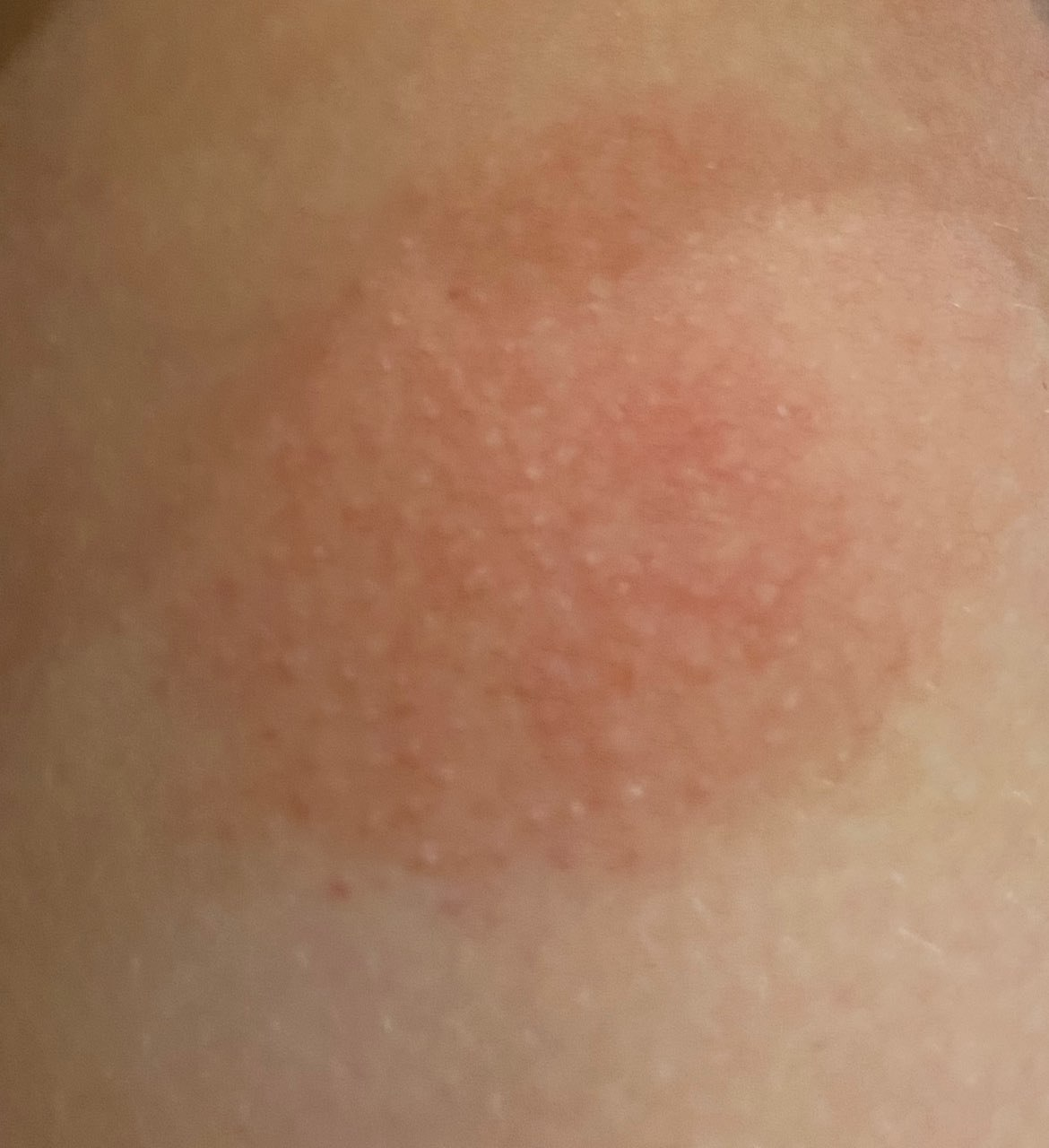
- Acute phase: Erythema (redness), edema, vesicles or papules, weeping (exudation), crust formation, burning sensation, soreness, occasional itching;
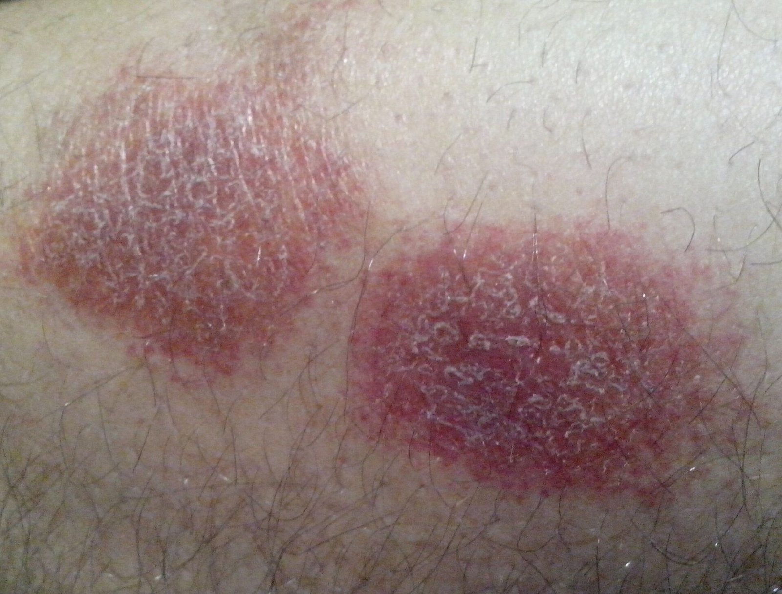
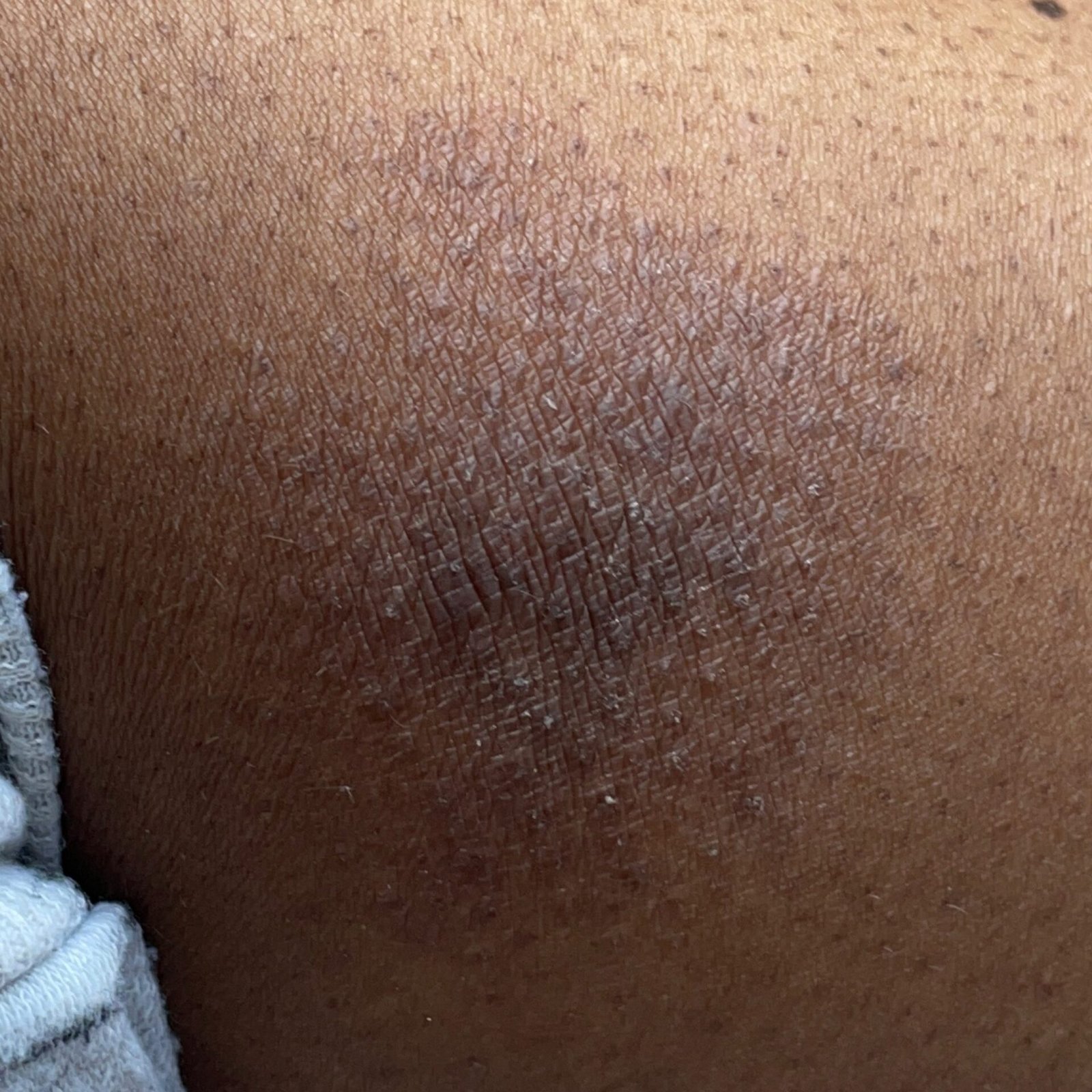
- Chronic phase: Lichenification (skin thickening), fissures, scaling, persistent erythematous plaques. Hyperkeratotic variants may occur in individuals with repetitive trauma (e.g., handling paper or tools).
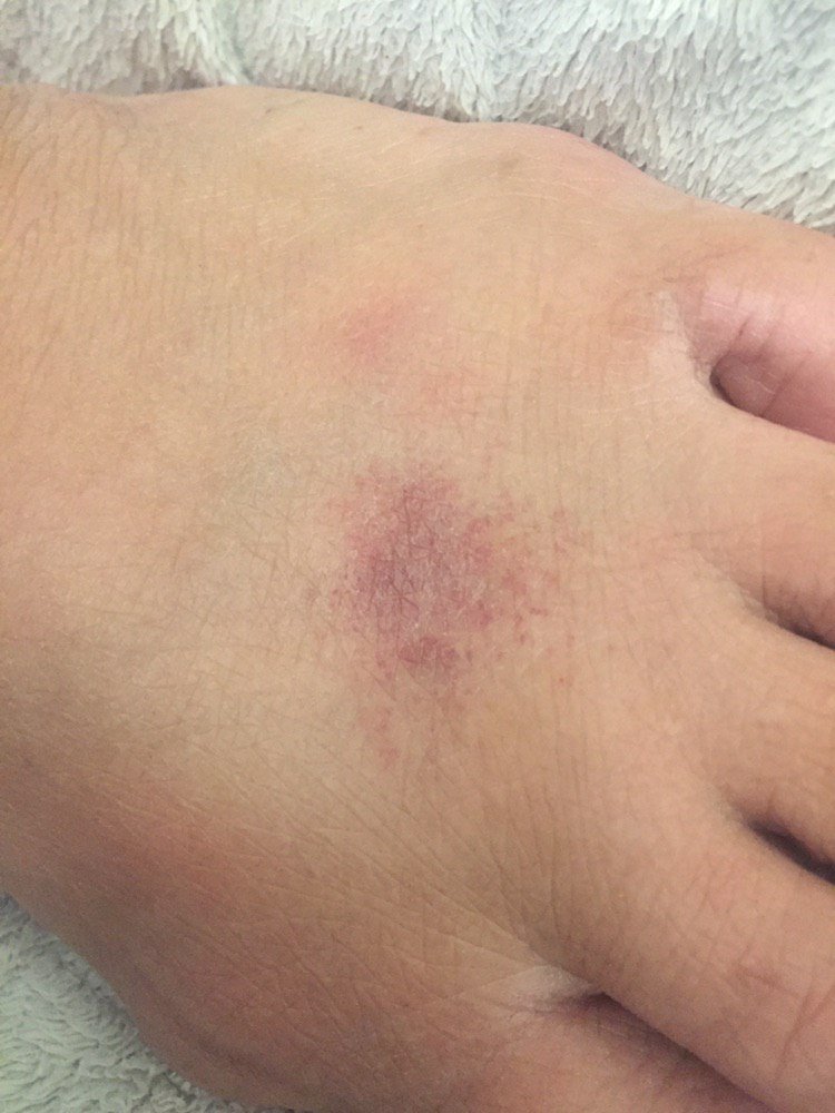
Common anatomical sites include:
- Dorsal and palmar surfaces of the hands and fingers
- Eyelids (from makeup, eye drops, airborne irritants)
- Lips (habitual licking causing wet-dry irritation)
Complications and Considerations
Low ambient humidity decreases the skin’s threshold for irritation, making it more susceptible to damage. Damaged skin also becomes more permeable to allergens, increasing the risk of developing secondary allergic contact dermatitis. Patients with an atopic background (e.g., hay fever, asthma, eczema) are more prone to severe and persistent forms.
Diagnosis
- KOH test: To exclude fungal infections in scaly or erythematous lesions;
- Patch testing: Used to exclude allergic contact dermatitis, especially in chronic, unresponsive cases or when history suggests allergen exposure;
- Skin biopsy: Rarely necessary, but may show epidermal spongiosis, dermal edema, and lymphocytic infiltrate.
Allergic Contact Dermatitis
Allergic contact dermatitis (ACD) is a delayed-type hypersensitivity reaction (Type IV), triggered by skin contact with a specific allergen. Unlike irritant dermatitis, ACD involves immune sensitization and develops after prior exposure to the allergen. Once sensitized, even minimal re-exposure may cause inflammation.
Common Allergens:
- Metals (nickel, chromium)
- Rubber additives (carbamates, thiurams, benzothiazoles)
- Cosmetics and preservatives (formaldehyde, fragrances, parabens)
- Topical medications (neomycin, bacitracin, corticosteroids)
- Hair dyes and nail products
- Plant allergens (e.g., poison ivy)
- Occupational chemicals and adhesives
Mechanism and Timeline:
- Initial sensitization takes 14–21 days after first contact;
- Inflammation in sensitized individuals develops 12–48 hours after re-exposure (range: 8–120 hours);
- Lesions may persist for up to 3 weeks after a single exposure;
- Photoallergic reactions require both allergen and sunlight to trigger inflammation;
- Systemic exposure to related allergens (e.g., oral medications) can trigger widespread eczema in sensitized patients.
Clinical Features:
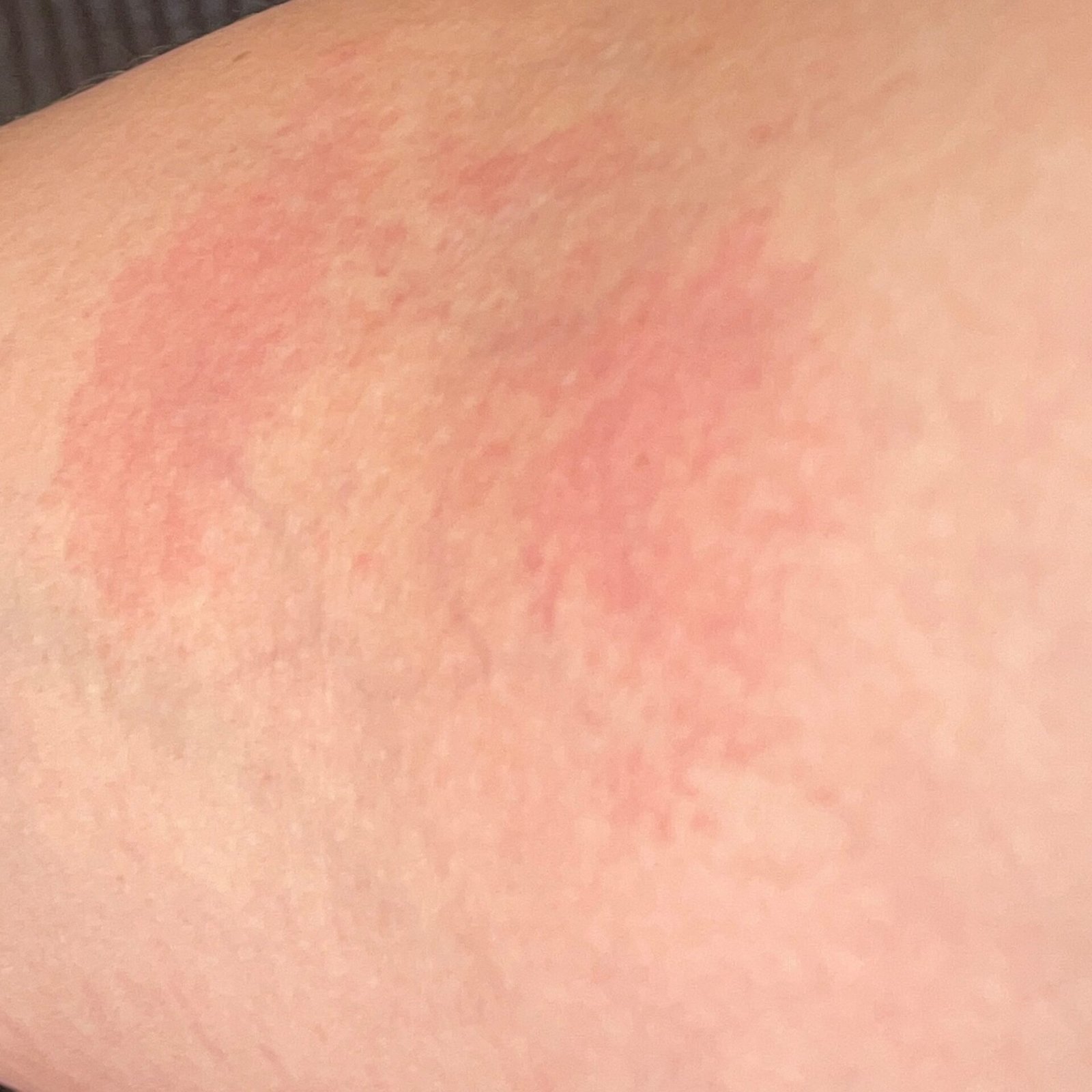
Subjective symptoms: Intense itching, sometimes burning or soreness.
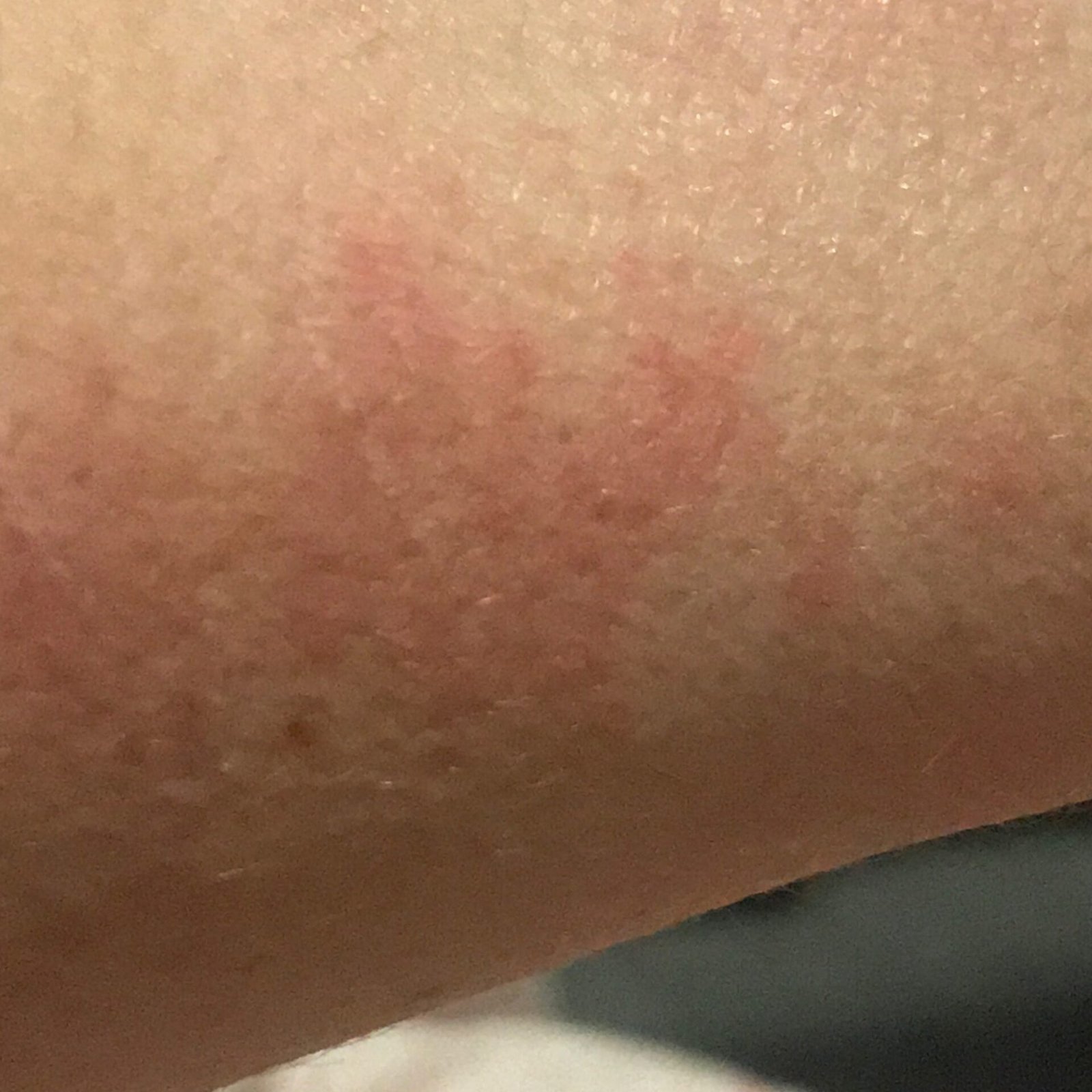
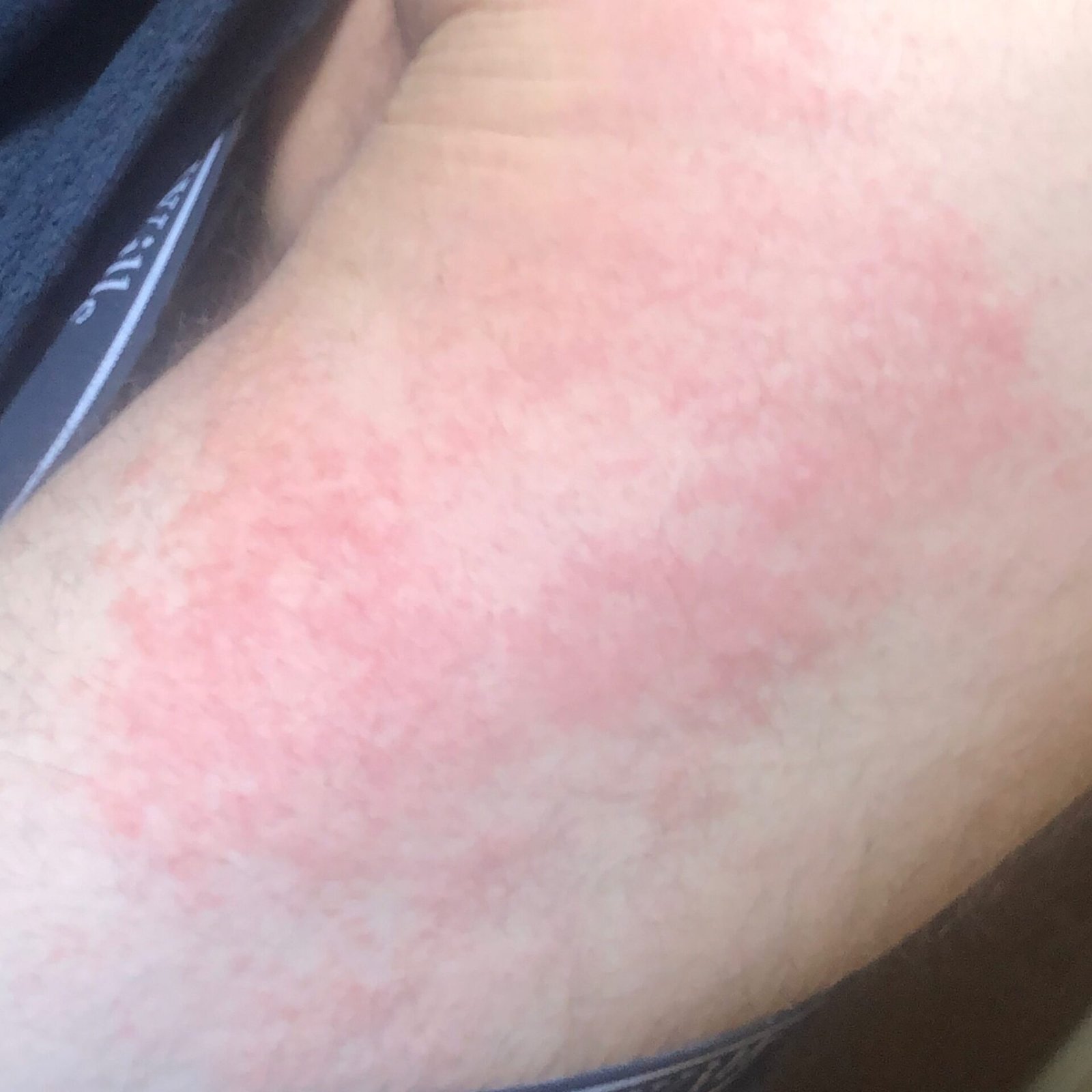
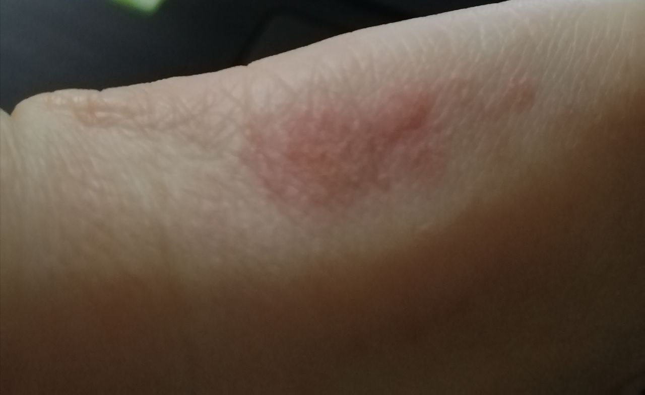
Objective signs: Vesicles, erythema, edema, scaling, crusting, in severe cases – bullae and erosions.
Common Sites:
- Hands, forearms, face, eyelids, lips
- Feet, genitals, scalp (depending on exposure source)
- Airborne allergens can affect exposed areas (e.g., face, neck)
- Photoallergic dermatitis often affects sun-exposed regions (sparing upper eyelids, under chin)
Diagnostics:
- Patch testing: Gold standard for allergen identification; panels include occupational, cosmetic, and drug-related allergens. Typically performed over 3 visits for application, intermediate reading, and delayed reaction assessment;
- Photopatch testing: For suspected photoallergens;
- Biopsy (if needed): Shows spongiosis, perivascular lymphocytic infiltrate, possible eosinophils.
Differential Diagnosis
- Irritant contact dermatitis: No allergen involvement; vesicles are rare and itching is often mild;
- Atopic dermatitis: Often coexists; distribution and chronicity help differentiate;
- Fungal infections: KOH microscopy helps exclude tinea corporis or candidiasis;
- Psoriasis: Sharply demarcated plaques with silvery scales;
- Rosacea or seborrheic dermatitis: Affect central face with less itching;
- Autoimmune and metabolic disorders: Rule out if systemic features or atypical distribution are present.
Treatment
General Principles:
- Avoidance of the trigger substance: Crucial for long-term control;
- Protective measures: Use appropriate gloves; note that some allergens (e.g., hair dye chemicals) may penetrate standard barriers;
- Discontinue all potential irritants and allergenic topical products;
Topical Therapy:
- Corticosteroid ointments: First-line treatment. Potency depends on location:
- Low potency for face and intertriginous areas
- Medium potency for limbs and trunk
- High potency for palms and soles
- Ointments are preferred over creams due to fewer sensitizing additives;
- Application: Twice daily for 2–3 weeks, followed by tapering;
- Topical calcineurin inhibitors: Tacrolimus or pimecrolimus for steroid-sensitive areas or long-term maintenance.
Systemic Therapy:
- Oral corticosteroids: Used for severe or generalized dermatitis, typically a tapering course over 2–3 weeks;
- Antihistamines: May help reduce itching;
- Immunosuppressive agents: For chronic refractory dermatitis (e.g., methotrexate, cyclosporine).
Prevention
Preventing contact dermatitis, especially allergic forms, involves identification and avoidance of sensitizers, coupled with protective skin care practices.
- Identify allergens through patch testing and eliminate contact when possible;
- Use protective clothing and gloves, especially in high-risk occupations;
- Moisturize regularly to strengthen the skin barrier;
- Avoid over-washing or exposure to harsh chemicals;
- Use fragrance-free, hypoallergenic skin care products and detergents;
- Educate patients on reading product ingredient lists and recognizing symptoms of flare-ups early.
With appropriate evaluation, allergen identification, skin care, and pharmacologic treatment, most cases of contact dermatitis can be effectively controlled, minimizing recurrences and improving quality of life.