Blue Nevus (ICD-10: D22) ⚠️
Blue Nevus
Blue Nevus (also known as the blue nevus of Jadassohn–Tièche, blue neuronevus, or dermal melanocytoma) is a benign skin growth that is primarily characterized by its distinct blue to dark blue coloration. This type of nevus typically appears during puberty, although it can manifest at any age, even at birth. Multiple blue nevi in one individual are rare. Statistically, blue nevi tend to occur more frequently in women compared to men.
Predisposing Factors
The exact cause of blue nevi remains unclear. However, several factors have been identified that may predispose individuals to develop these neoplasms. These factors may influence the likelihood of their appearance or growth:
- Genetic Factors: The development of blue nevi may be linked to an individual’s genetic background. In some cases, family history may play a role in their appearance.
- Ultraviolet Radiation: Exposure to both natural and artificial ultraviolet light can accelerate the multiplication of nevus cells (known as nevus melanocytes) and result in an excessive production of melanin. This pigment accumulates in the nevus, giving it its characteristic color.
- Hormonal Changes: Hormonal fluctuations in the body, especially those related to sex hormones, thyroid hormones, and adrenal hormones, can contribute to the formation of new nevi and the growth of existing ones.
- Ionizing Radiation, Viral Infections, and Physical Injuries: These environmental and health-related factors may also trigger the appearance or enlargement of blue nevi.
Diagnostics
The diagnosis of blue nevi typically involves a clinical examination, which includes a thorough visual inspection of the skin formation as well as dermatoscopy. If there are concerns about the possibility of malignant transformation, a biopsy may be performed for further analysis.
Symptoms
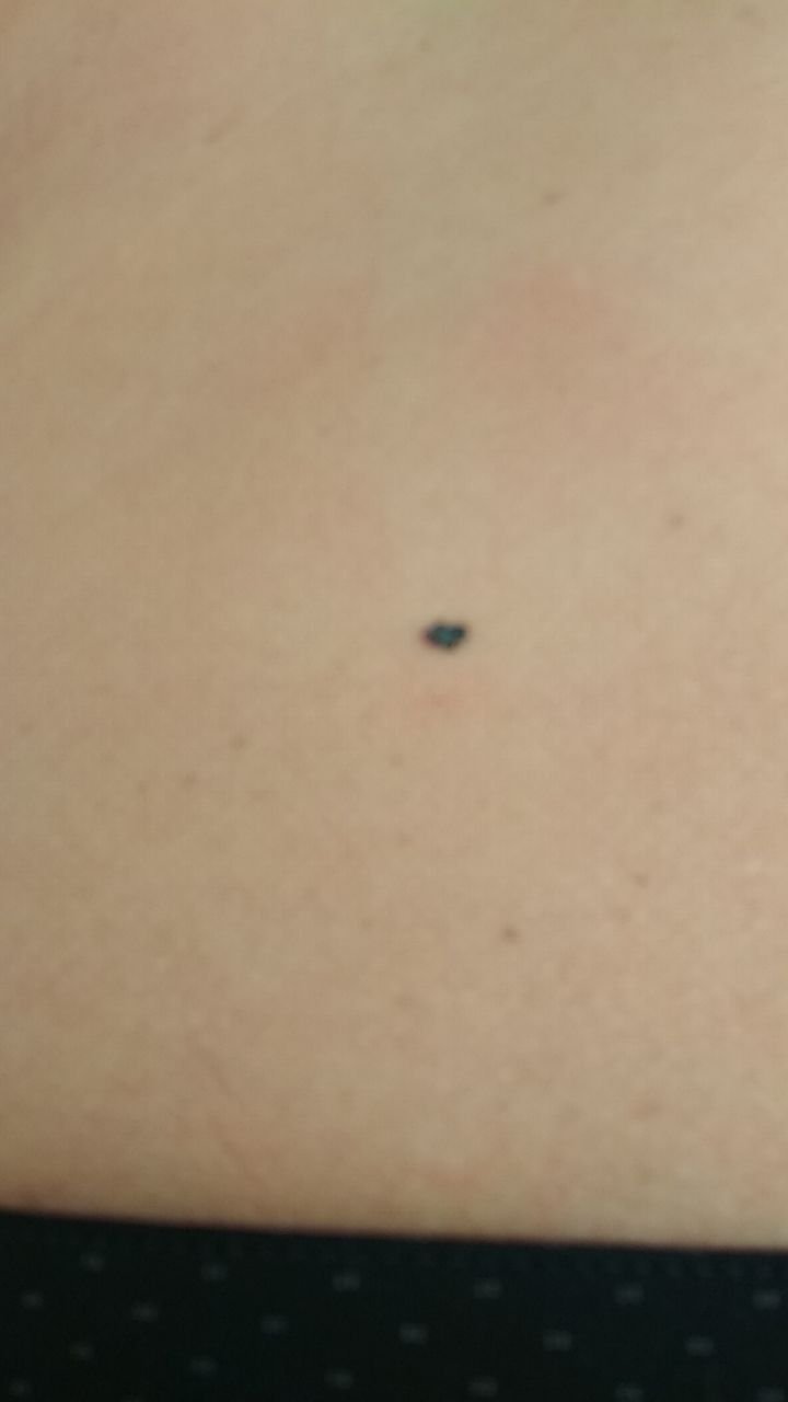
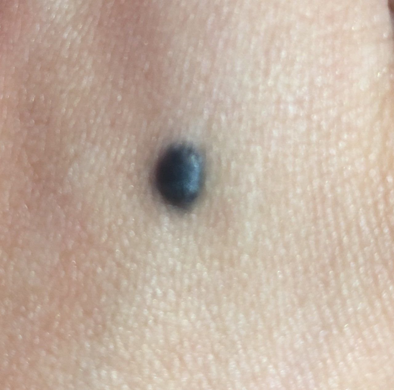
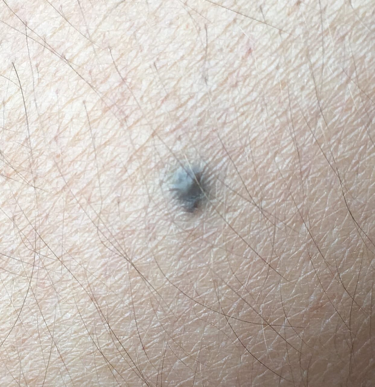
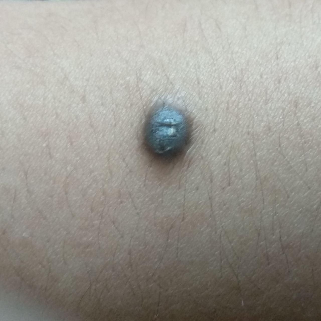
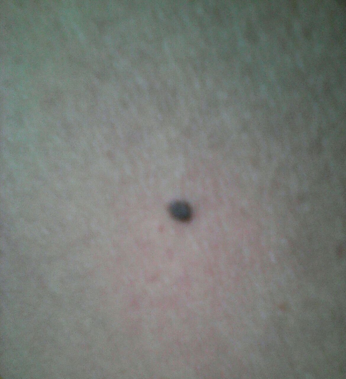
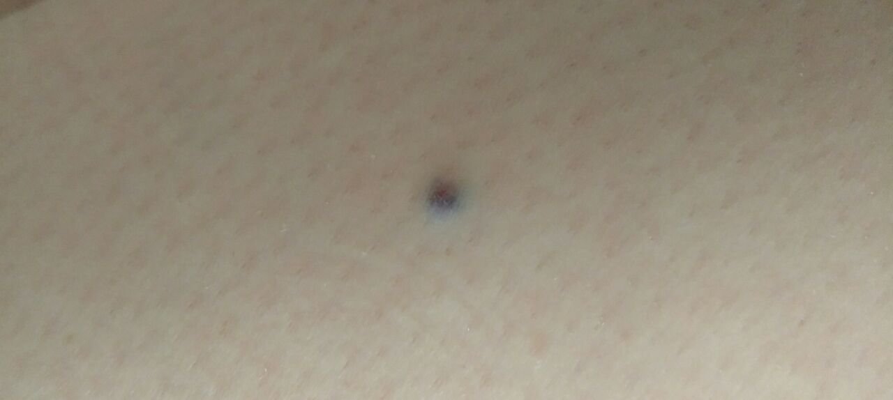
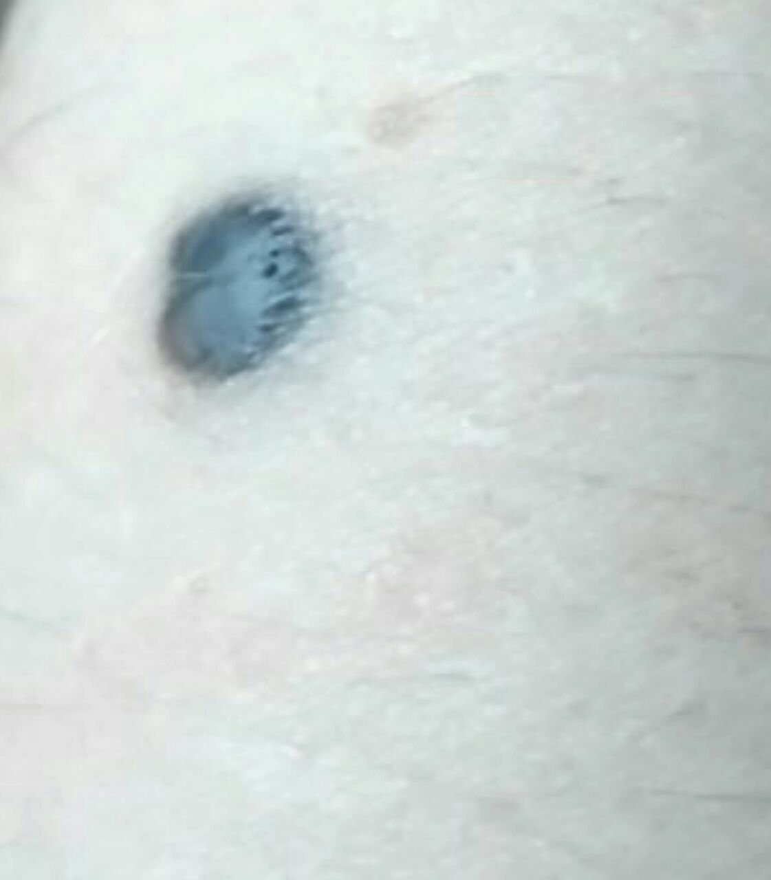
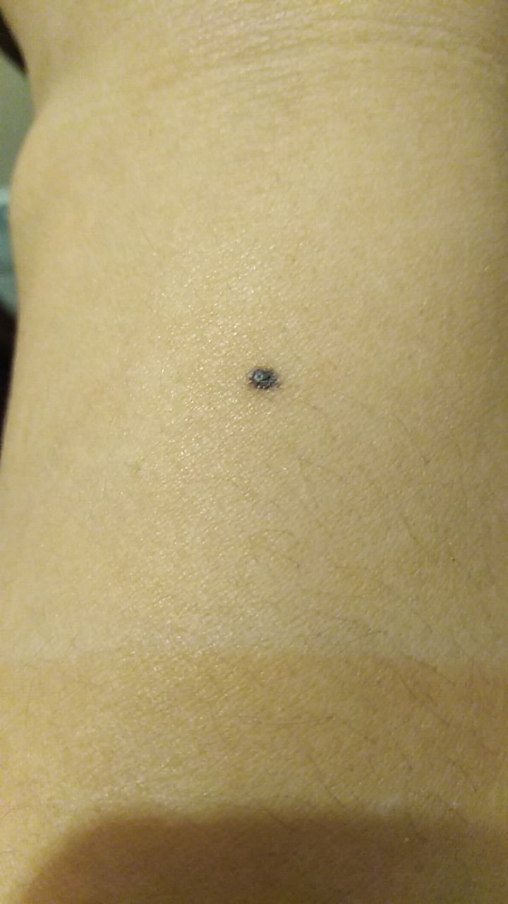
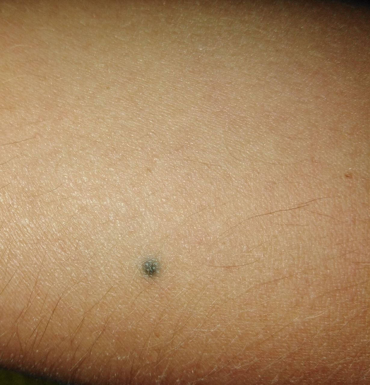
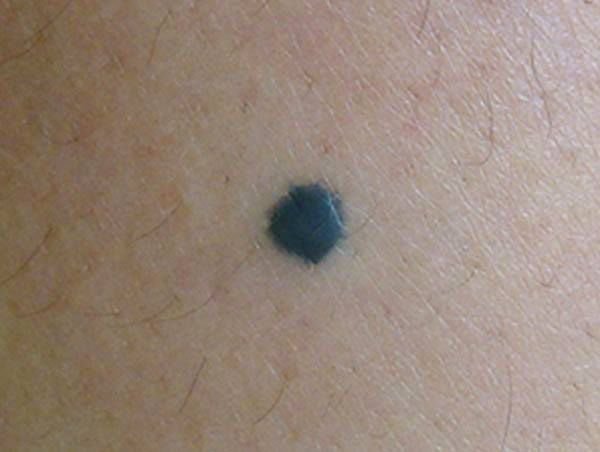
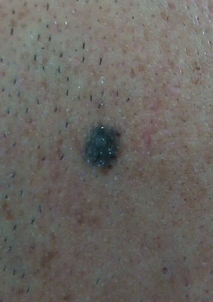
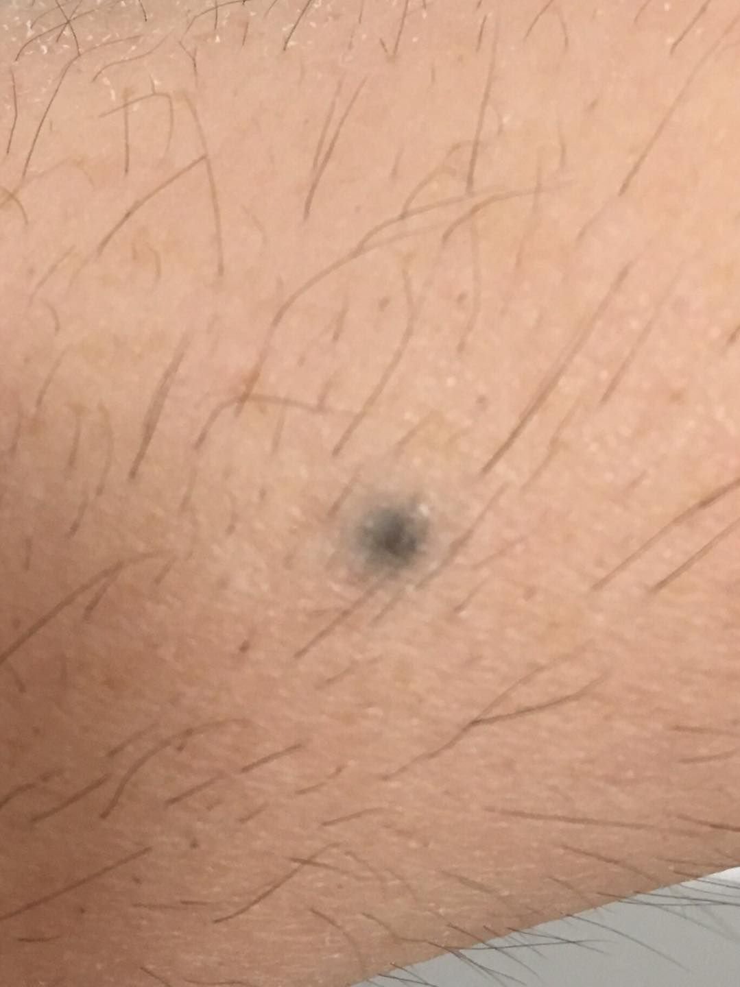
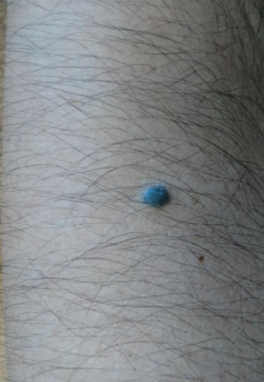
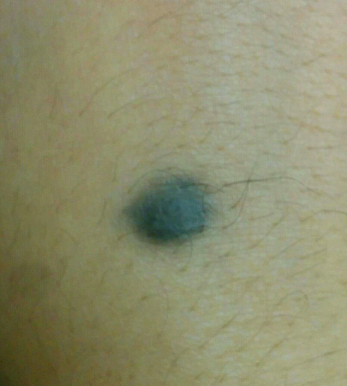
Visually, a blue nevus appears as a small spot or a slightly elevated nodule on the skin. It is typically symmetrical, with shapes ranging from oval or round to spindle-like. The surface texture of the nevus is usually similar to that of the surrounding skin, which may be smooth or even glossy, especially in smaller nevi. In larger nevi, over 10 mm in diameter, the surface may appear slightly more textured or tuberous.
The edges of the blue nevus are generally blurred or ill-defined, although they are usually smooth. In some cases, large blue nevi may have uneven or jagged edges, which could be an indication of potential malignancy. The color of the nevus ranges from blue to dark blue, with shades of gray-blue or blue-brown occasionally visible, reflecting the depth at which the pigment is deposited in the dermis. The intensity of the color tends to fade from the center towards the periphery, and in larger nevi, color heterogeneity or spotting may be present, resulting in a polychromatic appearance.
Hair is typically absent in blue nevi, although in some cases, coarse, dark hair may grow around the edges of larger nevi, particularly in congenital forms.
Blue nevi generally do not exceed 10 mm in size, and their growth is typically slow. Nevi larger than 1 cm are quite rare and are referred to as “blue cell nevi.” On palpation, these nevi feel similar to normal skin but may be slightly firmer, especially when they protrude above the skin’s surface. There are no subjective symptoms or sensations associated with these nevi.
Blue nevi most commonly occur on the dorsal surfaces of the hands and feet, the scalp, and the buttocks.
Dermatoscopic Description
Upon dermatoscopic examination, the following features are commonly observed in a blue nevus:
- Symmetry: The nevus tends to maintain a symmetrical appearance.
- Undefined Boundaries: The edges of the nevus are usually not sharply defined but remain relatively smooth.
- Gradual Color Transition: The color intensity diminishes progressively from the center of the nevus to the edges.
- Homogeneous Pigmentation: The pigmentation appears uniformly gray-blue in color. This is attributed to the presence of melanocytes in both the papillary (gray) and reticular (blue) layers of the dermis.
- Polychromatic Features: While less common, some blue nevi may exhibit color heterogeneity, including varying shades and the presence of vessels or globules within the pigmentation.
Differential Diagnosis
Blue nevi need to be distinguished from several other pigmented neoplasms, including:
- Post-inflammatory hyperpigmentation
- Congenital dermal melanocytosis
- Common pigmented nevi (simple or papillomatous)
- Hemangiomas
- Spitz nevi
- Dysplastic nevi
- Melanoma
Risks
In most cases, blue nevi are considered to be benign and pose no immediate risk. However, compared to common pigmented nevi, blue nevi do carry a slightly higher risk of transforming into melanoma. The risk of melanoma arising in a blue nevus is low. Signs that may indicate potential malignancy include changes in the appearance of the nevus or the development of new sensations such as itching or pain.
Melanoma arising in a blue nevus is rare.
Multiple or large congenital blue nevi may also be associated with certain genetic conditions or syndromes, making it essential for these individuals to receive thorough and ongoing medical evaluation.
Tactics
For blue nevi that show no signs of damage, changes in appearance, or new sensations, self-monitoring is typically sufficient. This should include periodic checks at least once a year, with assistance from others for areas that are difficult to inspect directly. If the nevus experiences mechanical damage, excessive exposure to ultraviolet or ionizing radiation, or noticeable changes in size or sensation, it is essential to seek medical attention from a dermatologist or oncologist.
A healthcare provider will assess whether continuous monitoring is required or if the nevus should be surgically removed. Nevi that are subjected to repeated physical trauma from clothing, jewelry, or occupational activities should be considered for removal.
Photographic documentation of the nevus can provide valuable records, allowing for the detection of even minor changes over time. For individuals with multiple blue nevi, it is highly recommended to create a map of their skin neoplasms, which will simplify future observations and the identification of any new or altered formations.
Patients with blue nevi should consult a dermatologist or oncologist at least twice a year (typically before and after the summer months) to assess any changes in the nevi’s appearance. Regular mapping of skin neoplasms can aid in tracking any developments or changes in existing lesions.
Treatment
The recommended treatment for blue nevi is surgical excision, typically using classic, electric, or radio scalpels. After removal, a histological examination is required to confirm the benign nature of the lesion.
Destructive treatments such as laser removal or cryotherapy are not advised for blue nevi due to potential risks and complications.
Prevention
The prevention of nevi formation and their potential for malignant transformation involves careful and considerate skin care:
- Limiting exposure to ultraviolet radiation, including avoiding tanning beds and excessive sun exposure.
- Using sunscreen and protective clothing during periods of peak sunlight.
- Avoiding chronic skin trauma from clothing, jewelry, or other external factors.
- Minimizing exposure to ionizing radiation and occupational hazards.
- Adhering to safety protocols when handling skin-damaging agents.
- Maintaining good personal hygiene and staying vigilant for any changes in skin health.
It is crucial to have regular exams of blue nevi, seek timely consultation with a specialist if any changes occur, and remove potentially harmful neoplasms as soon as possible to maintain skin health and prevent complications.