Papilloma (ICD-10: D23) 💚
Skin Papilloma (Viral Papilloma, Filiform Wart)
Skin Papilloma, also referred to as viral papilloma or filiform wart, is a benign neoplasm that rises above the surface of the skin. Viral papillomas typically begin to appear during adolescence, and as individuals age, these lesions tend to become more numerous. This type of neoplasm is characterized by its multiplicity, and the frequency of occurrence increases with age. Both congenital and acquired papillomas can be found, although in some cases, the viral etiology is absent.
Predisposing Factors
Many cutaneous papillomas and warts are associated with the human papillomavirus (HPV), though some benign papillomatous lesions are non-viral, which is generally associated with a low oncogenic risk. However, given that nearly 90% of the population carries the HPV virus but not everyone develops papillomas, it is evident that other factors contribute to the occurrence of these lesions on the skin. The following factors are known to increase the likelihood of developing papillomas:
- Immunodeficiency States: Weakened immune systems, whether due to conditions like HIV or immunosuppressive treatments, can predispose individuals to develop papillomas.
- Overweight: Being overweight or obese has been associated with an increased risk of developing skin lesions, including papillomas.
- Metabolic Disorders: Conditions such as diabetes mellitus and other metabolic issues may also increase the likelihood of papillomas appearing.
- Severe Infectious Diseases: Infections that compromise the immune system may trigger the development of papillomas, as the body becomes more susceptible to HPV.
- Poor Personal Hygiene: Inadequate hygiene can increase the risk of HPV transmission and the development of papillomas.
- Pregnancy: Hormonal changes during pregnancy can make the body more susceptible to developing papillomas, especially when immune defenses are temporarily weakened.
- Stress, Overwork, and Malnutrition: Any factors that decrease the body’s ability to fight infections or affect immune function can contribute to the development of papillomas.
- Chronic Skin Lesions: Areas of skin that are frequently injured, irritated, or damaged can serve as entry points for the HPV virus.
Diagnostics
The diagnosis of papillomas is based on a clinical examination, which includes a routine visual inspection of the lesions followed by dermatoscopy to examine the structure of the growths. In some cases, laboratory tests can be performed to detect HPV. If there is a concern that the papilloma may be malignant, a biopsy (excision biopsy) can be performed to confirm the diagnosis and rule out other conditions.
Symptoms
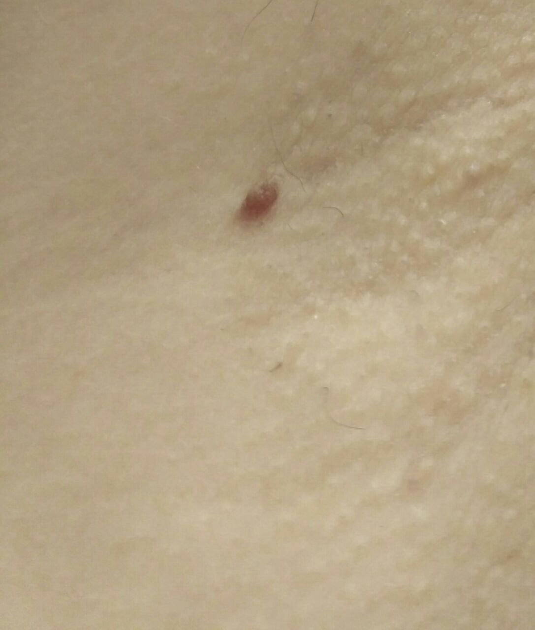
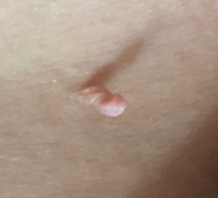
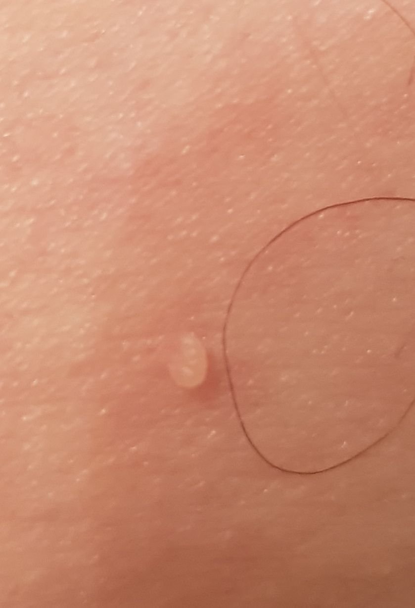
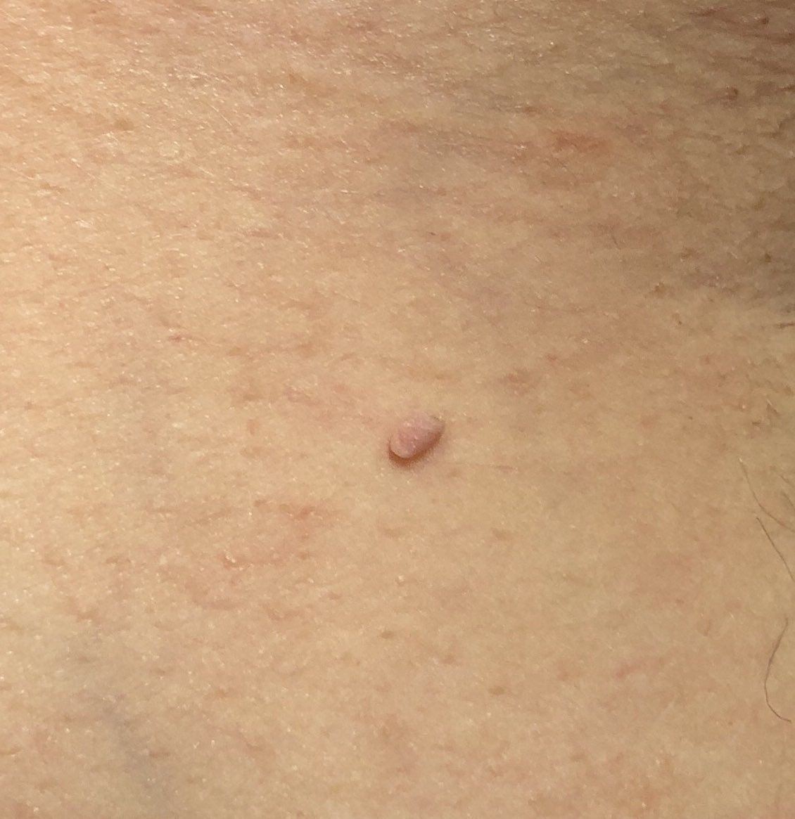
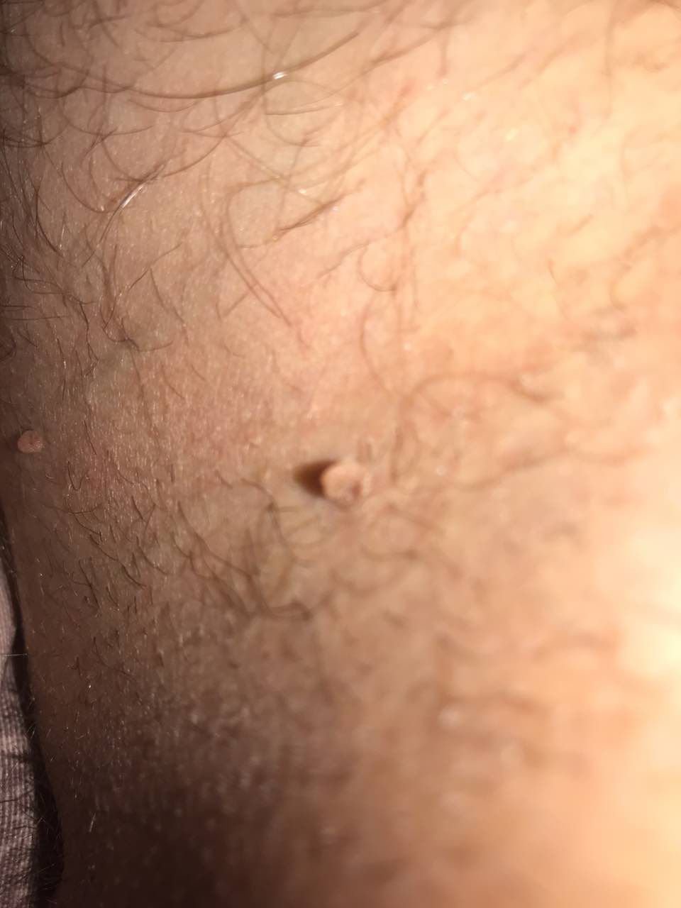
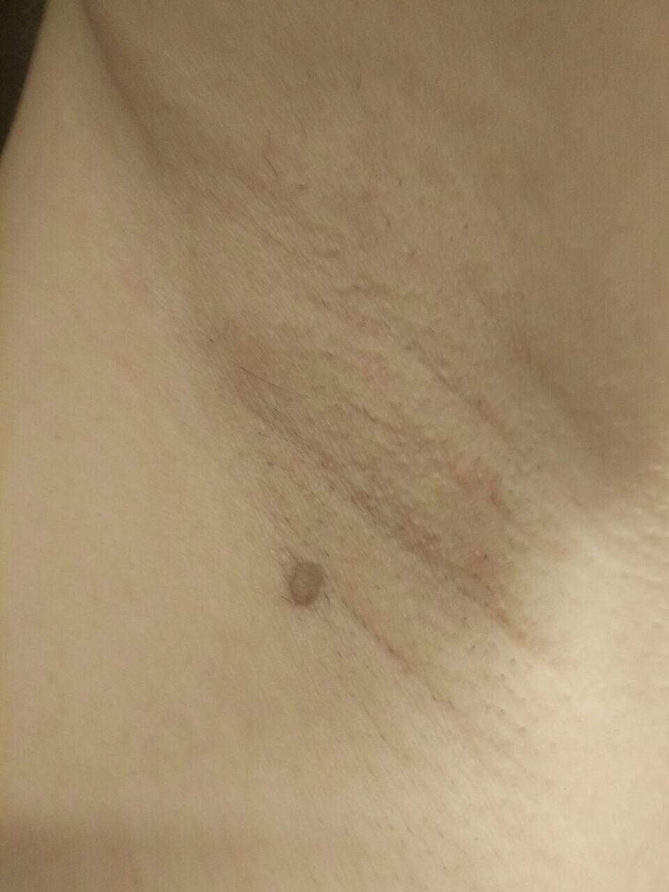
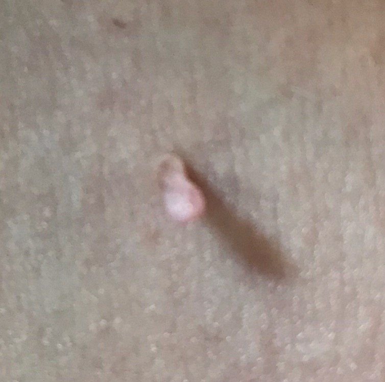
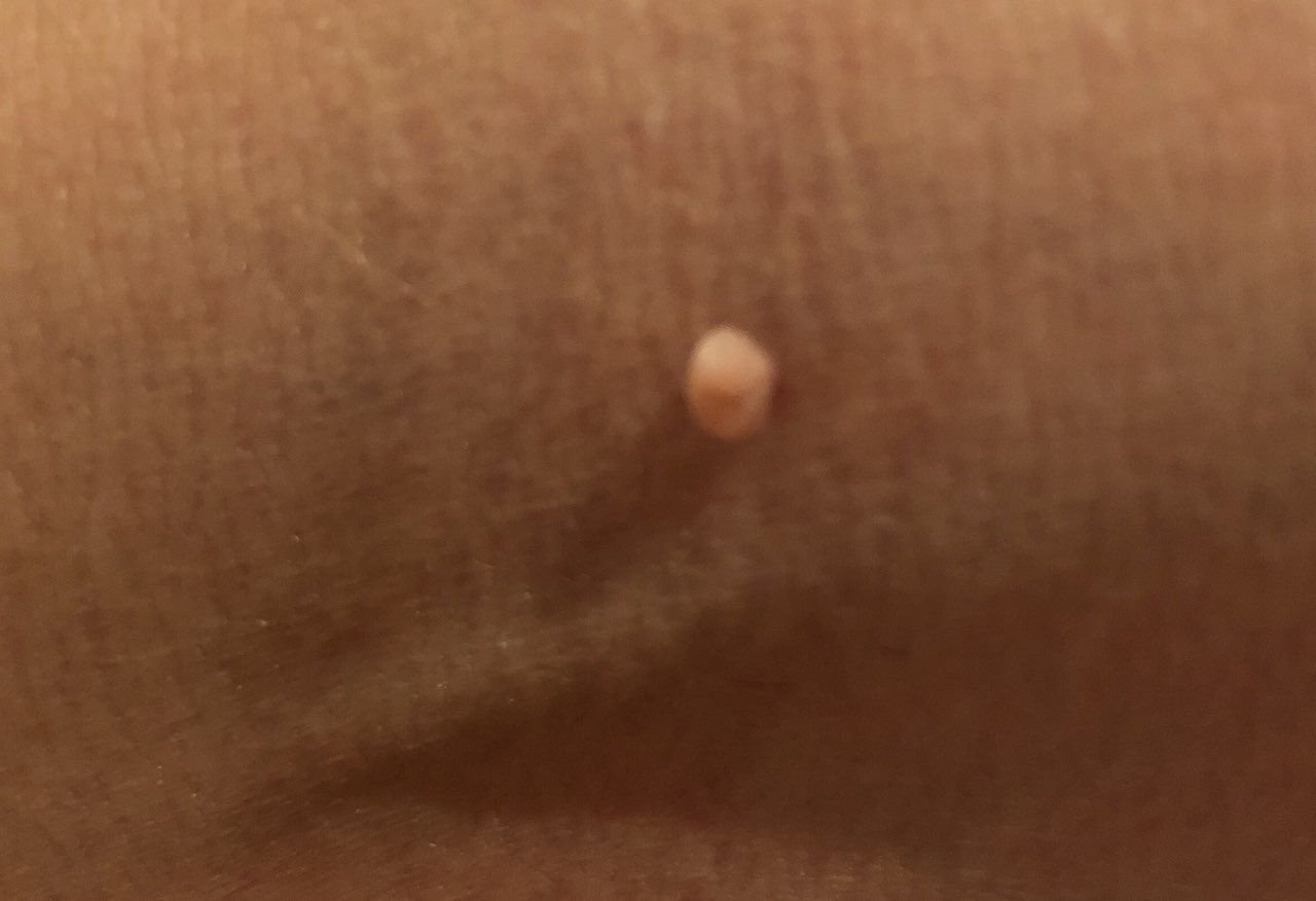
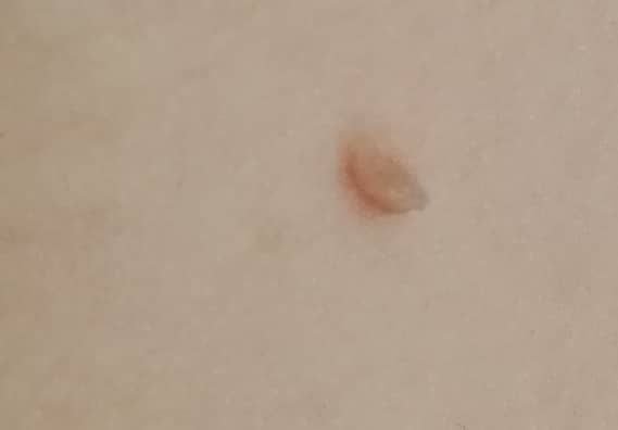
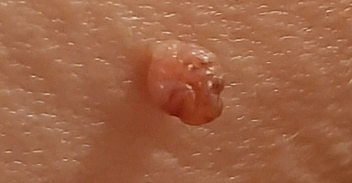
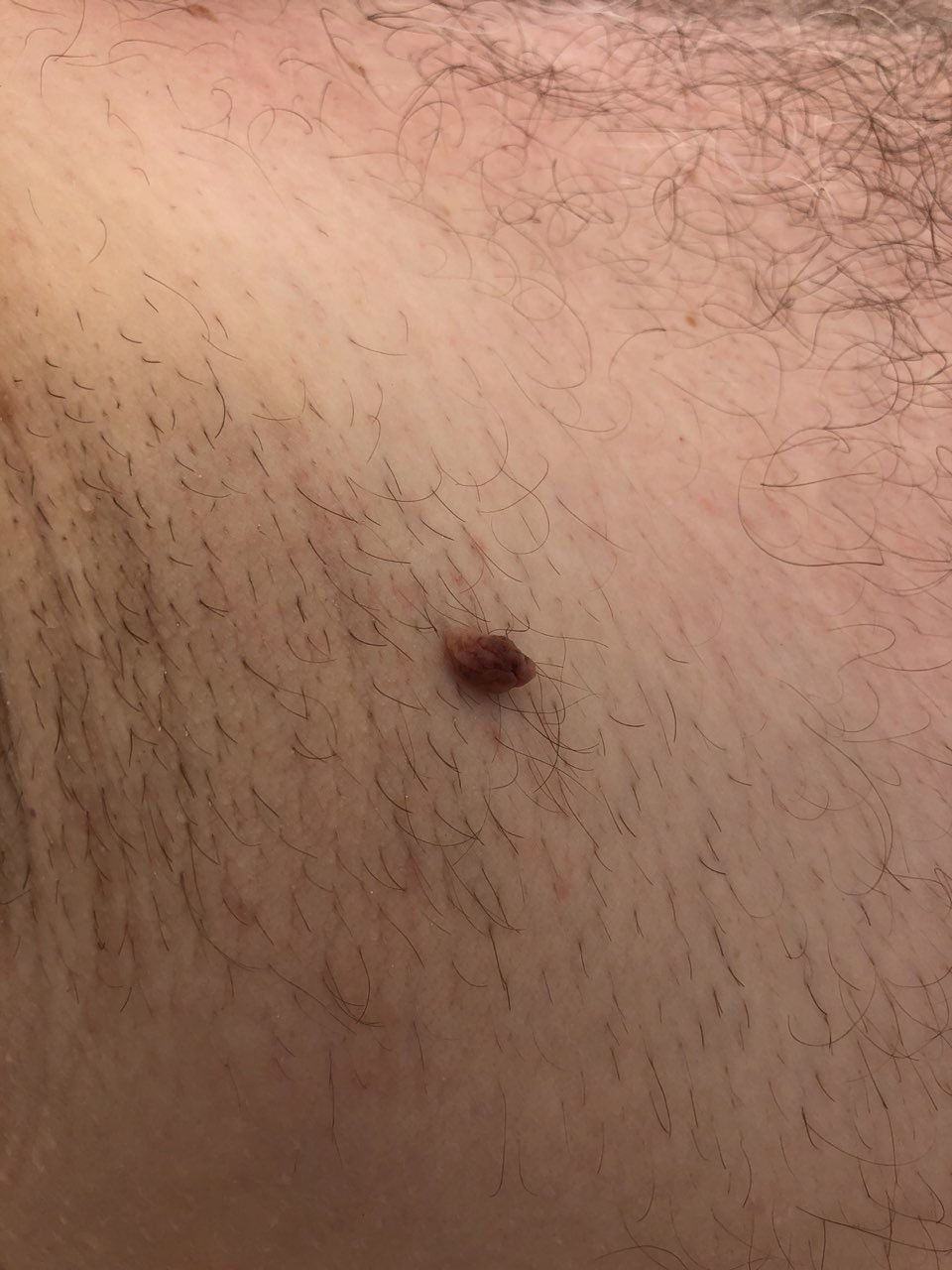
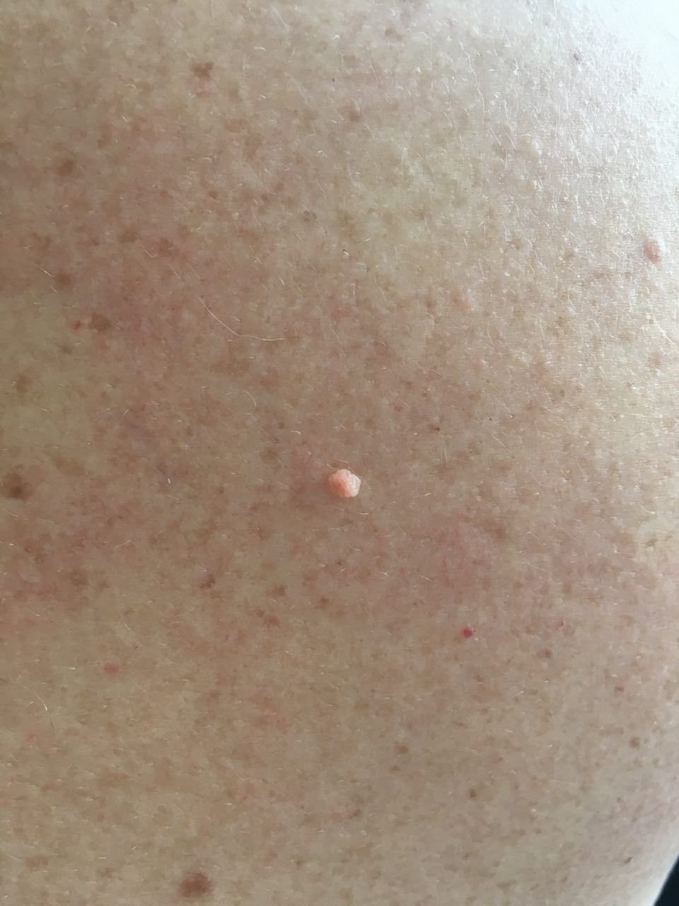
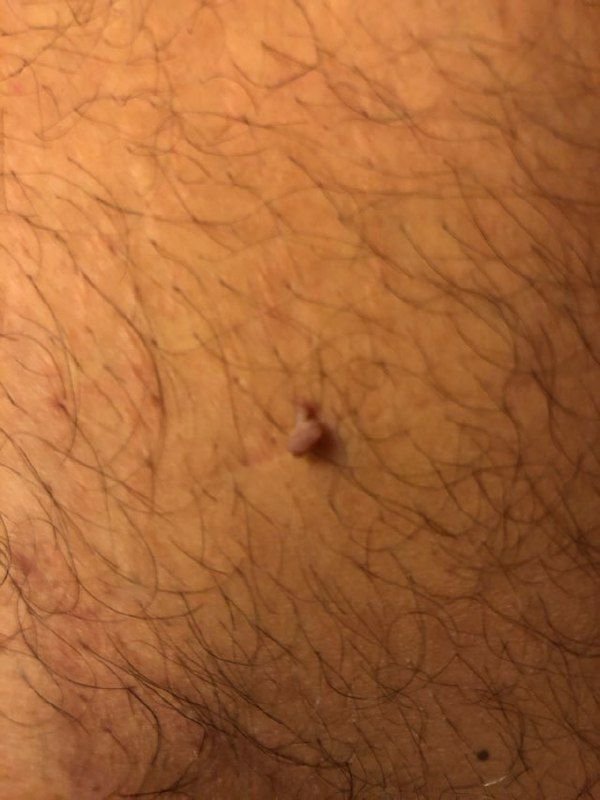
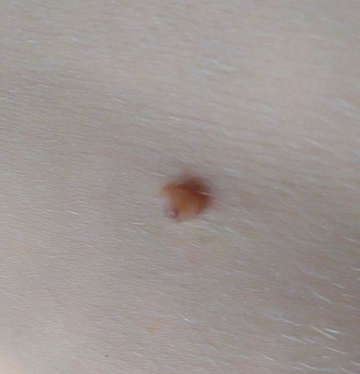
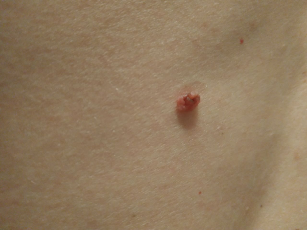
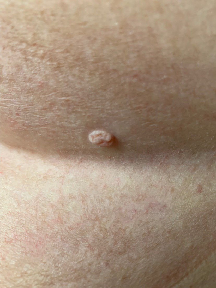
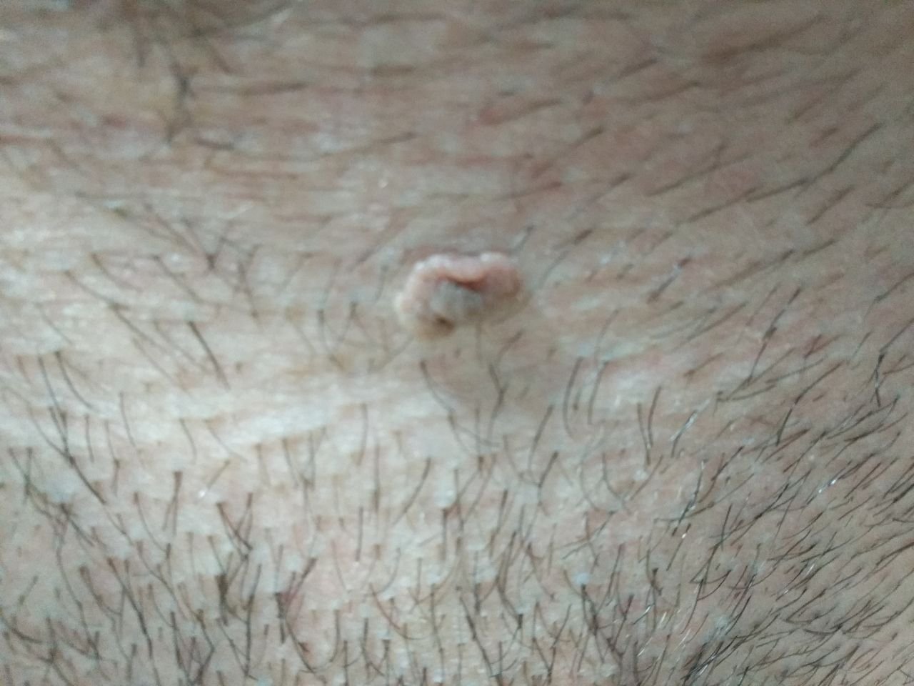
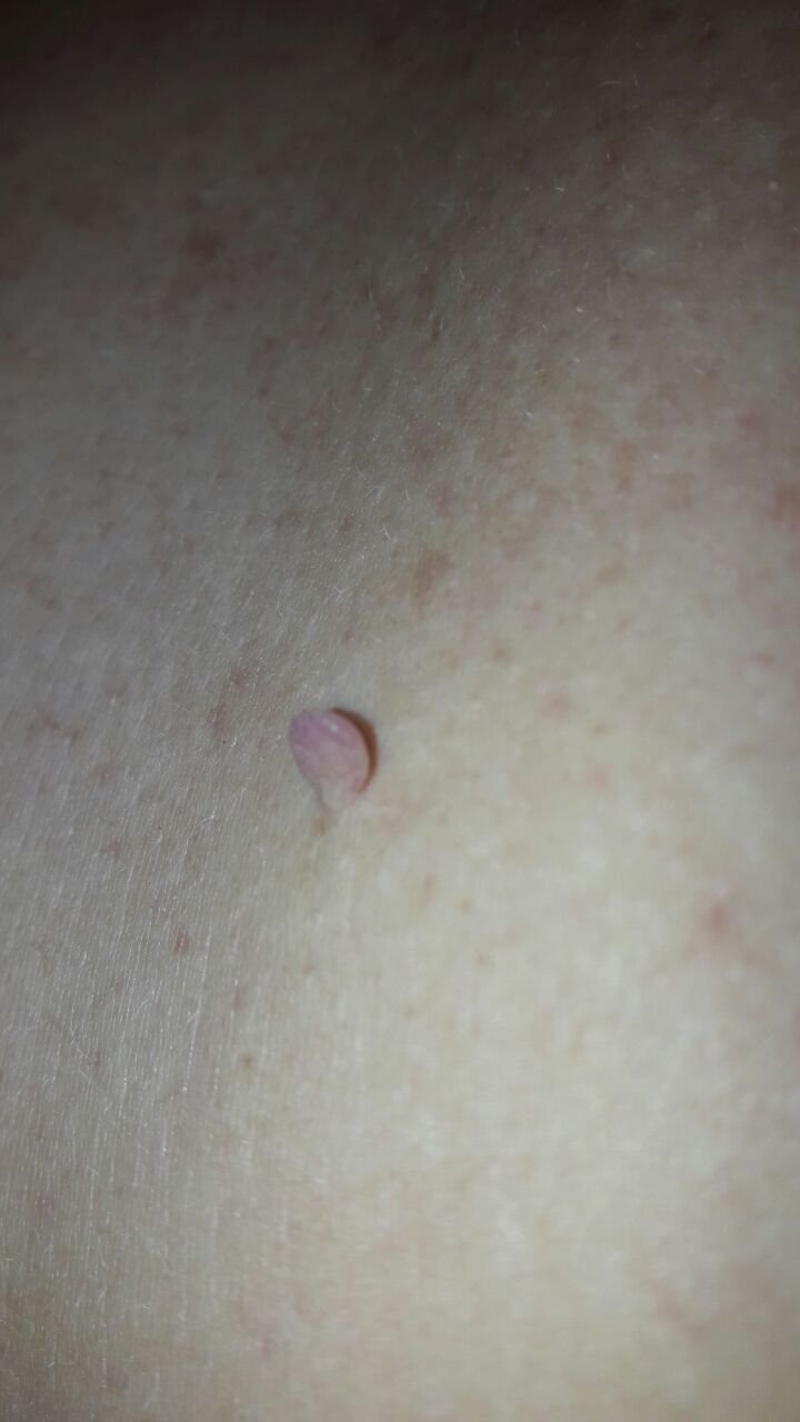
Upon visual inspection, a papilloma is recognized as an elongated formation that rises above the skin on a stem (pedicle). The stem can be as wide as the diameter of the papilloma or slightly narrower. The surface texture of the papilloma typically resembles that of normal skin, but larger papillomas may have a rough, warty surface with a “ragged” appearance.
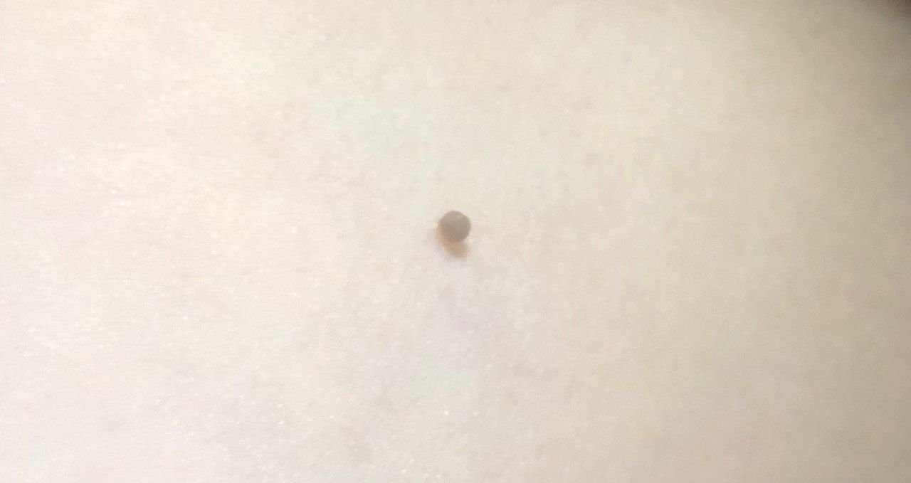
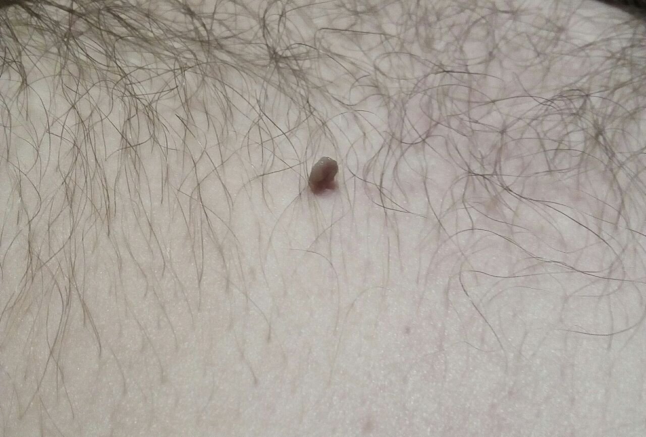
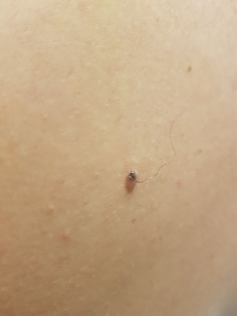
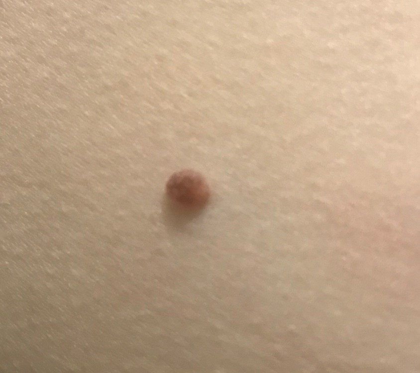
The borders of the papilloma are generally clear, although they can be uneven, especially in larger lesions. The color of the papilloma usually varies from flesh-colored (most common) to light brown. Darker colors are rare in these lesions. Papillomas do not typically affect hair growth. In some cases, coarse bristly or fluffy hairs may be observed growing in the central part of the lesion.
The size of papillomas is usually small, with typical dimensions being up to 2-3 mm in width and 3-5 mm in height above the skin’s surface. Larger papillomas are uncommon. On palpation, the papilloma feels similar to normal skin or slightly softer, particularly in the central portion. There are no subjective sensations associated with the papilloma, although mild itching can sometimes occur in long-standing cases.
Papillomas are most commonly found on the neck, axillary regions, inguinal regions, and the trunk (chest and back), though they can also appear on mucous membranes. These lesions are less frequently found on other parts of the body.
Dermatoscopic Description
During dermatoscopy, the following features of skin papillomas can be observed:
- Papillary Structure: The characteristic flattened elements of the papilloma, often visible due to the pressure applied during dermatoscopy.
- Elasticity and Deformation: Papillomas often exhibit elasticity and can deform when compressed, temporarily turning pale and decreasing in size.
- Diffuse Uniform Staining: The entire papilloma may appear uniformly pigmented under dermatoscopy.
Differential Diagnosis
When diagnosing papillomas, they must be differentiated from other similar skin lesions, including:
- Papillomatous nevus
- Nevus of the sebaceous glands
- Halo nevus
- Dermatofibroma
- Viral wart
- Molluscum contagiosum
- Nodular basal cell carcinoma
- Pigmentless melanoma
Risks
In general, papillomas are benign and do not pose an increased risk of malignancy. Persistent or changing lesions should be evaluated. However, if papillomas change in appearance, grow rapidly, or become denser, they should be evaluated by a dermatologist or oncologist, as these could be signs of malignant transformation.
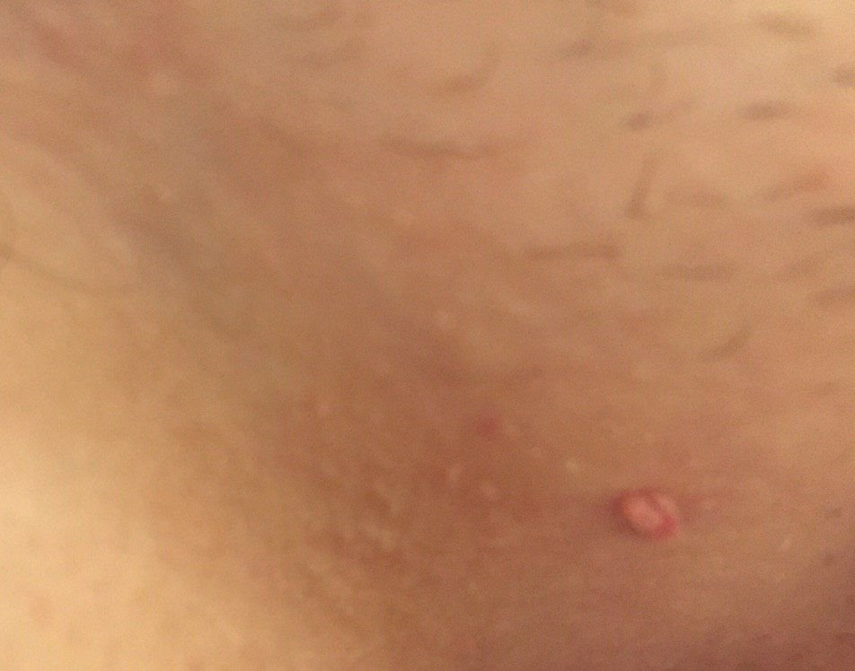
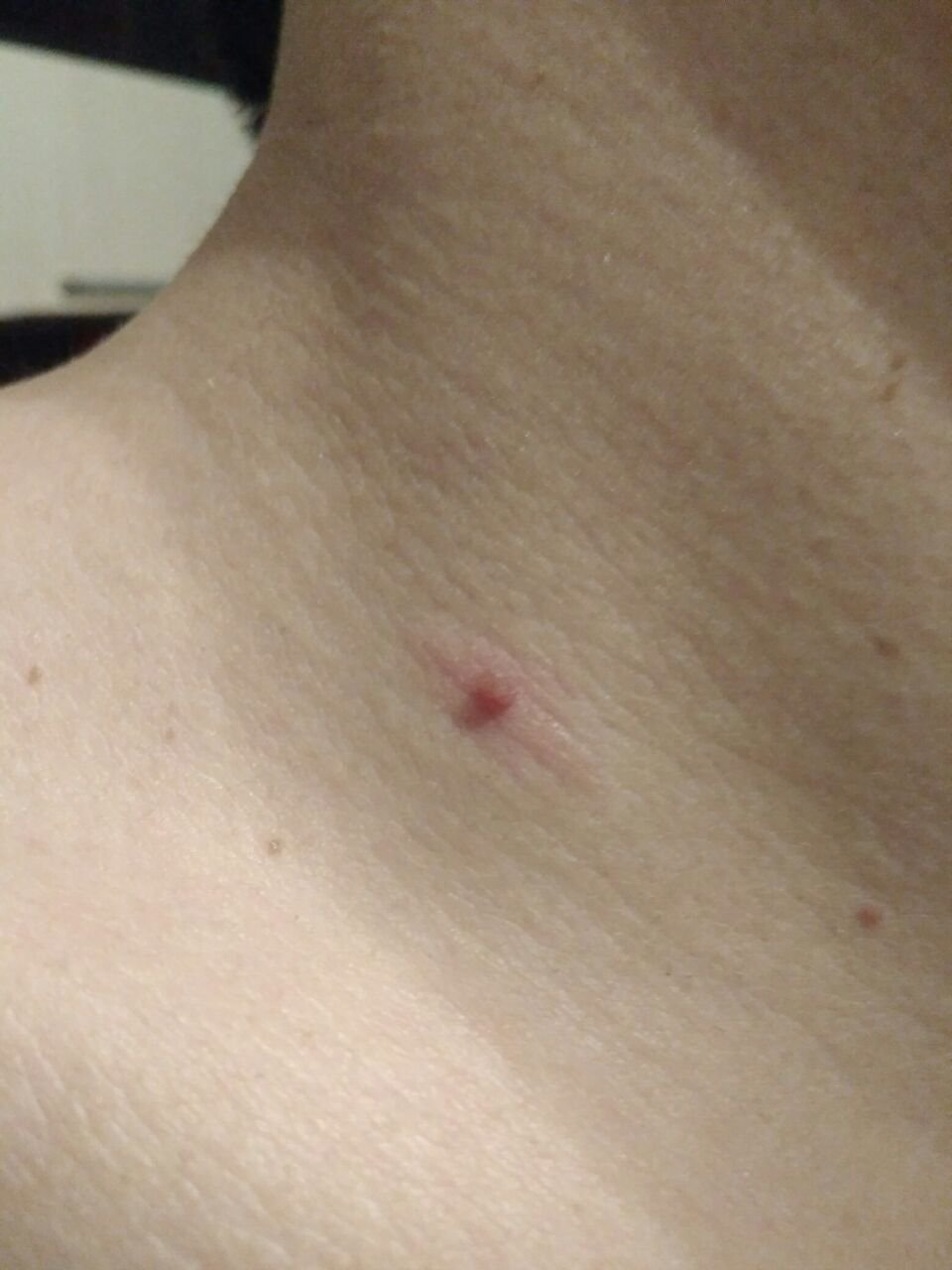
Papillomas are more dangerous due to their tendency to become easily injured because of their elongated shape and narrow stalk. This can result in bleeding, pain, and the potential for infection, making the wound an entry point for harmful microorganisms. In addition, papillomas can cause cosmetic and psychological discomfort, especially if they are located in visible areas.
Because of the viral nature of papillomas, and given that many individuals carry HPV without showing symptoms, it is important to be vigilant about one’s health and to undergo regular medical check-ups to detect any signs of malignancy. Routine oncological examinations by specialists are recommended.
Tactics
If the papilloma shows no signs of damage, change in appearance, or any symptoms, self-monitoring is typically sufficient. This should include an annual check-up or examination by another person for areas that are difficult to inspect. If mechanical injury, exposure to UV radiation, or ionizing radiation occurs, or if any changes are noticed, a visit to a dermatologist or oncologist is necessary.
The healthcare provider will assess whether ongoing monitoring or surgical removal of the papilloma is needed. Papillomas that experience constant trauma from clothing, jewelry, or from professional activities should be considered for removal to prevent further injury. In some cases, papillomas can be removed at the patient’s request, especially if they cause cosmetic concerns or psychological discomfort.
For dynamic observation, it is helpful to take photographs of the papillomas, as this allows for the detection of even minor changes over time. Patients with multiple papillomas should undergo regular dermatological examinations, particularly in the spring and autumn (before and after the summer sun exposure). Keeping a map of skin neoplasms can simplify the monitoring process and help identify new or changing lesions.
Treatment
For the treatment of papillomas, less invasive methods are typically preferred:
- Laser Removal: This is the safest and most effective method for removing papillomas, particularly those of varying shapes, sizes, and locations.
- Cryodestruction: Liquid nitrogen can be used to treat small, superficial papillomas, although there is a risk of scarring.
- Radio Wave Scalpel Removal: This method uses radio waves to remove the papilloma with minimal tissue damage.
- Electrocoagulation: This technique uses an electrical current to burn off the papilloma.
If these less invasive treatments are not suitable, or if there is uncertainty regarding the nature of the papilloma, surgical excision with histological examination may be required.
Self-removal of papillomas is not recommended due to the risk of complications such as bleeding, infection, and misdiagnosis of the lesion’s nature.
Because many cutaneous papillomas are HPV-related, recurrence is possible after removal. New papillomas may appear in the same or adjacent areas after removal. Preventative measures help reduce the likelihood of relapse.
Prevention
Preventing the appearance of papillomas involves a careful and proactive approach to skincare and overall health:
- Limit exposure to ultraviolet radiation, such as from tanning beds or prolonged sun exposure.
- Use sunscreen and protective clothing during periods of intense sunlight.
- Avoid chronic skin trauma that can lead to injury and create an entry point for HPV.
- Limit or eliminate exposure to ionizing radiation and occupational hazards.
- Follow safety protocols when handling skin-damaging chemicals or substances.
- Maintain good personal hygiene and stay vigilant for any changes in skin health.
It is also important to regularly inspect papillomas, seek timely consultation with a healthcare professional if any changes are observed, and remove potentially dangerous lesions to prevent complications.