New Study Shows ALA-PDT Effectively Treats Bowen’s Disease in Chinese Patients
Introduction
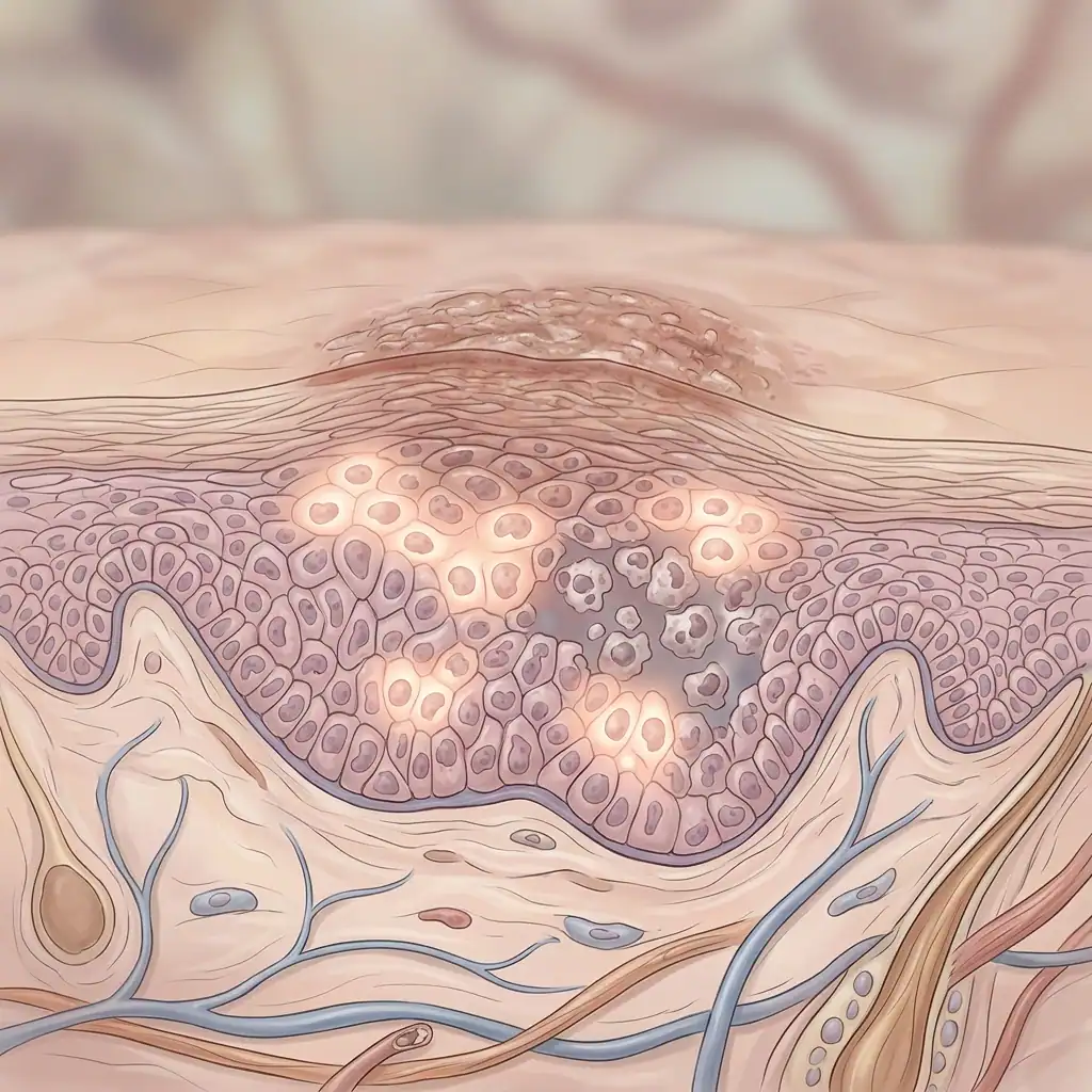
Bowen’s disease is an early form of skin cancer — specifically, an intraepidermal squamous cell carcinoma in situ — that most often appears on sun-exposed skin in older adults.
Although lesions usually grow slowly, untreated Bowen’s disease can progress to invasive squamous cell carcinoma in a small but meaningful number of cases, estimated at roughly 3% to 5%, which is why early treatment matters (Source: Xue WL et al., Efficacy of Photodynamic Therapy: a Meta-Analysis).
In China, surgical excision is still the most commonly used treatment, but surgery can bring pain, infection risk, scarring, and cosmetic concerns — especially when lesions are large, multiple, or sit on cosmetically sensitive areas such as the face, hands, or anogenital region.
Against that backdrop, a multicenter prospective clinical study recently evaluated how well 5-aminolevulinic acid–based photodynamic therapy (ALA-PDT) works for Bowen’s disease in Chinese patients, with a focus on both effectiveness and tolerability (Source: Ran M et al., 2026).
Why consider photodynamic therapy?
Photodynamic therapy (PDT) is a noninvasive treatment that uses a photosensitizing drug plus a specific light source to selectively destroy abnormal skin cells while sparing nearby healthy tissue.
For Bowen’s disease, the most commonly used photosensitizer is 5-aminolevulinic acid (ALA), which is converted inside target cells into the active compound protoporphyrin IX that reacts to light and generates cell-killing oxygen species (Source: Xue WL et al., Meta-Analysis).
Most of the published evidence for ALA-PDT comes from predominantly Caucasian populations, and while the results have been encouraging there, evidence in Asian populations has been more limited — which made the new multicenter trial in China important for filling that gap (Source: Ran M et al., 2026).
Study design and participants
Where the trial took place
The study was conducted at seven tertiary hospitals across China between April 2019 and April 2021, organized as a prospective, multicenter clinical trial to evaluate real-world performance of ALA-PDT for Bowen’s disease (Source: Ran M et al., 2026).
Who was enrolled
A total of 35 adult patients with histopathologically confirmed Bowen’s disease were enrolled, representing 44 individual lesions that were treated and followed (Source: Ran M et al., 2026).
The average age in the group was about 73 years, consistent with the older age profile typically seen in Bowen’s disease (Source: Ran M et al., 2026).
Most participants — roughly 88.5% — had a single lesion, while a small number presented with multiple lesions. Lesion size varied, averaging about 4.58 cm², and lesions were located across a range of anatomical sites including trunk, extremities, head and neck, hands and feet, and the anogenital area (Source: Ran M et al., 2026).
Treatment protocol
Preparation and application
All lesions were treated with a standardized topical 20% ALA gel or solution. Prior to application, clinicians gently removed scales and crusts to improve photosensitizer penetration into the lesion (Source: Ran M et al., 2026).
The ALA was applied to the lesion and extended roughly 1 cm beyond the visible margin, and the treatment area was then occluded for an incubation period of 3 to 4 hours to allow accumulation of protoporphyrin IX in abnormal cells (Source: Ran M et al., 2026).
Fluorescence diagnosis and illumination
After incubation, clinicians used a handheld light source for photodynamic fluorescence diagnosis to visualize protoporphyrin IX accumulation; lesions showed the characteristic brick-red fluorescence, confirming selective photosensitizer uptake (Source: Ran M et al., 2026).
Treated areas were illuminated with 635‑nm red LED light at an energy density of 80–120 J/cm². Red light is chosen because it penetrates deeper into tissue than shorter wavelengths, helping reach the full thickness of epidermal lesions (Source: Ran M et al., 2026).
Patients were reassessed every 7 to 14 days, and additional sessions were given depending on clinical response. Across the study group, patients received between 3 and 6 treatment sessions (Source: Ran M et al., 2026).
Results
Primary endpoint: complete response
The primary outcome was the complete response rate measured three months after the final treatment, defined as the disappearance of the lesion with only residual pigmentation or hypopigmentation remaining (Source: Ran M et al., 2026).
At three months, the patient-level complete response rate was 97.1% (34 of 35 patients), and the lesion-level complete response was 97.7% (43 of 44 lesions), indicating a high short-term effectiveness for ALA-PDT in this population (Source: Ran M et al., 2026).
Subgroup analysis
Investigators analyzed whether outcomes differed by sex, age, lesion number, lesion size, or lesion location and found that none of these factors significantly altered the treatment response, suggesting broad effectiveness across typical clinical subgroups (Source: Ran M et al., 2026).
Recurrence and durability
Patients were followed for 12 months to assess recurrence. Of the 33 patients available for that follow-up window, only one had a recurrence, corresponding to a 3.0% recurrence rate — a figure comparable to recurrence rates reported after surgical excision in earlier studies (Source: Ran M et al., 2026; Fang S et al., 2024).
Cosmetic outcomes and patient satisfaction
Cosmetic results were formally assessed at 12 months using a standardized grading system. Overall, 93.1% of treated lesions were rated as having excellent or good cosmetic outcomes, with minimal persistent erythema, pigmentation change, or scarring (Source: Ran M et al., 2026).
Patient satisfaction mirrored the objective cosmetic outcomes: at 12 months, 92.6% of participants reported being satisfied or very satisfied with their treatment, reflecting the combination of effectiveness and the minimally invasive nature of PDT (Source: Ran M et al., 2026).
Safety and tolerability
The most commonly reported adverse effect was pain during illumination, which was experienced by all patients but tended to be mild to moderate and transient (Source: Ran M et al., 2026).
Pain intensity typically peaked in the first minutes of illumination and then declined. The mean visual analog scale (VAS) score reported was 4.56 at 3 minutes after treatment initiation and 2.61 five minutes after treatment completion (Source: Ran M et al., 2026).
Other side effects included transient erythema in 66.7% of patients, itching in 27.7%, and occasional pigmentation changes in 2.8% of treated sites; these reactions were generally mild and resolved without additional treatment (Source: Ran M et al., 2026).
Importantly, no systemic photosensitivity reactions occurred and no participants discontinued treatment because of adverse events in this trial, supporting the overall tolerability of ALA-PDT in this setting (Source: Ran M et al., 2026).
Study limitations and context
While the trial’s prospective, multicenter design strengthens the findings, the study had limitations: a relatively small sample size and no randomized control arm comparing ALA-PDT head-to-head with surgery or other therapies.
Because of those limitations, the results should be interpreted as strong supportive evidence rather than definitive proof that ALA-PDT is superior to other modalities in all settings; larger randomized controlled trials would help refine protocols, optimal dosing regimens, and long-term outcomes (Source: Ran M et al., 2026).
That said, the observed high response rates, low 12-month recurrence, favorable cosmetics, and good patient satisfaction align with previous reports and meta-analytic findings supporting ALA-PDT as an effective, tissue-sparing option for Bowen’s disease (Source: Xue WL et al., Meta-Analysis; Fang S et al., 2024).
Practical takeaways for clinicians and patients
-
ALA-PDT offers a noninvasive option for patients who want to avoid surgery, especially when cosmetic outcome is a priority or when lesions are multiple or in sensitive locations (Source: Ran M et al., 2026).
-
Expect a protocol that includes lesion preparation, application of 20% ALA, an incubation of several hours, fluorescence confirmation of uptake, and red light illumination (Source: Ran M et al., 2026).
-
Typical treatment courses in this trial ranged from 3 to 6 sessions, with follow-up visits every 7–14 days to guide additional therapy as needed (Source: Ran M et al., 2026).
-
Patients should be counseled that some pain during illumination is common but usually transient, and that most cosmetic results are excellent or good at 12 months (Source: Ran M et al., 2026).
Conclusion
This multicenter prospective study adds important prospective data that ALA-PDT is a highly effective and well-tolerated treatment option for Bowen’s disease in Chinese patients, yielding very high short-term complete response rates, low 12-month recurrence, and excellent cosmetic outcomes (Source: Ran M et al., 2026).
While larger randomized trials will help define its role relative to surgery and other therapies, the current evidence supports ALA-PDT as a valuable tissue-sparing alternative, particularly when cosmesis and preservation of function are priorities (Source: Xue WL et al., Meta-Analysis; Fang S et al., 2024).
Sources
- Ran M, Tang Y, Wu W, Wan M, Zhang L, Zhang J, Xue S, Li H. 5-Aminolevulinic Acid–Based Photodynamic Therapy (ALA-PDT) for Bowen’s Disease in Chinese Patients: A Multicenter Prospective Study. Dermatologic Therapy. 2026;9662750. https://doi.org/10.1155/dth/9662750 (Source: Ran M et al., 2026).
- Xue WL, Ruan JQ, Liu HY, He HX. Efficacy of Photodynamic Therapy for the Treatment of Bowen’s Disease: A Meta-Analysis of Randomized Controlled Trials. Dermatology. doi:10.1159/000519319 (Source: Xue WL et al., Meta-Analysis).
- Fang S, Zhang L, Wang P, et al. Real-world data of 5-aminolaevulinic acid-mediated photodynamic therapy for Bowen disease: a 10-year retrospective study in patients with darker-coloured skin (2011-2021). Clin Exp Dermatol. 2024;49(10):1190-1196. doi:10.1093/ced/llae139 (Source: Fang S et al., 2024).