Simple Nevus (also known as benign nevus, pigmented nevus, mole, or birthmark) is a benign skin growth that appears as a small spot or slightly raised nodule. Simple nevus can be either congenital (present at birth) or acquired at any age. Around 3% of newborns have multiple simple nevi, with the frequency increasing with age. Simple nevi are slightly more common in women than in men, with a ratio of 3:2, respectively.
There is no definitive cause for the formation of simple nevi. However, several predisposing factors may increase the risk of their appearance:
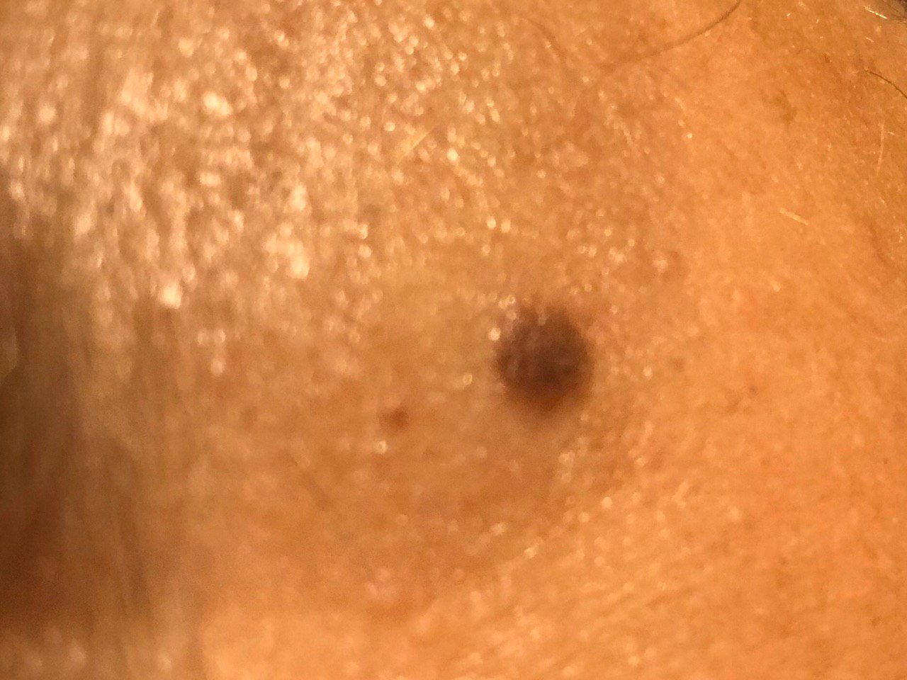
The diagnosis of simple nevi is based on clinical examination, which includes routine visual inspection and dermatoscopy. If there are concerns about malignant growth, a biopsy may be performed for further examination.
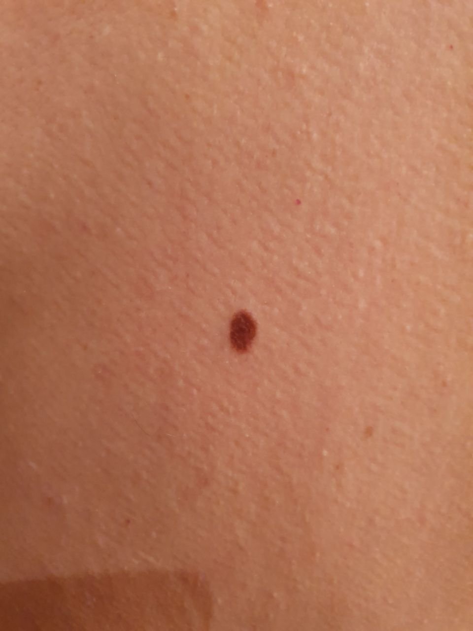
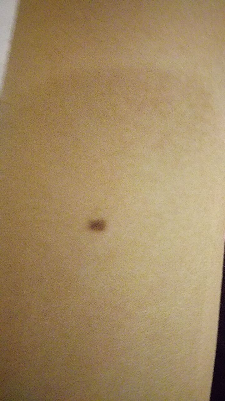
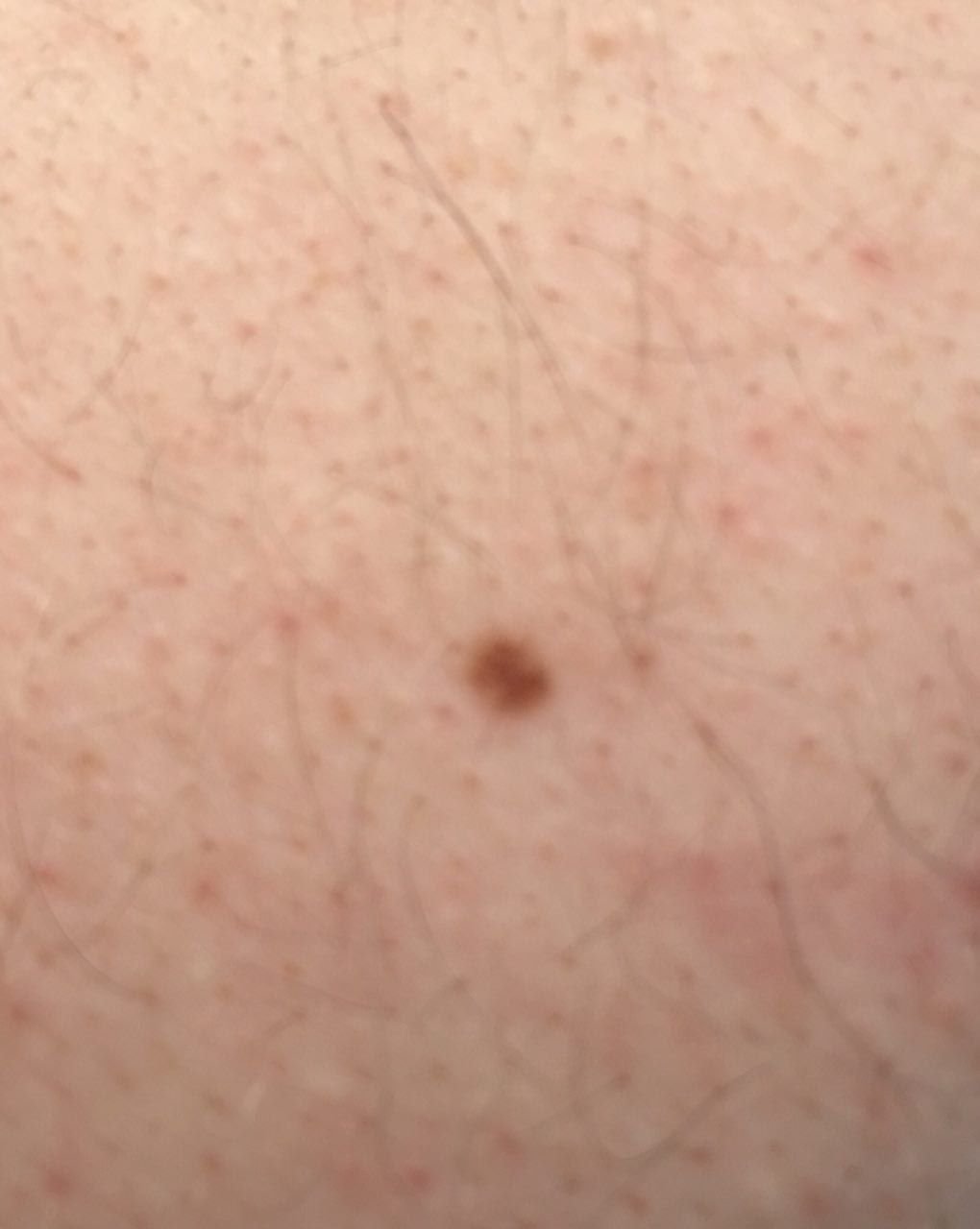
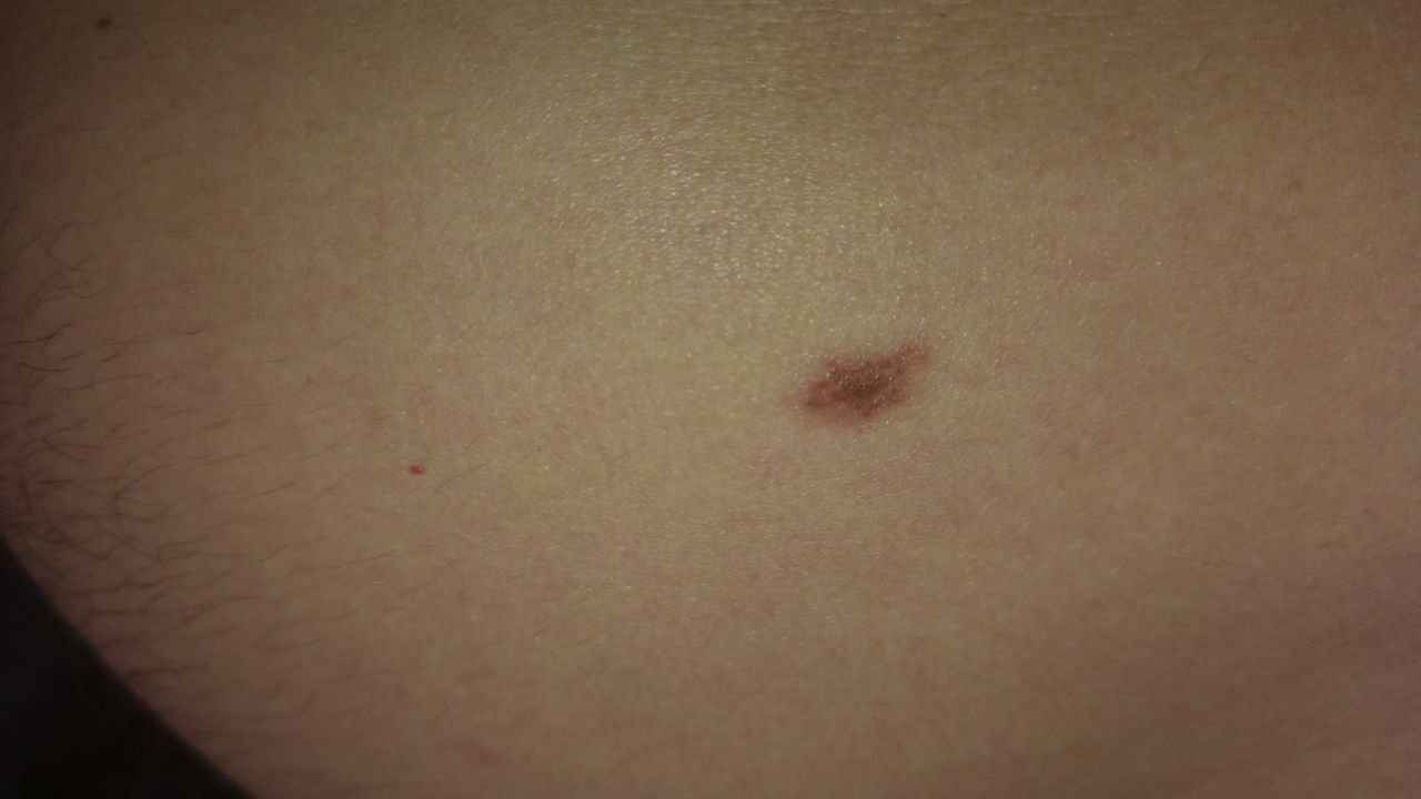
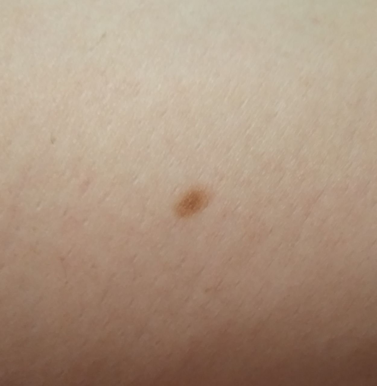
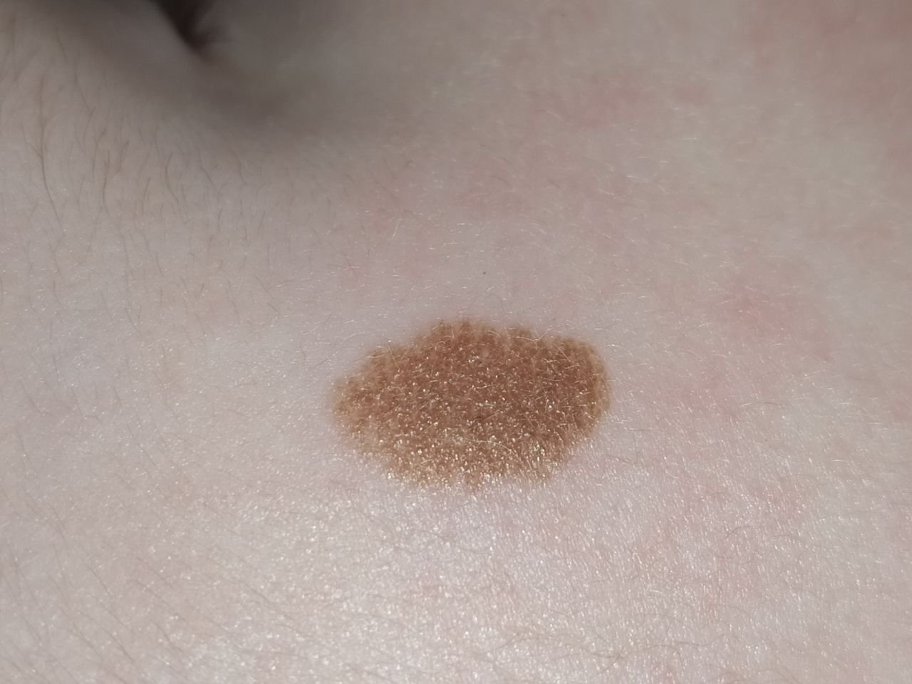
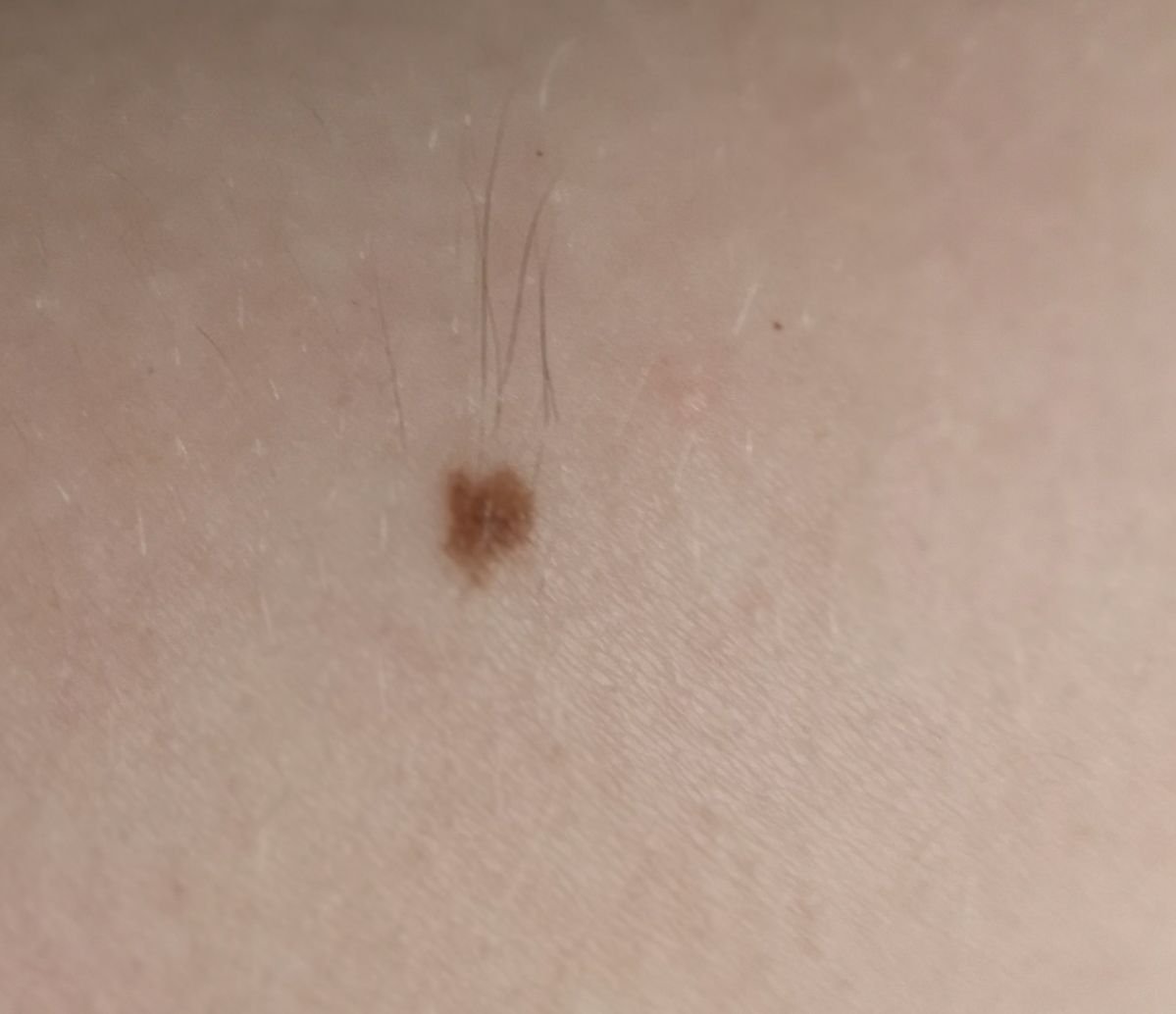
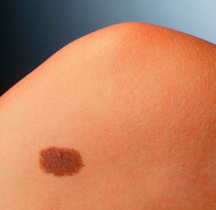
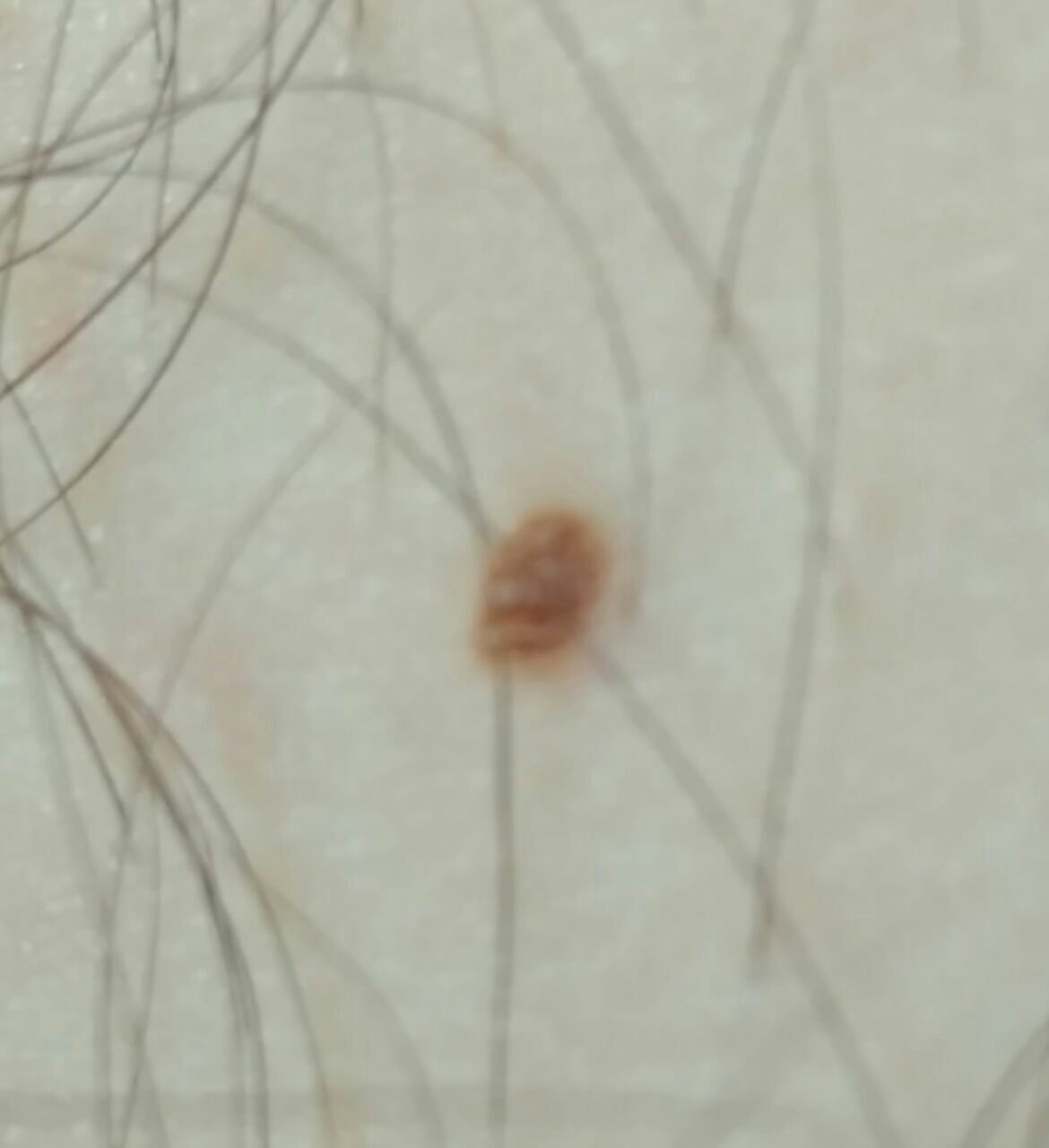
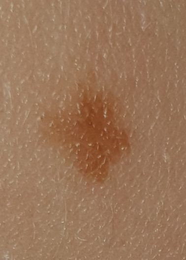
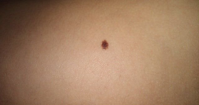
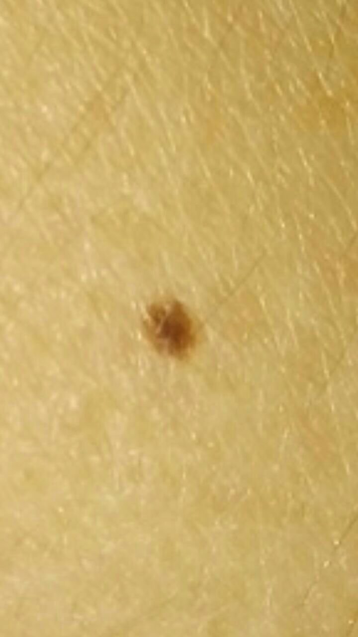
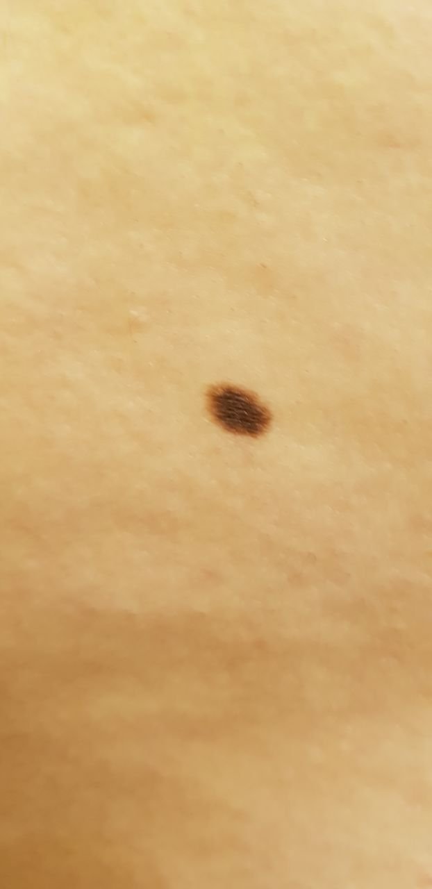
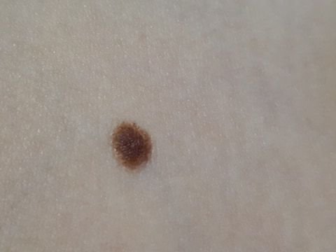
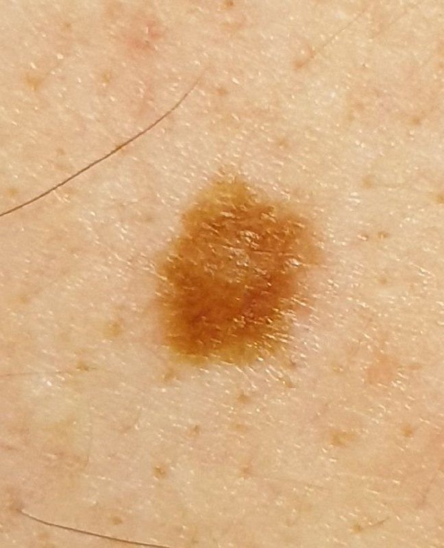
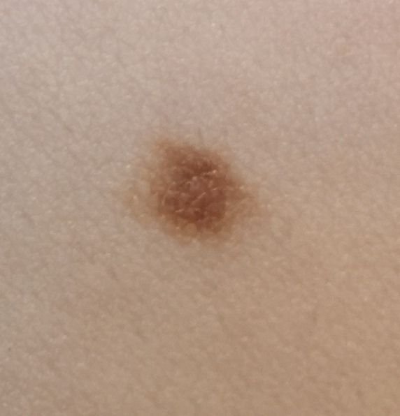
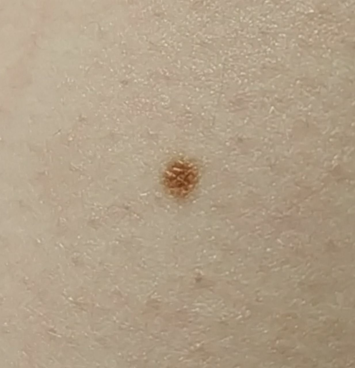
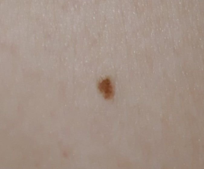
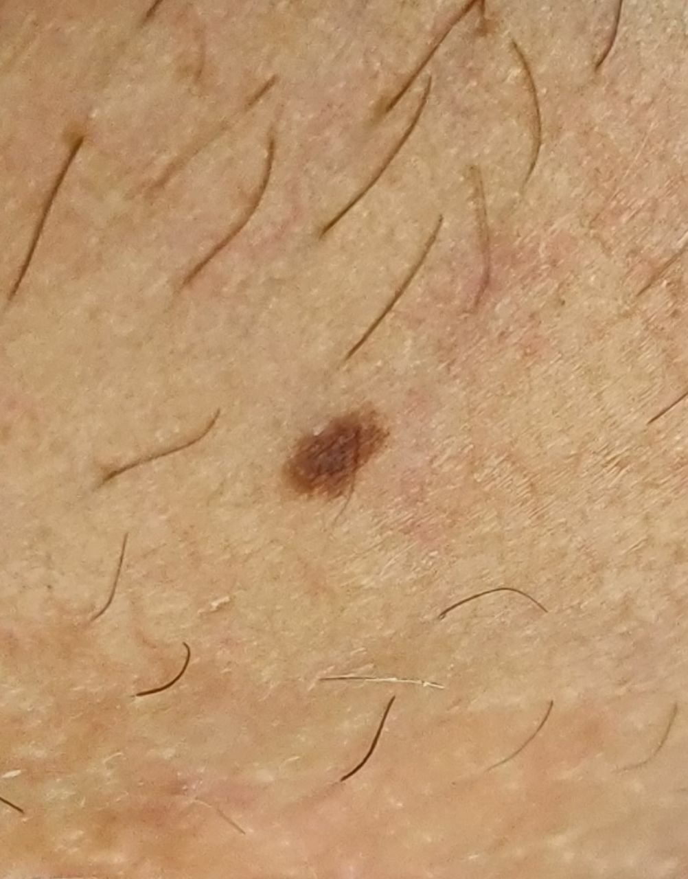
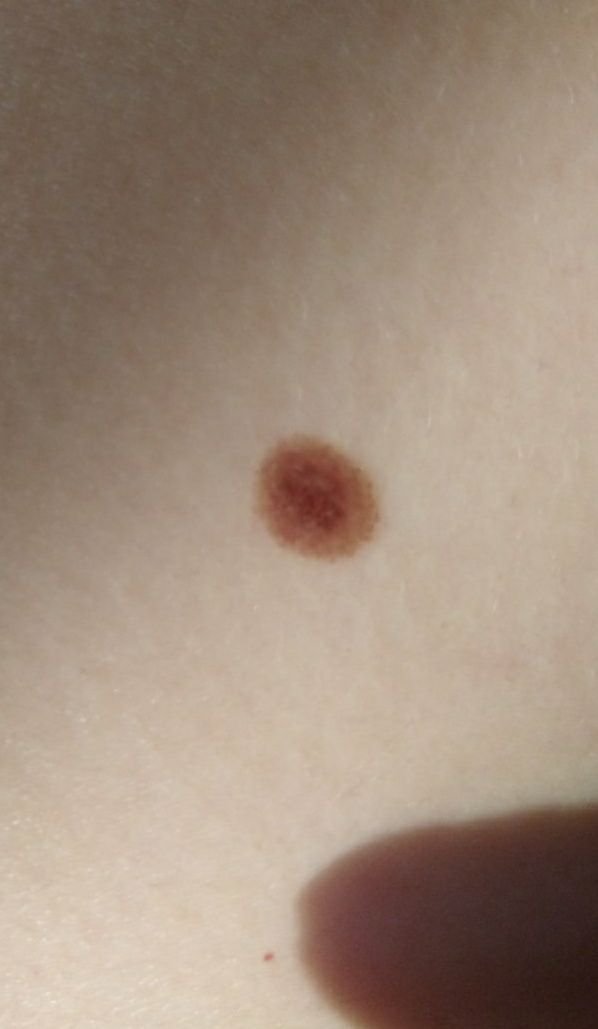
A simple nevus typically appears as a small spot or slightly raised nodule. It is usually symmetrical (oval or round), though large congenital nevi may be irregularly shaped. The surface texture of the nevus is similar to that of normal skin, but it may sometimes differ slightly from the surrounding skin pattern.
The edges of benign nevi are well-defined and smooth. Large congenital nevi, however, may have uneven borders. The color of a simple nevus ranges from light brown to dark brown, with uniform pigment distribution. In some cases, the color may gradually fade from the center to the outer edges. The color of congenital nevi may change during the first few years of life.
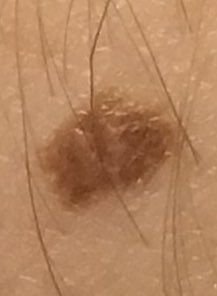
Simple nevi do not typically affect hair growth. However, some congenital nevi may show increased growth of coarse, dark hair, often accompanied by noticeable pigmentation.
Simple nevi can vary greatly in size, but most commonly they measure up to 10 mm. Nevi larger than 10 mm are usually congenital and are rare, though they can grow to be as large as 20 cm or more (giant congenital nevi).
When palpated, simple nevi feel like regular skin and do not cause any discomfort.
Nevi are mostly found on the trunk (~38%) or limbs (~48%), with fewer located on the head and neck (~14%).
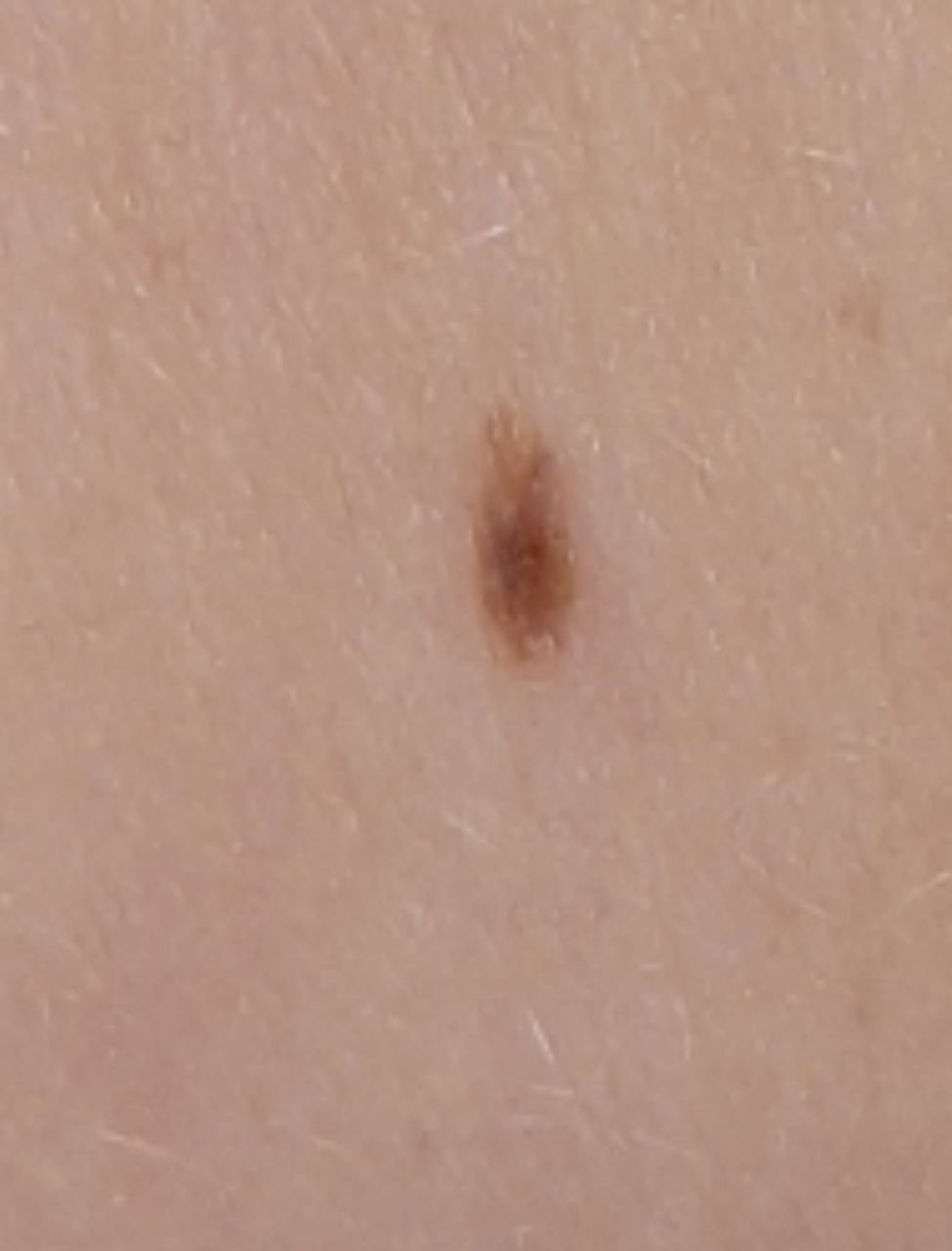
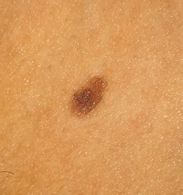
Acral nevi (on the palms and soles) differ slightly in shape, border, and pigment distribution due to the characteristic skin pattern of these areas (“fingerprints”). These nevi are often elongated, with irregular borders, darker pigmentation, and pigment distribution in parallel stripes.
On dermatoscopy, a simple nevus exhibits the following characteristics:
Acral nevi have specific dermatoscopic features:
Simple nevi should be differentiated from other pigmented lesions, such as:
A simple nevus is typically harmless and does not pose an increased risk of melanoma. In the absence of external factors like trauma, ultraviolet radiation, or ionizing radiation, the risk of malignant transformation is comparable to that of normal skin. Signs of malignancy may include changes in appearance or new sensations in the nevus.
Congenital nevi, especially large ones (larger than 20 cm in diameter), show a slightly higher risk of developing melanoma, but the risk for nevi smaller than 20 cm is less than 1%.
Large and multiple congenital nevi may be associated with certain genetic syndromes or diseases, so individuals with such nevi require careful observation and medical evaluation.
If there is no damage to the simple nevus and no changes in appearance or sensation, self-monitoring (or having others check hard-to-see areas) is sufficient at least once a year. However, if the nevus experiences mechanical trauma, excessive UV exposure, or ionizing radiation, or if any changes or new sensations appear, it is essential to consult a dermatologist or oncologist.
The doctor will determine if dynamic monitoring is necessary, or if the nevus should be removed. Nevi subject to constant irritation from clothing, jewelry, or occupation should also be removed.
For dynamic monitoring, it is useful to take photographs of the skin neoplasm, which will help detect even minor changes in appearance over time.
Patients with large congenital or multiple acquired nevi should be examined by a dermatologist or oncologist at least twice a year (before and after the summer months). It is also recommended to create a map of skin neoplasms, which can greatly simplify monitoring and identification of new or changing lesions.
Only surgical removal (using a classic scalpel, electric, or radio scalpel) is recommended, with mandatory histological examination of the excised tissue.
Destructive methods, such as laser removal or cryodestruction, are not advised for treating pigmented nevi.
Preventing the appearance of nevi and their malignant transformation involves gentle skin care:
Regularly examine pigmented nevi, seek timely consultation with a specialist if any changes occur, and remove potentially dangerous nevi when necessary.