Keratoacanthoma is a rapidly growing, non-pigmented, and typically benign skin neoplasm that often resembles squamous cell carcinoma in both clinical and histological appearance. Despite its malignant-like features, keratoacanthoma frequently undergoes spontaneous regression within several months of its initial appearance. This tumor usually arises in adulthood, predominantly after the age of 35–40 years, and is more common in men than in women.
While no definitive cause has been identified for keratoacanthoma, several contributing factors are believed to increase the risk of its development. These predisposing factors include:
Diagnosis of keratoacanthoma is based on thorough clinical evaluation, which includes physical inspection of the lesion and dermatoscopic examination. Due to its striking similarity to basal cell carcinoma and especially squamous cell carcinoma, a biopsy is typically performed to confirm the diagnosis and rule out malignancy. Histopathological analysis is essential to distinguish keratoacanthoma from more aggressive forms of skin cancer.
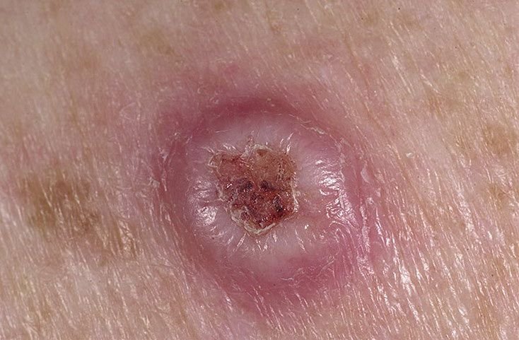
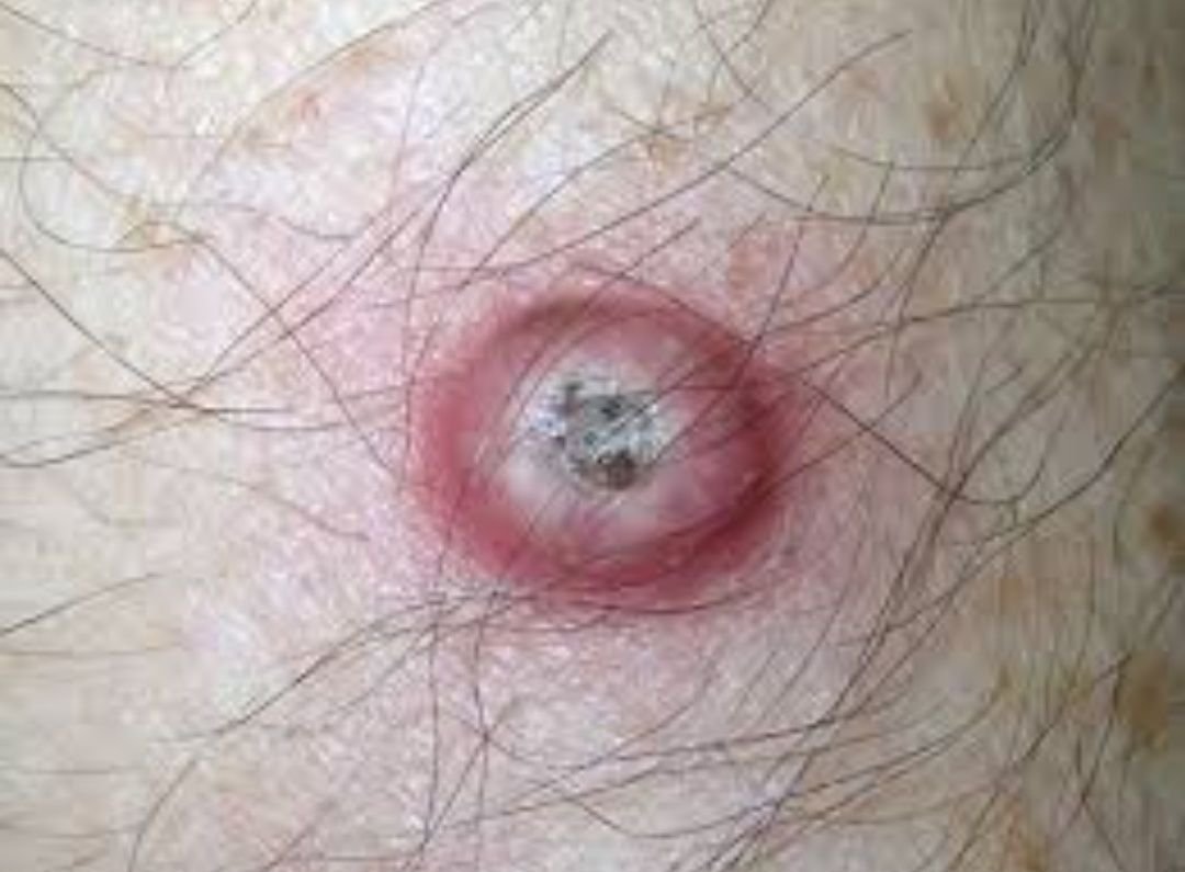
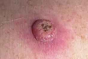
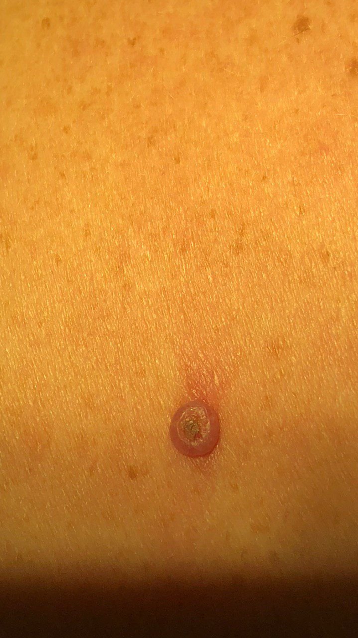
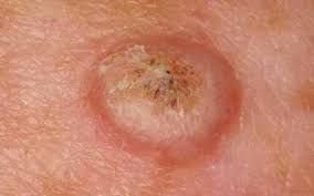
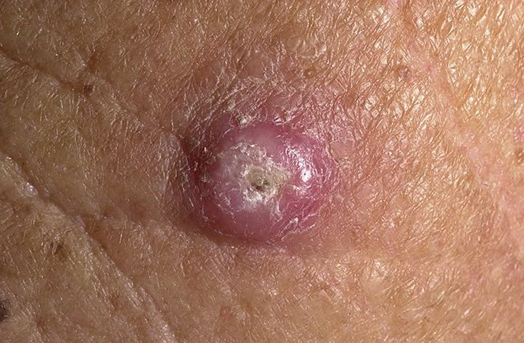
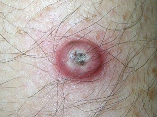
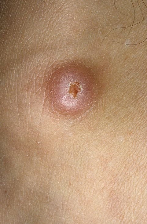
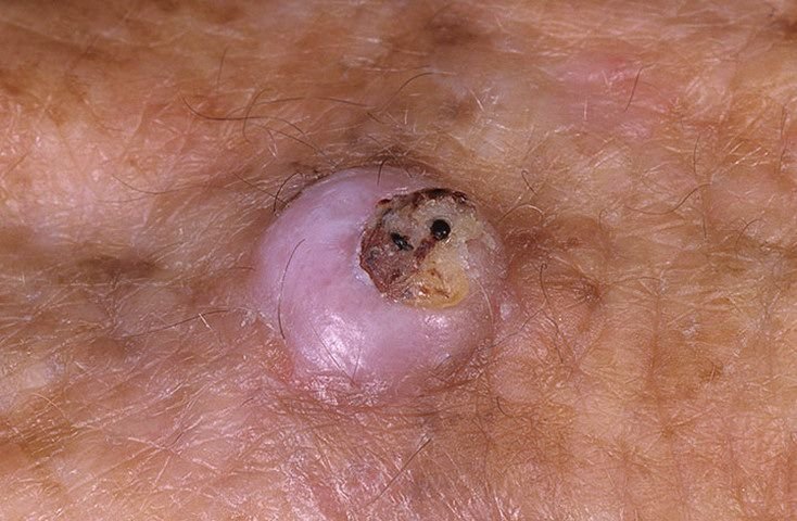
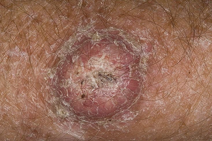
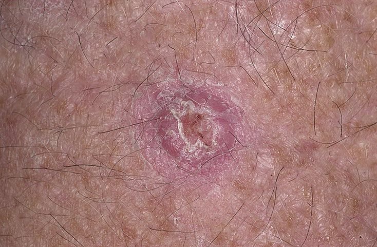
Keratoacanthoma often appears as a raised, dome-shaped lesion with a central crater or keratin-filled core. The peripheral surface is usually smooth and devoid of normal skin lines. In some cases, the center is ulcerated or crusted. The tumor may initially grow rapidly over weeks, reaching a size of 10–20 mm, after which growth typically slows. Lesions larger than 20 mm may develop bleeding or pain with minor trauma.
The borders of the lesion are usually symmetrical and regular, although some cases may show diffuse, ill-defined edges with surrounding erythema. The central area may appear grayish due to keratinization, while the periphery is often pink, red, or yellowish. Hair does not grow on the lesion’s surface. On palpation, the lesion feels firm yet mobile relative to deeper tissues. In smaller lesions, there are usually no subjective symptoms, but larger tumors may cause tenderness or discomfort.
Keratoacanthomas most frequently occur on sun-exposed areas of the body. Common sites include the forearms, hands, face, neck, back, and lower legs. Less commonly, lesions may arise on the chest, abdomen, or thighs.
Dermatoscopic examination of keratoacanthoma may reveal the following features:
Keratoacanthoma must be distinguished from several other dermatologic conditions, some of which are malignant. These include:
Although keratoacanthoma is generally benign and may undergo spontaneous regression, it is still considered an optional precancerous condition. The risk of malignant transformation into squamous cell carcinoma is relatively low but increases in the presence of additional risk factors, such as chronic trauma, burns, or chemical exposure.
Furthermore, individuals with a history of keratoacanthoma may have an elevated risk of developing other skin cancers elsewhere on the body. This necessitates careful follow-up and regular skin examinations to ensure early detection and differentiation of new lesions.
Upon suspicion or confirmation of keratoacanthoma, referral to a dermatologist or oncologist is recommended. Due to its clinical resemblance to invasive carcinomas, histological confirmation is essential. Even with a confirmed benign diagnosis, surgical excision is generally recommended due to the potential for growth, discomfort, bleeding, and malignant transformation.
If surgery is declined, the patient must undergo active dynamic observation, including photographic documentation of the lesion to monitor for subtle changes. Monitoring is especially important for tumors exceeding 20 mm or those that exhibit new symptoms.
Routine skin examinations are advisable in the spring and autumn, particularly in individuals with a history of skin cancer or keratoacanthoma. Full-body skin mapping aids in long-term surveillance and allows for early detection of suspicious changes in skin morphology.
The gold-standard treatment is surgical excision with a margin of healthy tissue, ensuring complete removal and minimizing recurrence risk. Excision should be full-thickness to capture the entire lesion.
Flat plane excision or superficial removal techniques are discouraged, as they increase the chance of recurrence. Similarly, methods such as laser ablation or cryodestruction are not recommended for keratoacanthoma due to poor histological control and a higher rate of local relapse.
Preventive strategies aim to reduce the likelihood of developing keratoacanthoma and limit environmental and physical insults to the skin:
Early identification and timely management are critical to minimizing complications and improving outcomes in patients with keratoacanthoma.