Lentigo, also known as melanin hyperpigmentation, actinic lentigo, or sunspots, is a benign skin growth characterized by a light brown spot or multiple smaller spots of the same type. Lentigo typically appears in individuals over the age of 35, usually as a result of prolonged exposure to solar ultraviolet radiation. It is rare to see lentigo in younger individuals (less than 20% of cases), and when it does occur, it is often associated with metabolic or hormonal disorders. As people age, the risk of developing lentigo increases, with 90% of individuals over 60 years old having at least one lentigo.
The exact cause of lentigo is not fully understood, but several predisposing factors have been identified that may increase the likelihood of developing hyperpigmentation. These factors can influence the onset and growth of lentigo:
The diagnosis of lentigo is based on a clinical examination, which includes visual inspection of the lesion and dermatoscopy to evaluate the characteristics of the pigmentation. If there is any suspicion that the lentigo may be malignant or showing abnormal growth patterns, a biopsy (such as excision biopsy) may be recommended to rule out other conditions.
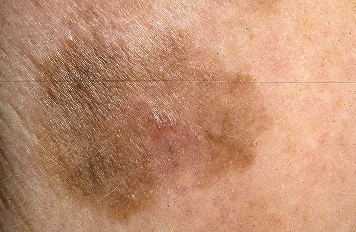
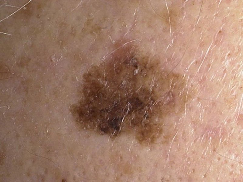
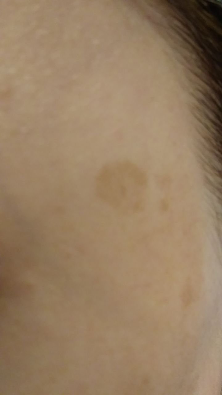
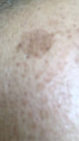
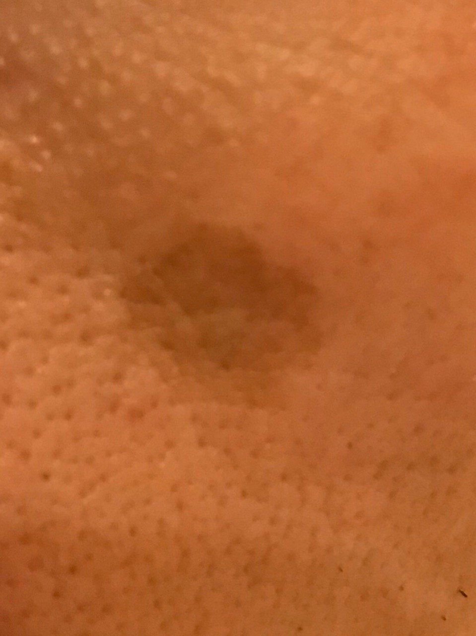
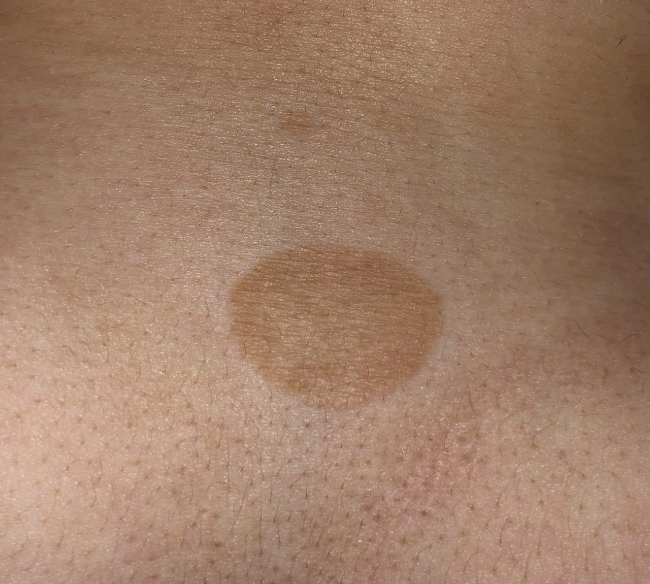
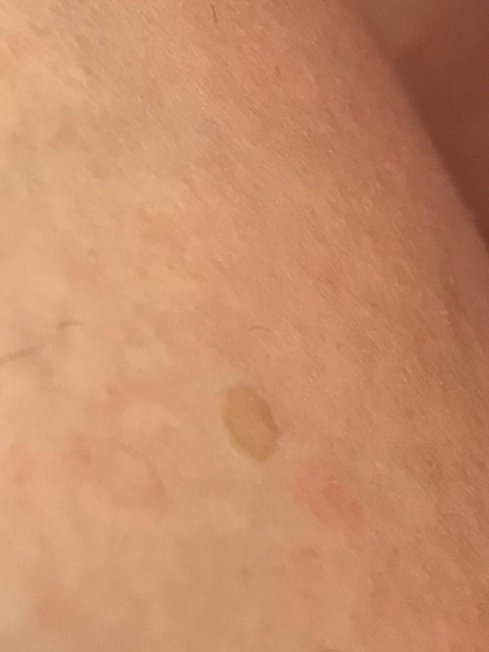
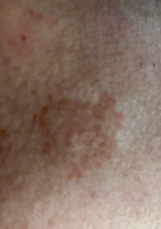
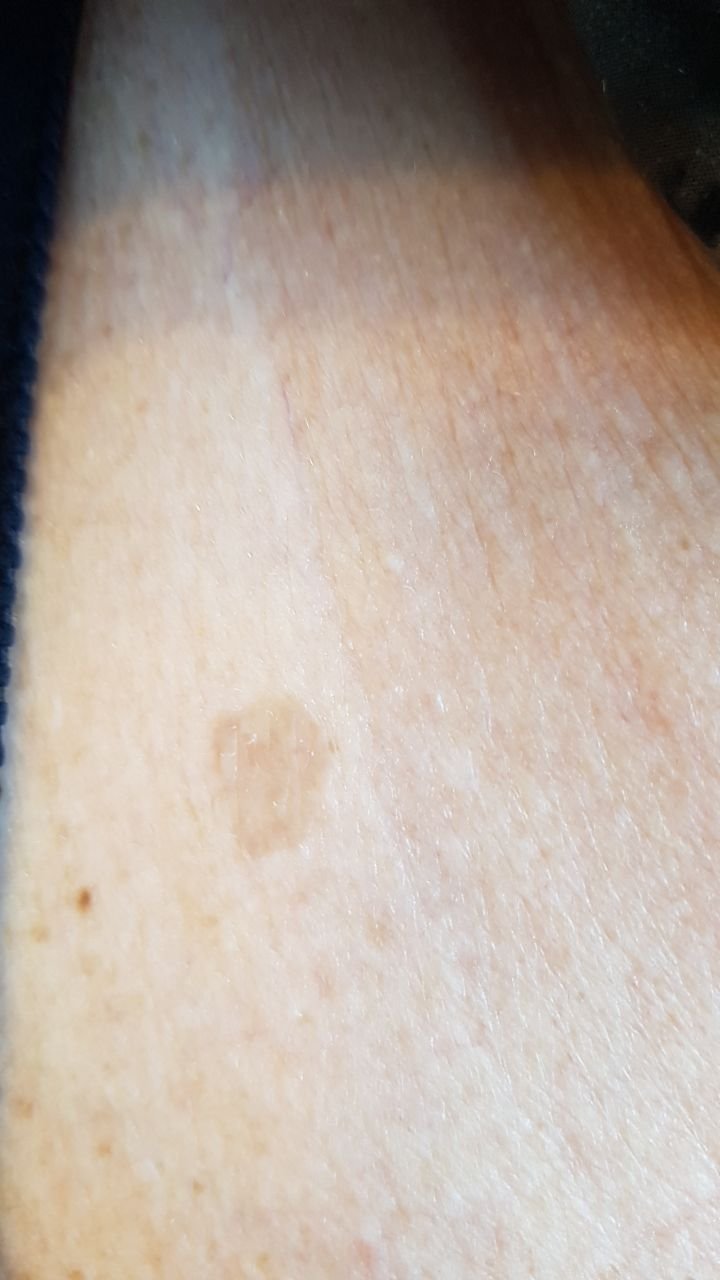
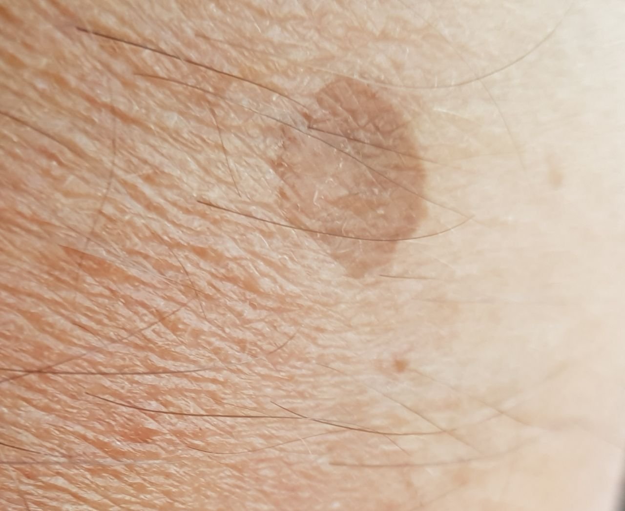
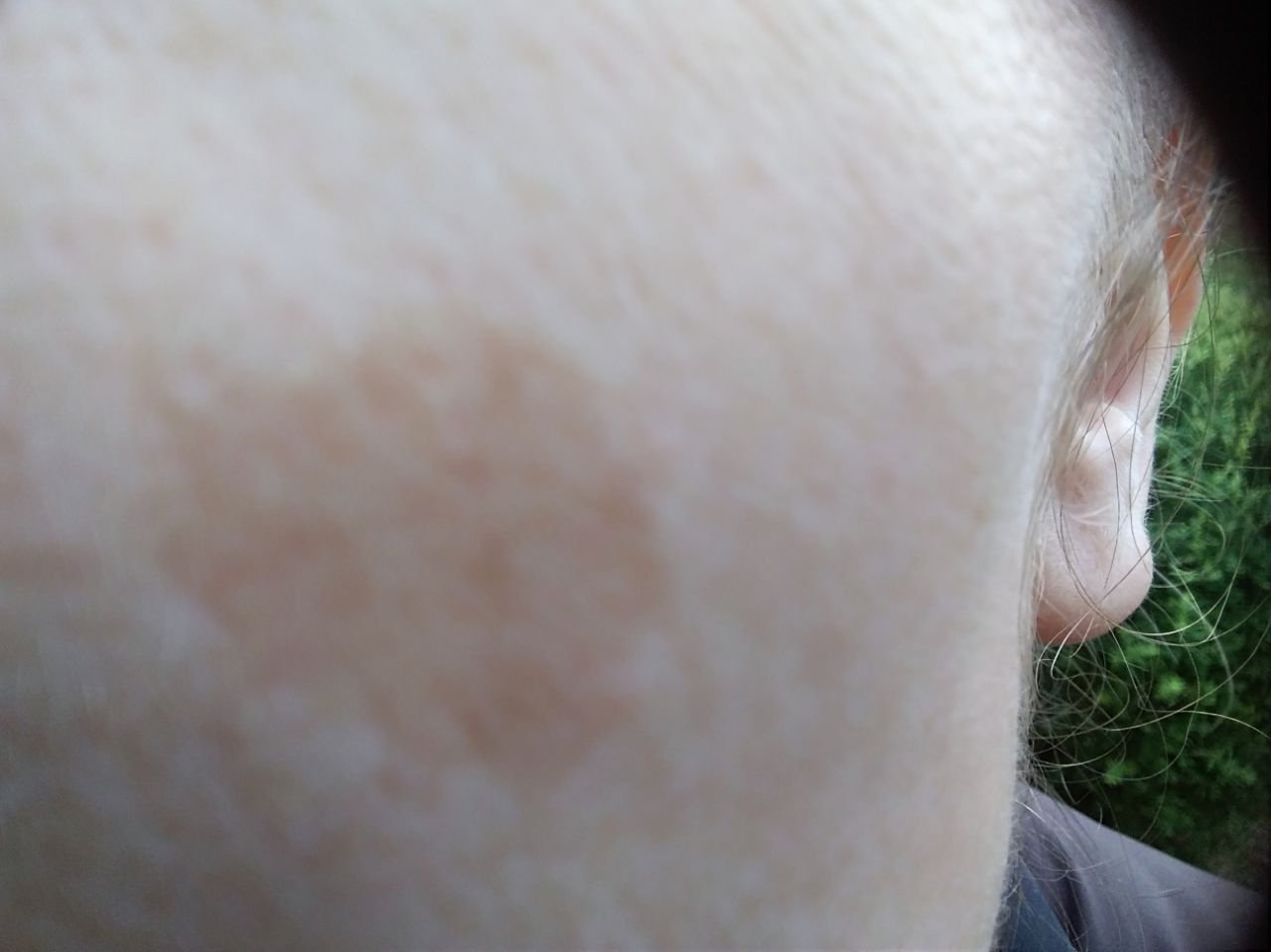
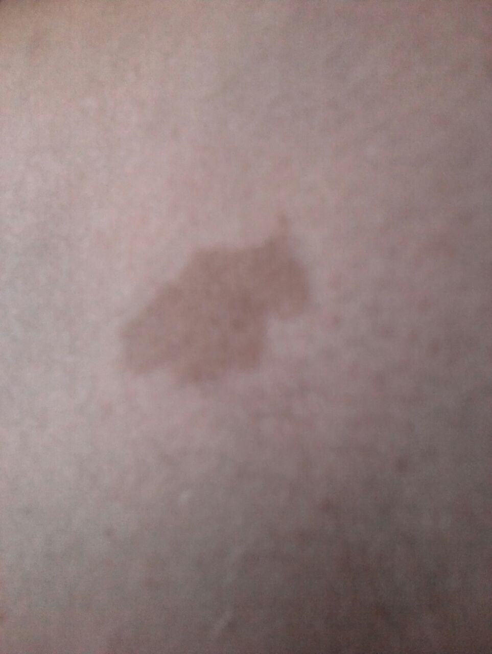
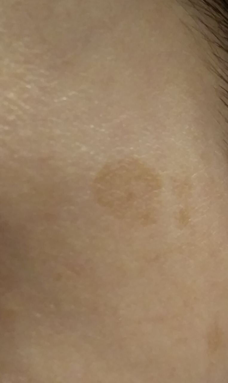
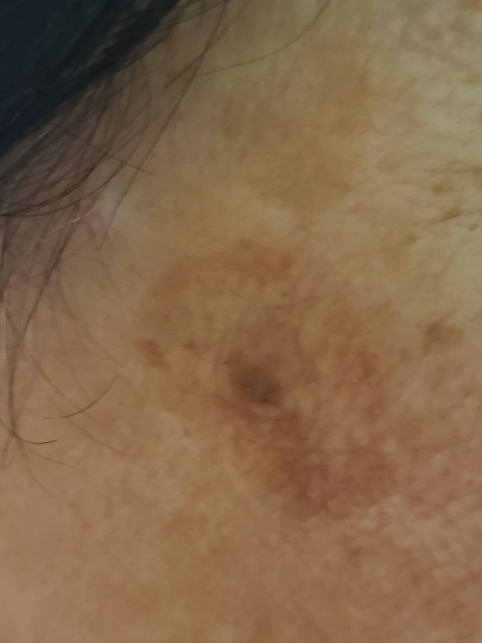
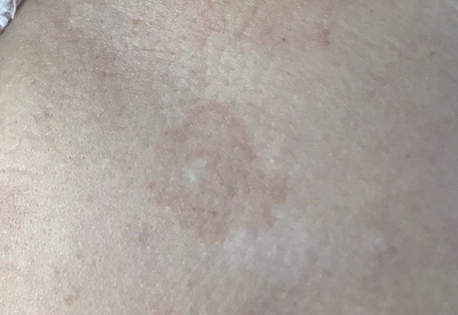
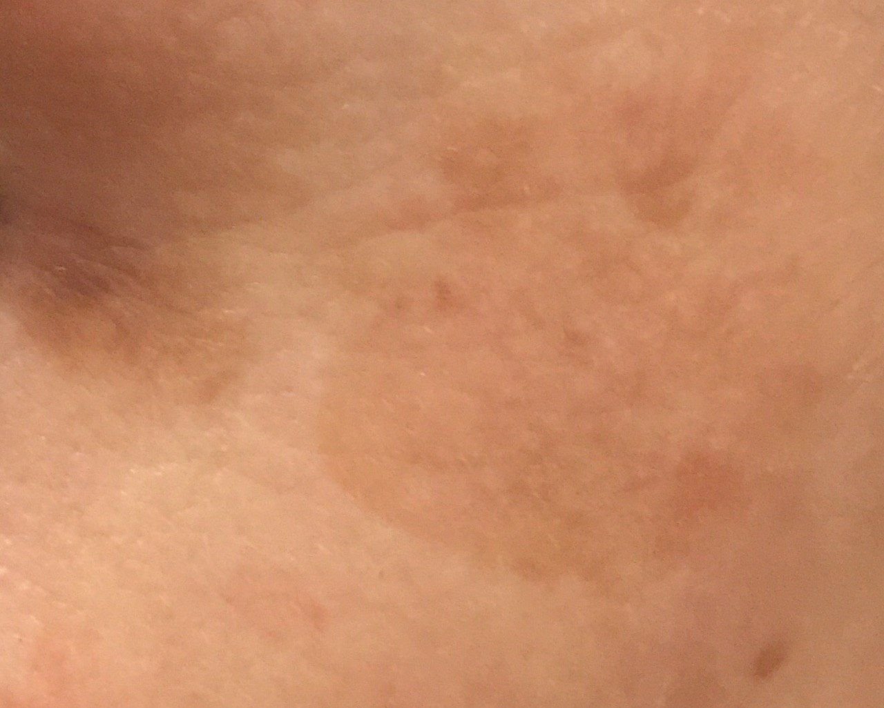
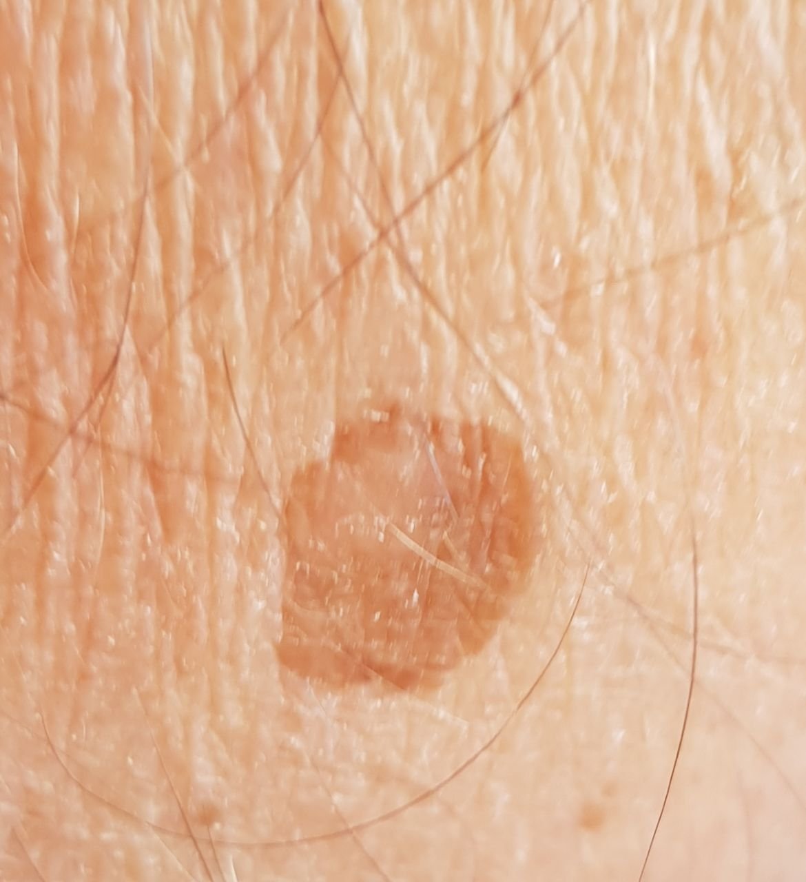
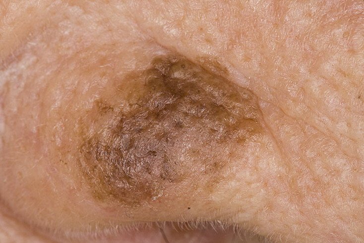
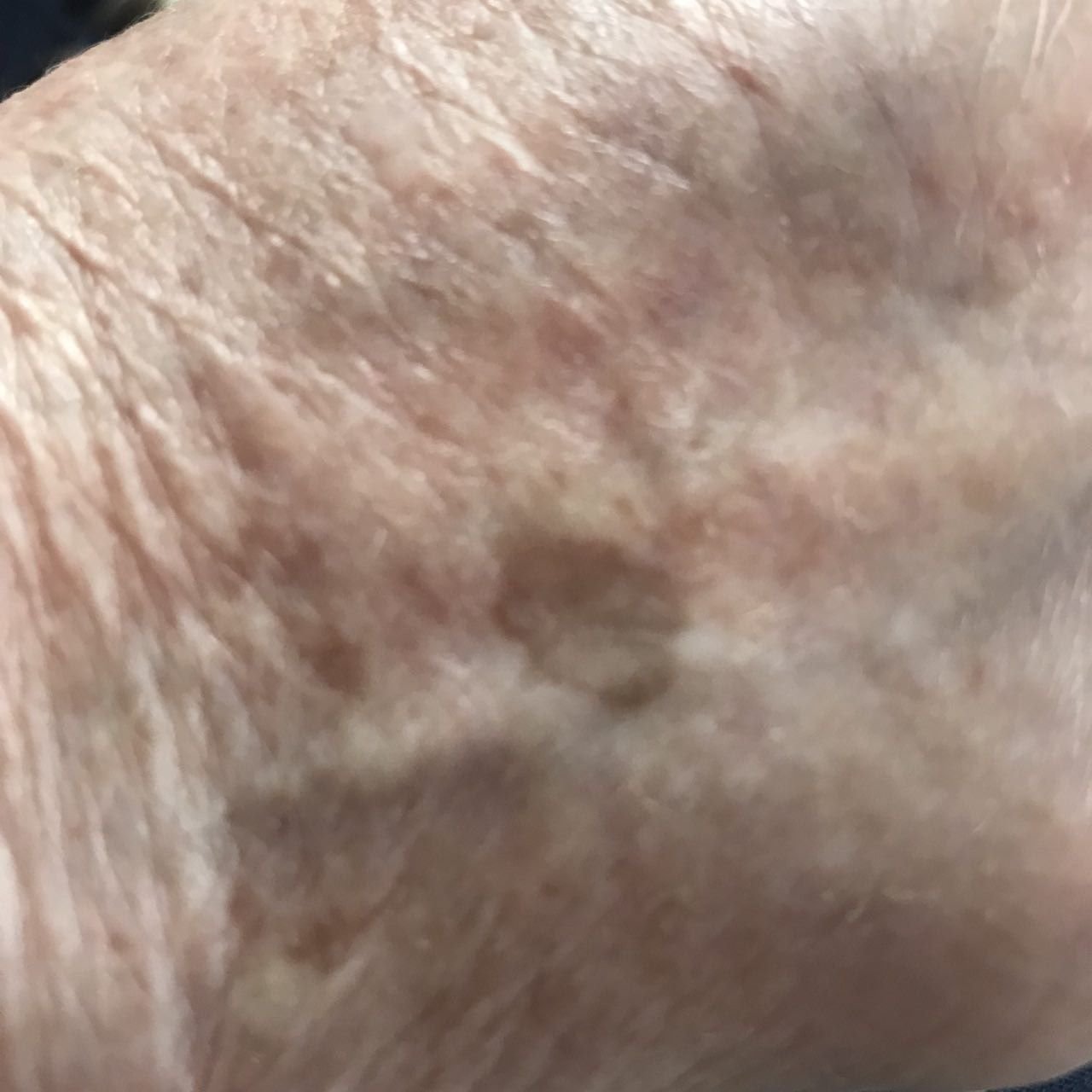
During a visual examination, lentigo appears as a flat spot or a group of spots that are slightly elevated above the skin’s surface (usually no more than 1 mm). The lesions may be symmetrical or irregular in shape, such as when several spots merge together or form a group. Lentigo can appear as a collection of multiple spots, sometimes covering entire anatomical areas. The surface of lentigo typically resembles the texture of normal skin, though small rough areas or peeling may sometimes be observed.
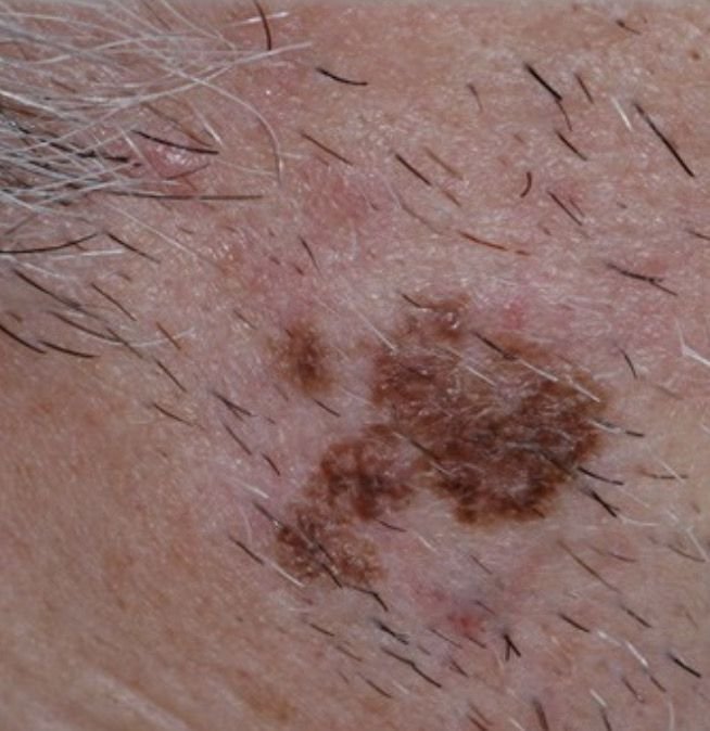
The boundaries of lentigo are usually clear but can often be uneven, especially with larger or multifocal spots. The coloration ranges from light brown to dark brown, and the pigment is usually evenly distributed throughout the lesion. Occasionally, there is a gradual decrease in color intensity from the center towards the edges, or an irregular change in shade within the area of pigmentation. Over time, the color of lentigo may become more intense. In some cases, shades of gray may appear due to keratinization in the upper layers of the epidermis.
Lentigo does not typically affect hair growth. However, in some cases, coarse or fluffy hair may grow in the central area of the lesion.
The size of lentigo can vary widely. Individual spots can range from 2-3 mm in diameter to much larger spots, up to 3-4 cm. When lesions are grouped together, they may cover several areas, sometimes extending to tens of centimeters. On palpation, lentigo feels like normal skin, though in older lesions, roughness may be noted, and mild itching can occasionally occur.
Lentigo is most commonly found on sun-exposed areas, such as the face, neck, and hands. In elderly individuals, lentigo may also appear on other areas of the body.
Dermatoscopic examination of lentigo reveals the following features:
It is important to differentiate lentigo from other pigmented neoplasms or conditions, including:
Lentigo is generally considered safe and does not have a significantly increased risk of malignant transformation. In the absence of external influences such as trauma or ultraviolet radiation, the risk of malignancy remains low, comparable to the risk of melanoma in unchanged skin. However, signs of potential malignancy include changes in the appearance of the lesion, such as increased size, irregular shape, or the appearance of subjective sensations such as itching or bleeding.
Malignant lentigo (also known as Dubreuilh melanosis) is a precancerous condition, and individuals with this form of lentigo have a significantly higher risk of developing melanoma.
If lentigo shows no signs of damage or significant changes in appearance, self-monitoring is usually sufficient. This should include annual checks for changes, particularly in hard-to-see areas. If mechanical damage to the lesion occurs, or if any changes in appearance or new sensations develop, a dermatologist or oncologist should be consulted immediately.
A healthcare provider will determine whether further monitoring or removal is required based on the lesion’s characteristics. Nevi that are subject to chronic trauma (due to clothing, jewelry, or professional activities) should be considered for removal to prevent further irritation.
For individuals undergoing dynamic observation, it is helpful to photograph the lentigo to document any changes over time. Patients with multiple lentigo lesions should be evaluated by a dermatologist or oncologist, ideally before and after the summer months (to assess sun exposure). Creating a map of skin neoplasms is also a useful tool for monitoring changes in existing lesions and detecting new ones.
Lentigo is typically considered a cosmetic issue, and treatment options are usually discussed on an individual basis. If the cosmetic appearance of lentigo is not a concern, treatment may not be necessary. However, for those seeking removal, small lesions can be removed surgically. For multiple lentigo lesions that are similar in appearance, conservative treatments such as cosmetic procedures may be used.
Any treatment of lentigo with destructive methods (laser treatment, cryodestruction, or cosmetic procedures) should be done under the supervision of a dermatologist or oncologist, preferably after dermatoscopic evaluation. Destructive methods are generally not recommended for pigment lesions, as it may be difficult to identify malignant degeneration in a timely manner through clinical examination alone.
If surgical removal is not possible, or if cosmetic improvement is a priority, careful monitoring of the area where the lentigo was located is crucial after treatment.
The prevention of lentigo and its malignant degeneration involves gentle and consistent care of the skin:
Regular examination of lentigo spots, timely consultation with a specialist if any changes are noticed, and removal of potentially dangerous lesions are essential for maintaining skin health.