Lichen nitidus is a rare, chronic, non-infectious dermatological condition that manifests as numerous tiny, shiny papules on the skin. The disease is typically asymptomatic, benign in nature, and usually self-limiting. Although its pathogenesis remains poorly understood, lichen nitidus is classified among inflammatory papular dermatoses and often requires only observation.
The disease can affect individuals of any age or sex but is more commonly seen in children and young adults, especially boys aged 3–10 years. In most cases, no specific treatment is needed, although topical or systemic therapy may be considered in cases involving widespread lesions or symptomatic discomfort such as itching.
The precise causes of lichen nitidus are still unknown. Several theories suggest that it may be an immune-mediated or autoimmune skin reaction triggered by environmental, infectious, or internal factors. In some cases, it may be associated with other inflammatory skin diseases, such as atopic dermatitis, psoriasis, or vitiligo.
Lichen nitidus has also been described as a potential infectious-allergic reaction, as some patients demonstrate improvements with antibiotic therapy or have a history of focal bacterial infection. A possible genetic predisposition or association with allergic sensitivity is also under consideration due to overlaps with atopic backgrounds.
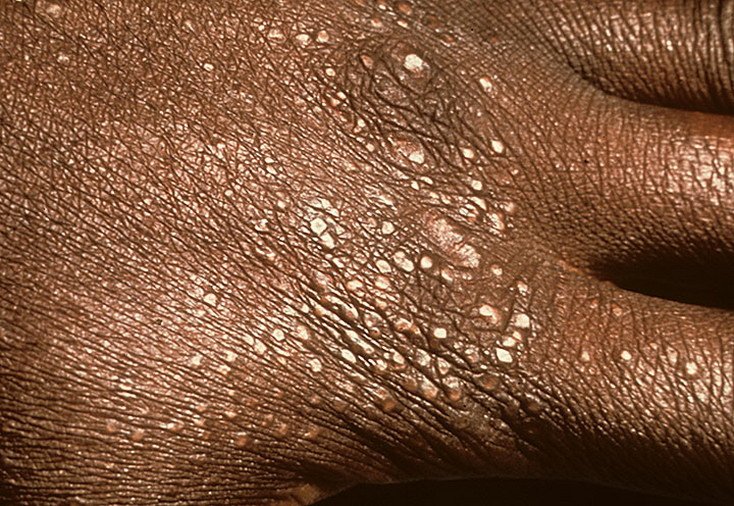
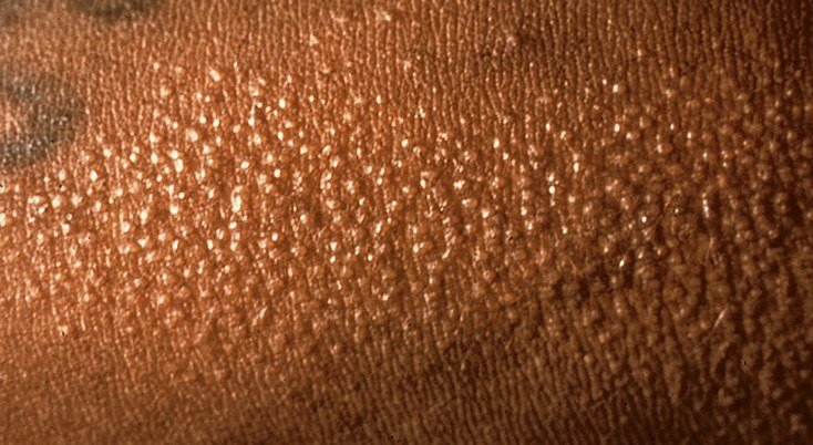
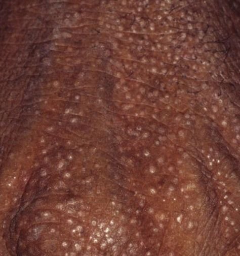
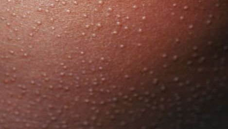
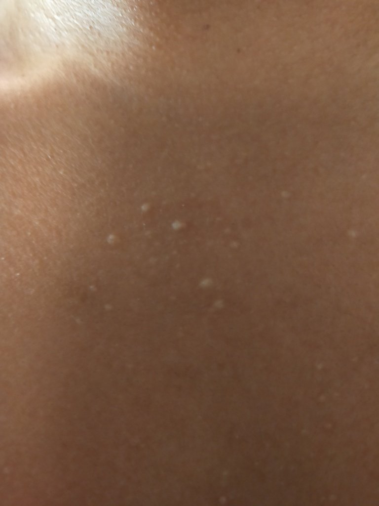
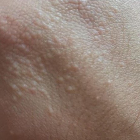
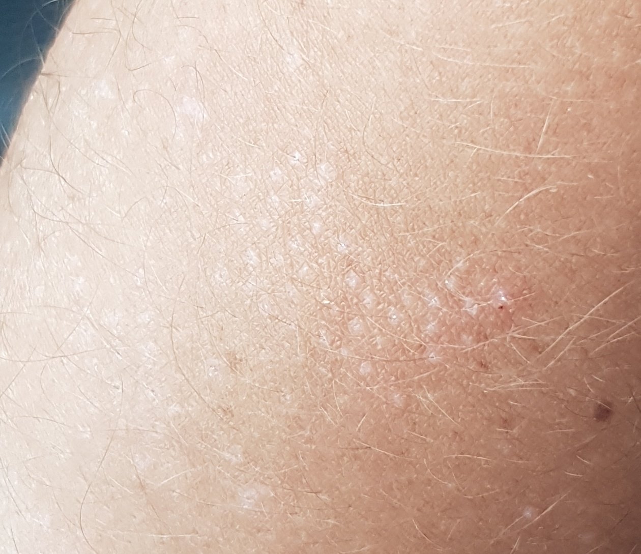
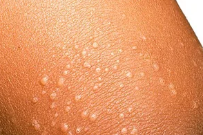
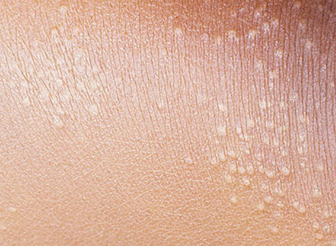
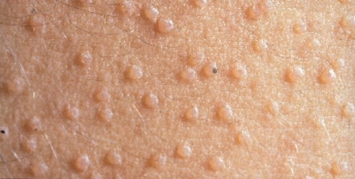
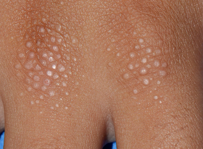
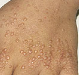
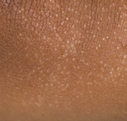
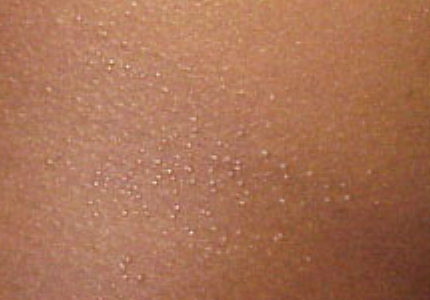
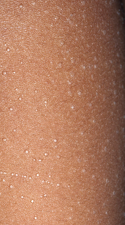
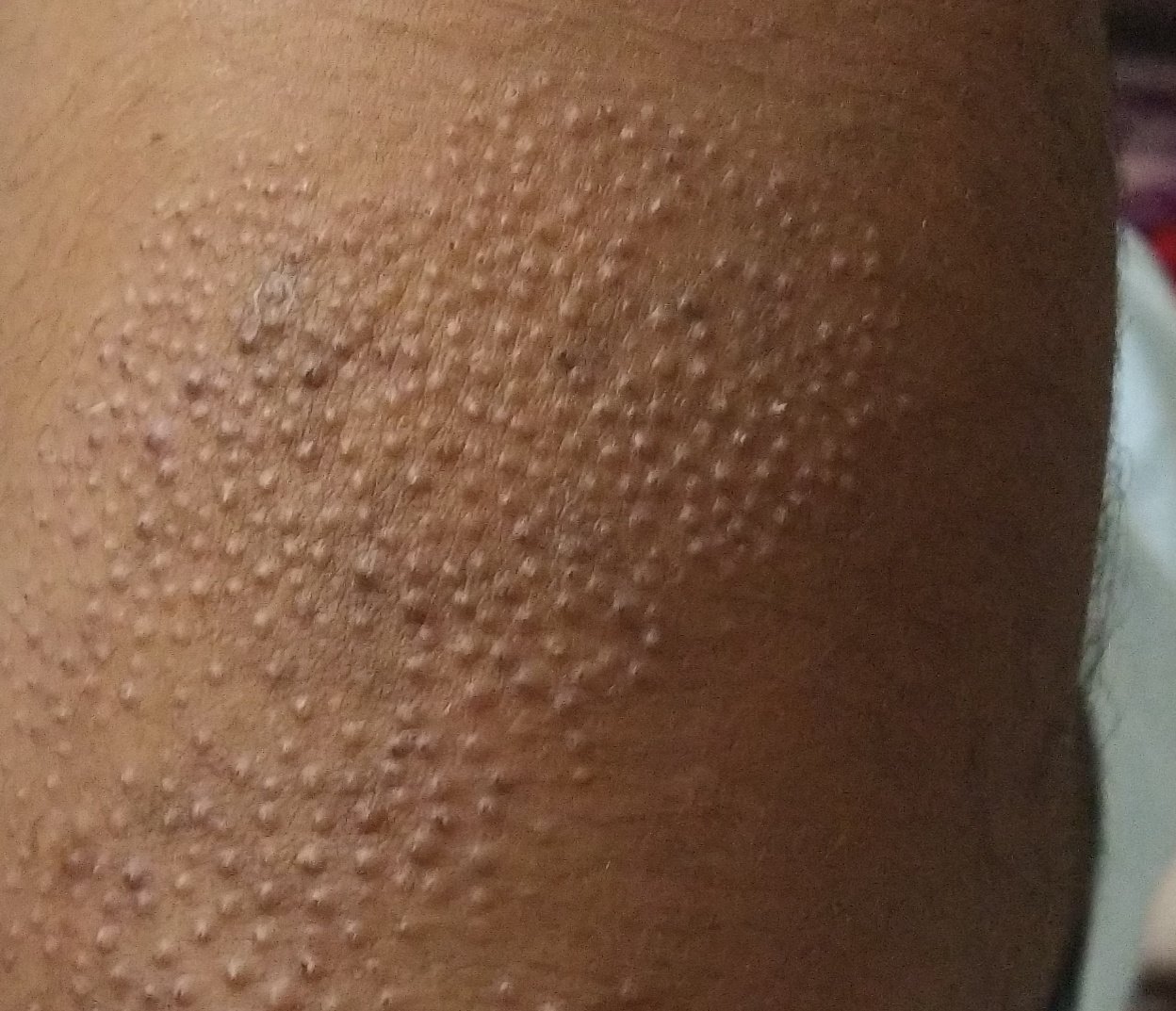
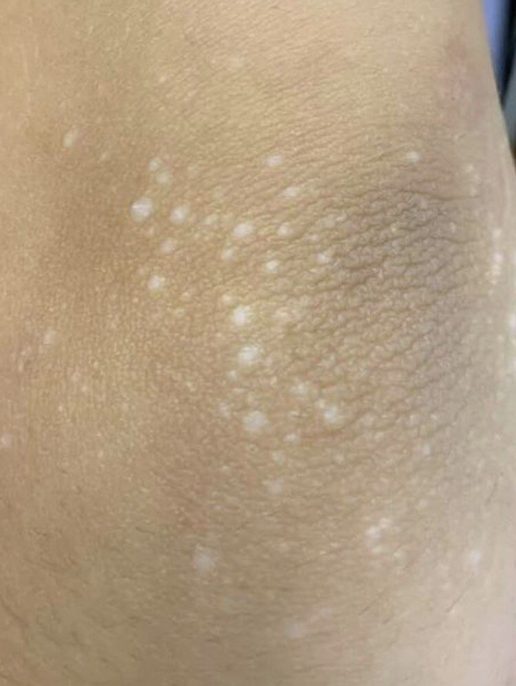
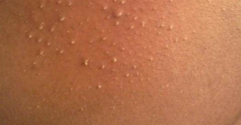
The main symptom of lichen nitidus is the appearance of small, flesh-colored or slightly pink papules, generally 1–3 mm in diameter. These papules have a shiny, smooth surface and a flattened top. They may remain isolated or cluster together in groups, forming larger patches of densely packed lesions, but do not merge into plaques like in other dermatoses.
Lesions most commonly appear in the following locations:
In widespread cases, papules may affect large portions of the trunk or limbs, potentially leading to cosmetic concern or mild discomfort.
The diagnosis of lichen nitidus is primarily clinical and based on the characteristic appearance of the papules and their distribution pattern. In most cases, a dermatologist can confirm the diagnosis through visual examination and patient history.
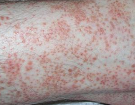
Several dermatoses can resemble lichen nitidus, making differential diagnosis essential in ambiguous cases:
In most cases, lichen nitidus resolves spontaneously within several months and does not require specific treatment. However, if lesions are extensive, persistent, cosmetically concerning, or associated with pruritus, the following treatment options may be considered:
Lichen nitidus is generally considered a benign and self-limiting disease with a favorable outcome. Serious complications are rare. However, in some individuals, visible lesions may cause:
The prognosis for lichen nitidus is excellent. In most cases, the rash resolves on its own within months to a couple of years. Generalized forms may take longer to remit, and isolated cases may persist intermittently. The condition does not increase the risk of malignancy or systemic disease.
Lichen nitidus is a rare, non-contagious, chronic papular dermatosis of unknown origin. Despite its benign nature and usually asymptomatic course, it may pose diagnostic and cosmetic challenges. Most patients do not require treatment, but topical or systemic therapies may be used when lesions are symptomatic or widespread.
Timely consultation with a dermatologist ensures accurate diagnosis, monitoring for atypical evolution, and the exclusion of more serious skin diseases that may mimic lichen nitidus.