Papillomatous Nevus, also known as benign nevus, pigmented nevus, or mole, is a benign skin growth that typically rises above the skin’s surface. This type of nevus is generally acquired, and its occurrence tends to increase with age, peaking between the ages of 15 and 30. Papillomatous nevi are often characterized by their multiplicity, and the number of such lesions tends to increase over time. In terms of gender, papillomatous nevi are somewhat more common in women compared to men, with a ratio of 3:2, respectively.
The exact cause of papillomatous nevi remains unclear. However, several predisposing factors have been identified that may increase the risk of developing these skin neoplasms. These factors can influence the onset and growth of papillomatous nevi:
The diagnosis of papillomatous nevi is primarily based on a clinical examination, which includes visual inspection of the lesions and dermatoscopy to assess the structure of the growths. If malignant growth is suspected, a biopsy may be conducted to obtain a definitive diagnosis and rule out other skin conditions.
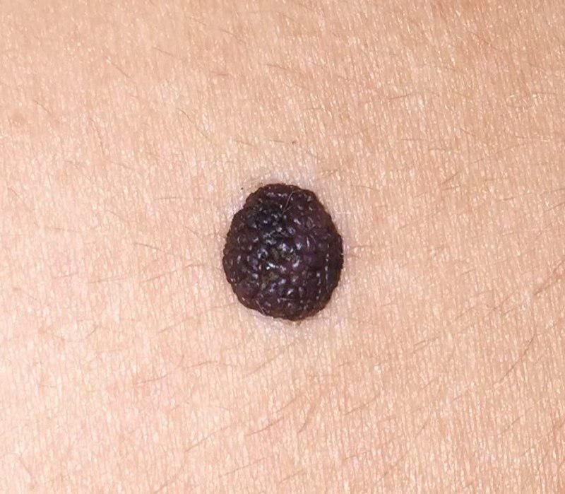
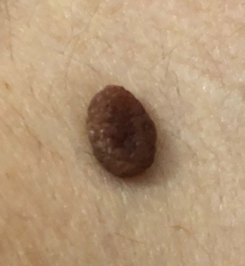
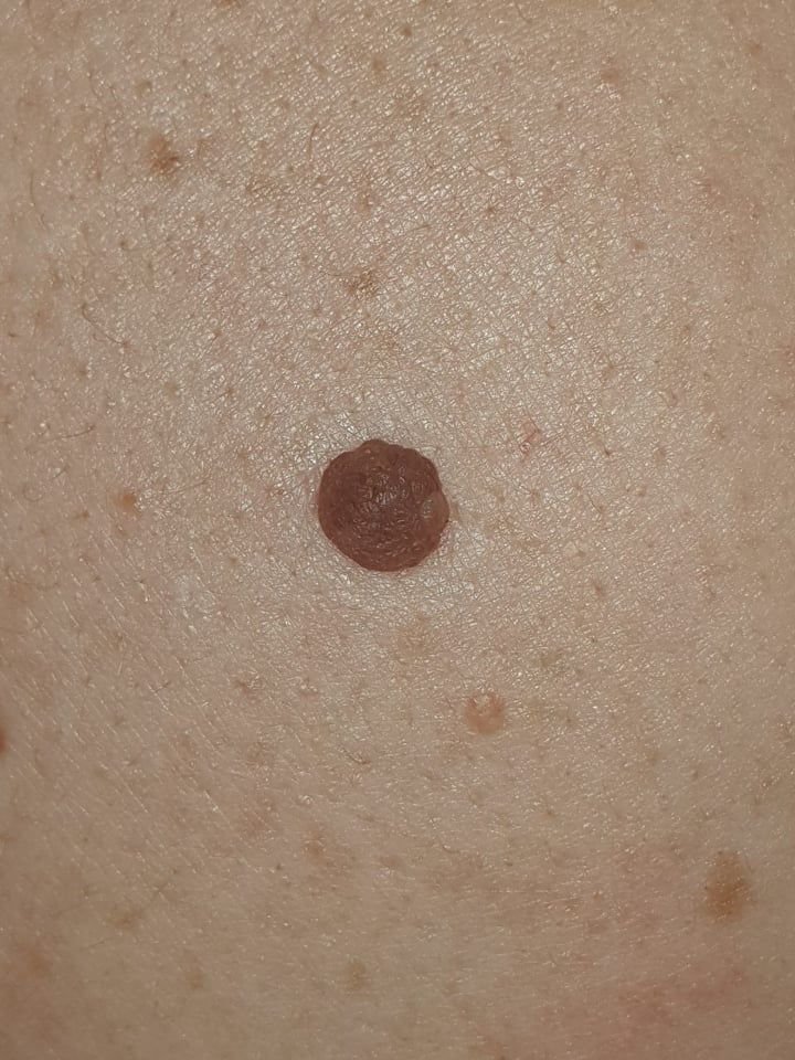
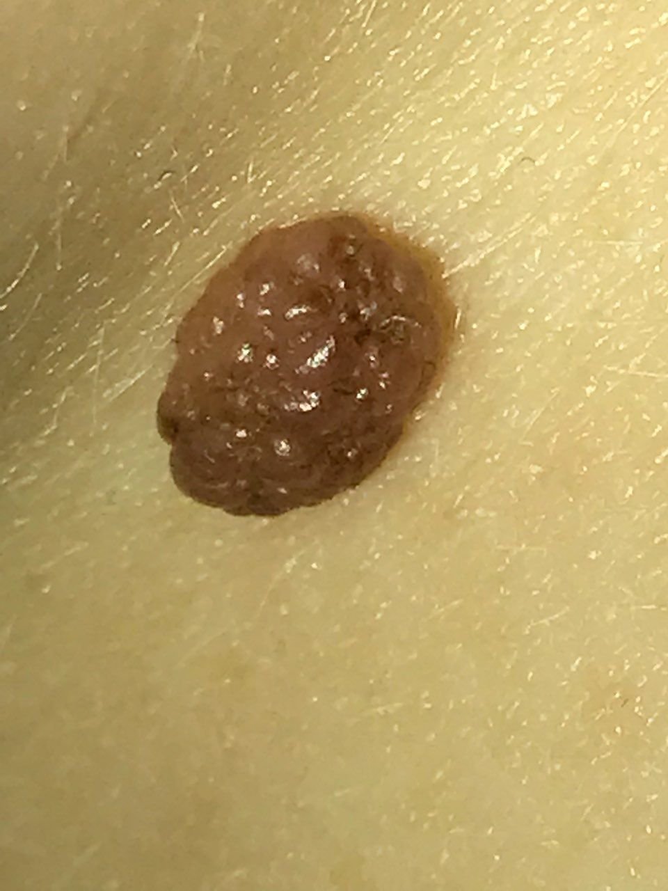
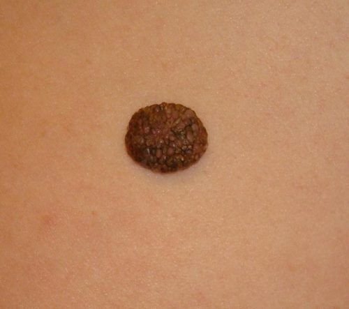
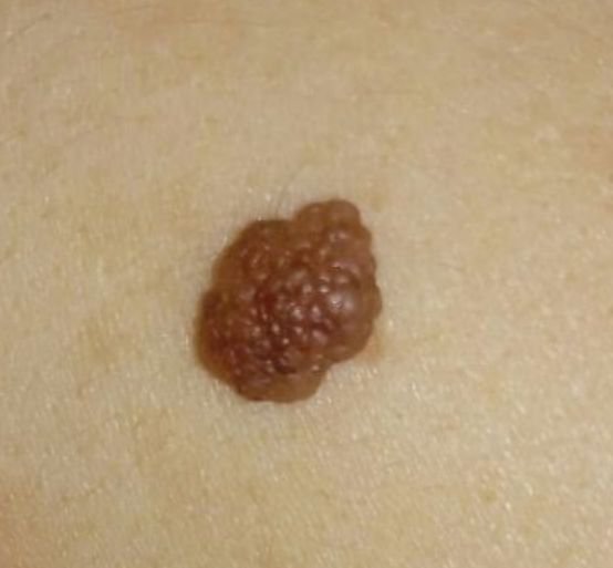
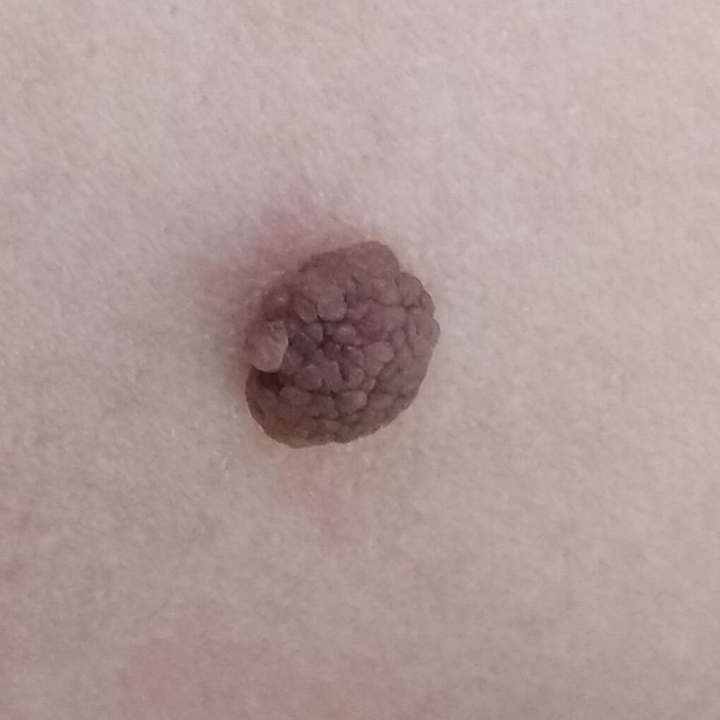
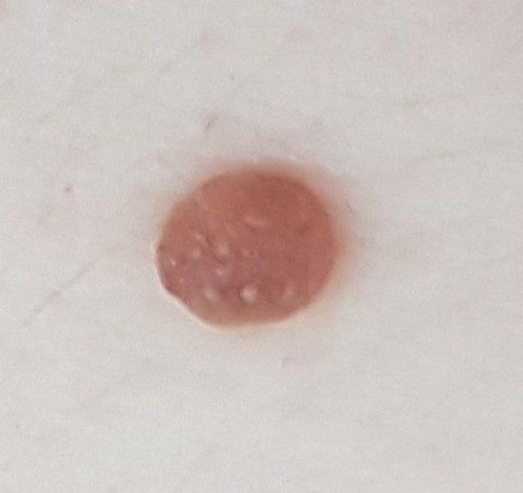
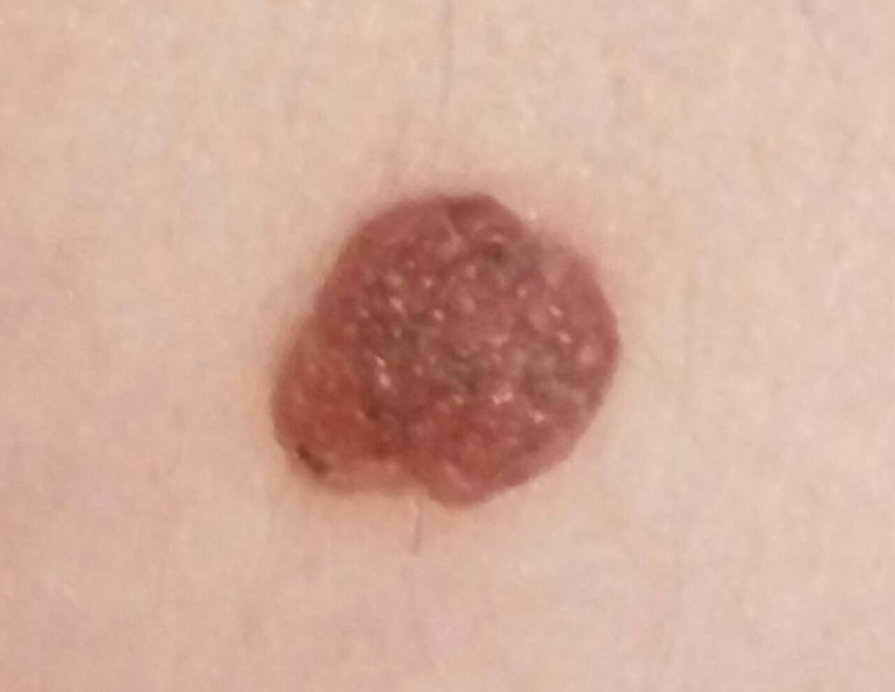
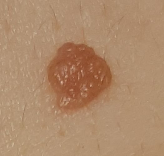
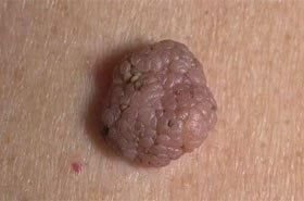
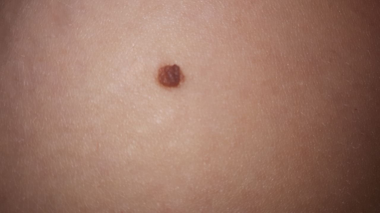
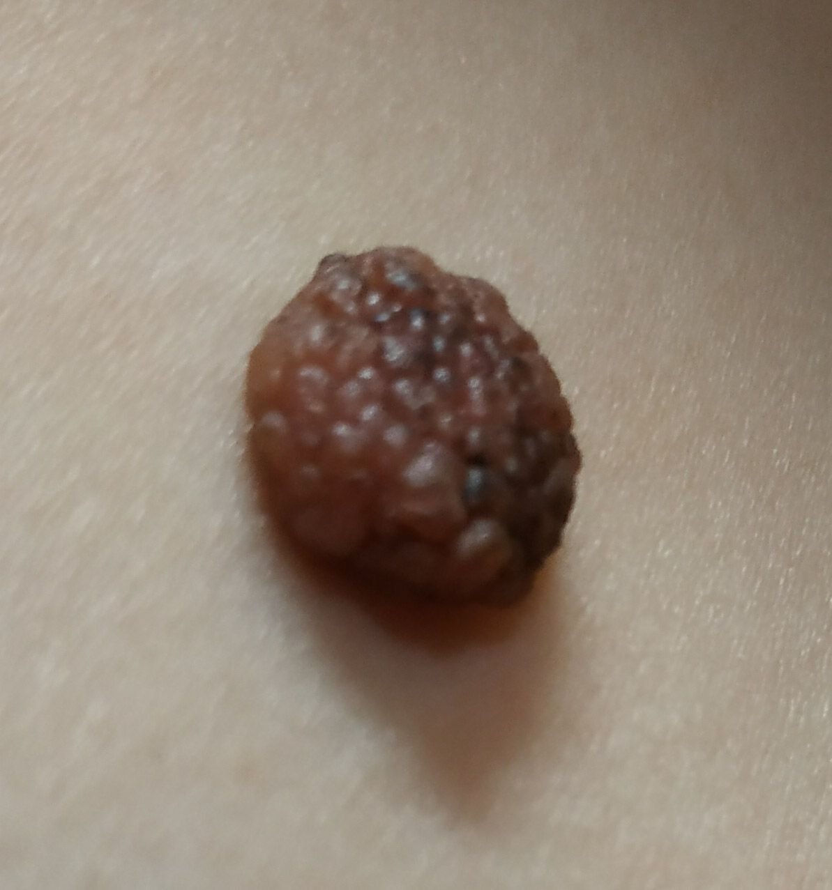
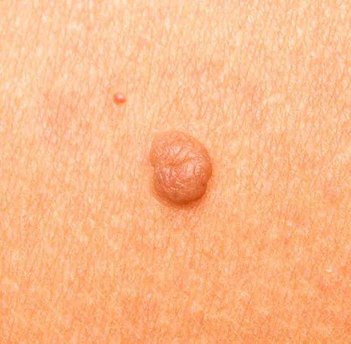
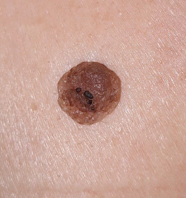
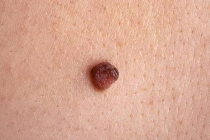
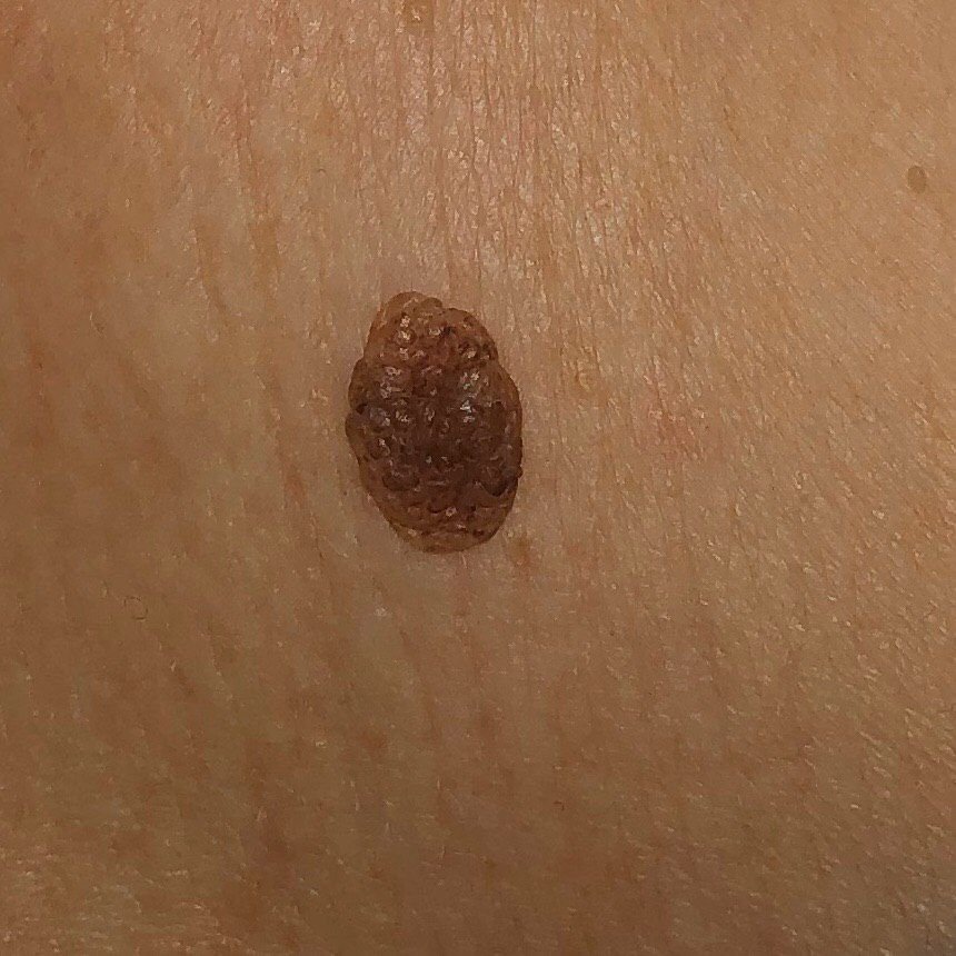
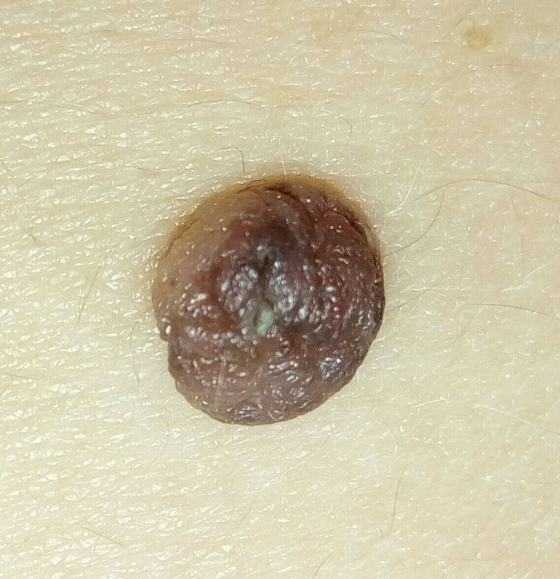
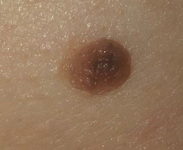
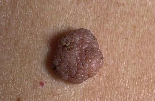
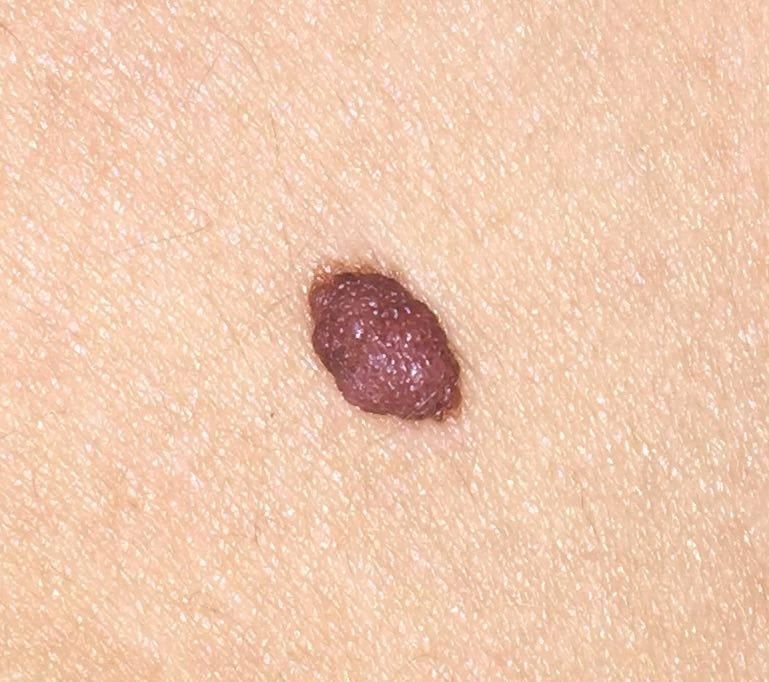
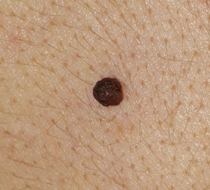
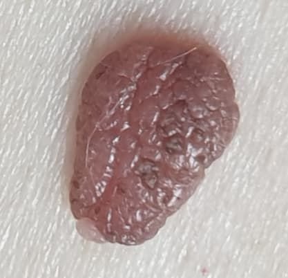
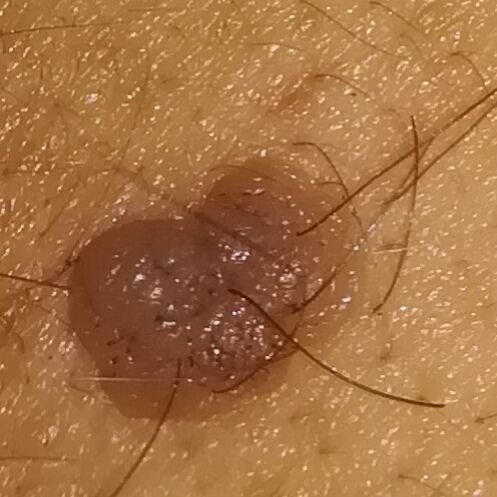
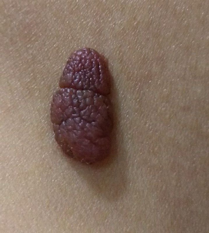
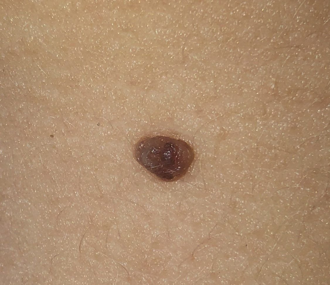
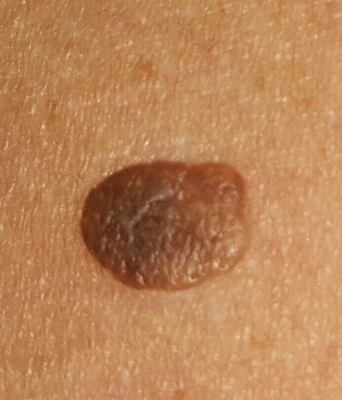
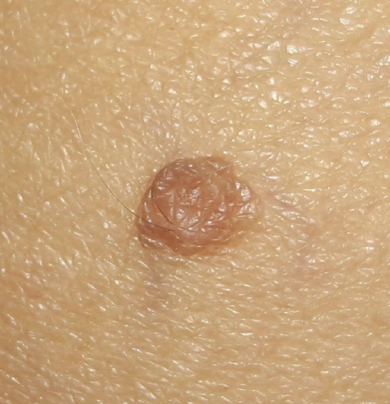
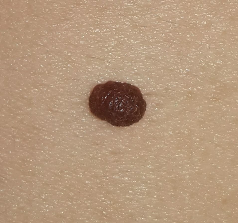
Upon visual examination, a papillomatous nevus typically presents as a hemispherical or slightly elevated growth that rises above the skin on a short, wide stalk (pedicle). The shape of the lesion is most often symmetrical (oval or round), though large nevi may have irregular shapes. The surface of the nevus may vary, with smaller papillomatous nevi exhibiting a smooth texture resembling normal skin, while larger nevi may appear slightly tuberous or even rough. Larger papillomatous nevi (over 8 mm) may have a coarse, warty surface, which is characteristic of verrucous nevi.
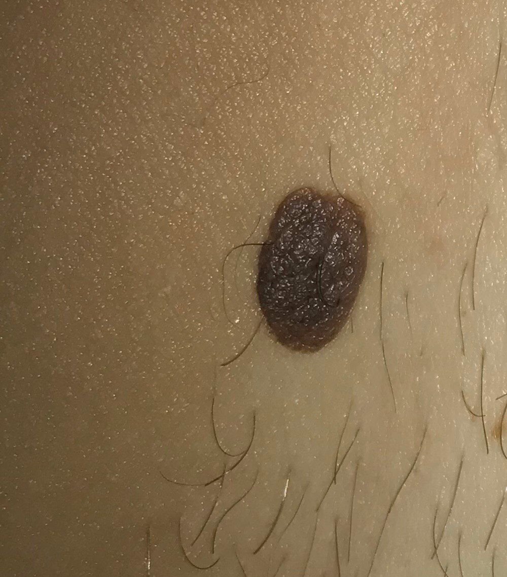
The borders of papillomatous nevi are typically clear and even, although larger nevi may have uneven edges. The color of the nevus can range from flesh-colored to tan and dark brown, and the pigment distribution is typically uniform. Occasionally, there is a gradual decrease in color intensity from the center to the periphery or slight variations in shade within the same lesion (which is typical for verrucous nevi).
Hair growth in the area of a papillomatous nevus is typically unaffected. In some cases, coarse bristly hair may develop in the center, especially in congenital papillomas, or downy hair may appear in areas of hypopigmented papillomatous nevi.
The size of papillomatous nevi can vary widely, with most lesions measuring up to 15 mm in diameter. Nevi larger than 15 mm are rare. The height of these nevi above the skin level is usually less than 10 mm. Large papillomatous nevi that resemble cauliflower are quite rare.
On palpation, papillomatous nevi have a consistency similar to that of normal skin, though larger lesions may feel slightly softer. There are typically no subjective symptoms associated with papillomatous nevi, although mild itching may occasionally occur in long-standing forms.
These neoplasms are most commonly located on the face, scalp, neck, and trunk (including the chest and back), though they can occasionally appear on other areas of the body.
When examining a papillomatous nevus under dermatoscopy, the following features are typically observed:
It is important to differentiate papillomatous nevi from other pigmented or nodular skin lesions, such as:
Papillomatous nevi are benign and do not carry an increased risk of melanoma or other malignancies. In the absence of external influences such as trauma, ultraviolet radiation, or ionizing radiation, the risk of malignant degeneration is low and comparable to the risk associated with normal skin. However, signs of potential malignancy include a change in the appearance of the nevus (such as rapid growth or irregular shape), an increase in its density, and the appearance of subjective sensations like pain or itching.
The primary risk associated with papillomatous nevi is their tendency to become easily injured due to their elongated shape and narrow stalk. When this occurs, the lesion can bleed, become painful, and create an opening for pathogenic microorganisms, leading to infection. Papillomas can also cause psychological discomfort, especially when located in visible areas.
Since papillomatous nevi are often viral in origin and are typically multiple, the presence of these lesions suggests a decreased immune defense against the human papillomavirus (HPV). Although HPV has a relatively low oncogenic risk, it is important for individuals with multiple papillomas to undergo routine oncological evaluations.
If there are no signs of damage, changes in appearance, or symptoms in the papillomatous nevus, self-monitoring is usually sufficient. This should include periodic checks, at least once a year, to track any changes. If mechanical damage occurs, if the nevus is exposed to ultraviolet or ionizing radiation, or if any changes are observed, a consultation with a dermatologist or oncologist is necessary.
The healthcare provider will determine whether dynamic monitoring is sufficient or if removal of the lesion is recommended. Nevi that are subject to constant or chronic trauma, such as from clothing, jewelry, or professional activities, should be removed to prevent further injury. Additionally, some individuals may wish to remove papillomatous nevi for cosmetic or psychological reasons.
For dynamic observation, it is useful to take photographs of the papillomatous nevi to monitor any changes over time. Patients with multiple papillomas should undergo a dermatologist examination, ideally in the spring and autumn (before and after the summer sun exposure). It is also recommended to compile a map of skin neoplasms to assist with further monitoring and to identify new or altered lesions.
Treatment of papillomatous nevi depends on their size, location, and whether they cause any discomfort. Less invasive methods are preferred when possible:
If less invasive methods are not suitable, or if there is any uncertainty about the nature of the nevus, surgical excision with histological examination is the next step.
Self-removal of papillomatous nevi is strongly discouraged due to the risk of complications, such as bleeding, infection, and misdiagnosis of the lesion’s nature. If a papilloma is removed surgically, careful monitoring is necessary to ensure the area heals properly and to detect any recurrence.
The prevention of papillomatous nevi involves proper care of the skin and addressing underlying health factors:
Regularly inspecting papillomatous nevi, consulting a healthcare professional if any changes occur, and removing potentially dangerous lesions are essential for maintaining skin health.