Pyogenic Granuloma is a benign neoplasm, characterized by the localized proliferation of blood capillaries, often in response to an external injury. This type of neoplasm appears as a small, raised, bright red lesion, which can vary in size and shape. Pyogenic granulomas are commonly found in various parts of the body, including mucous membranes, conjunctiva, and even the cornea. These lesions are more prevalent in young individuals and pregnant women due to the changes in immune and hormonal functions that can influence their formation.
The exact cause of pyogenic granulomas is not well-defined. While injuries were previously considered the primary cause, recent research shows that only 25% of all pyogenic granulomas are linked to injuries. There are numerous potential predisposing factors, including:
Diagnosing a pyogenic granuloma is typically based on a thorough clinical examination, which includes visual inspection of the lesion and dermatoscopic evaluation. If there is any suspicion that the growth may be malignant or if the lesion is unusually large, a biopsy may be necessary for further examination.
For congenital or large pyogenic granulomas that may occupy critical areas, such as near vital organs or blood vessels, an ultrasound and a multidisciplinary evaluation by specialists may be required to assess the extent of the lesion and determine appropriate treatment.
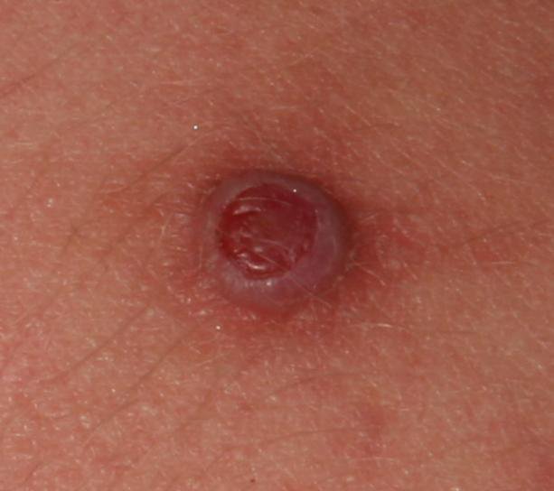
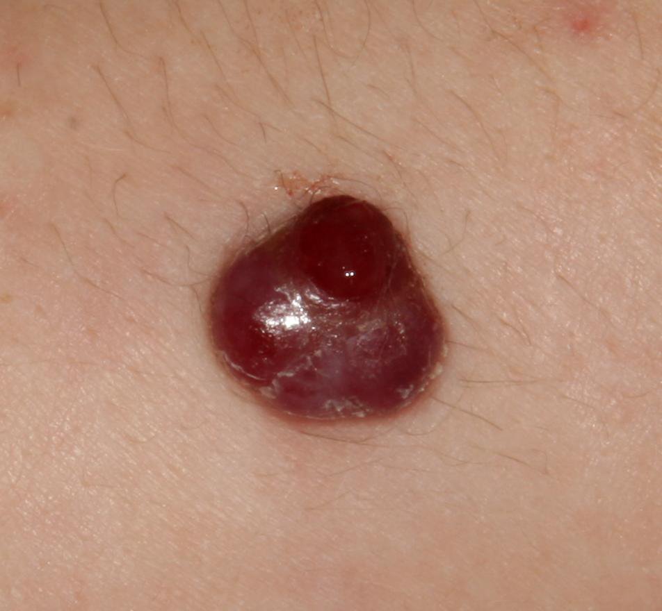
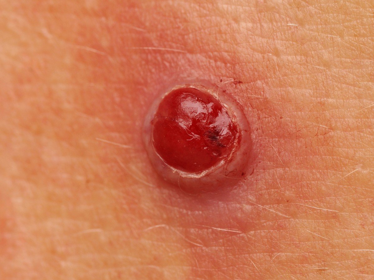
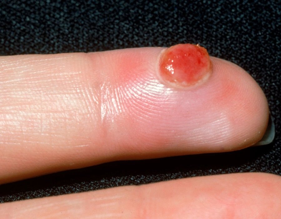
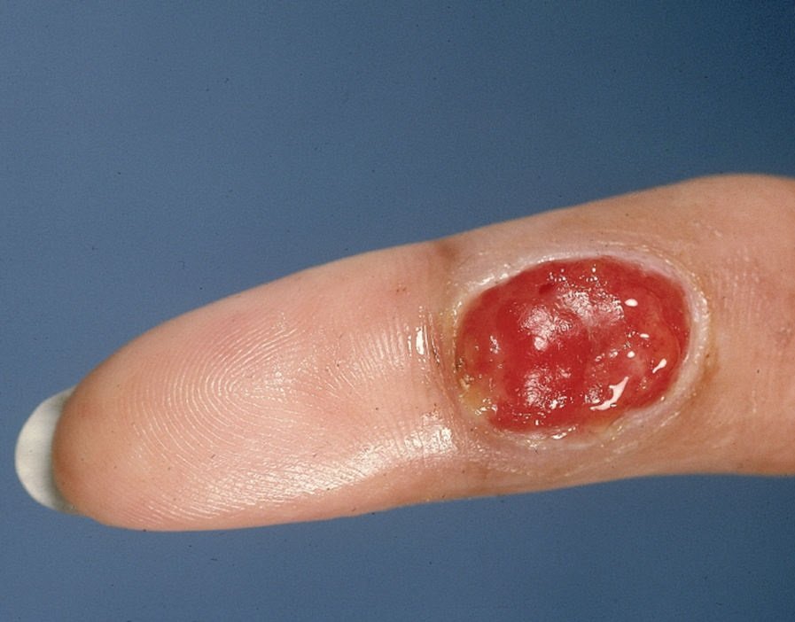
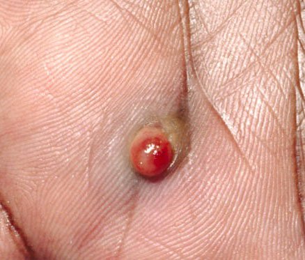
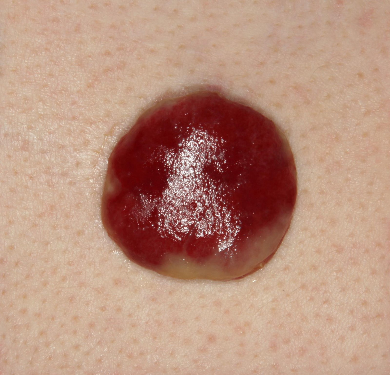
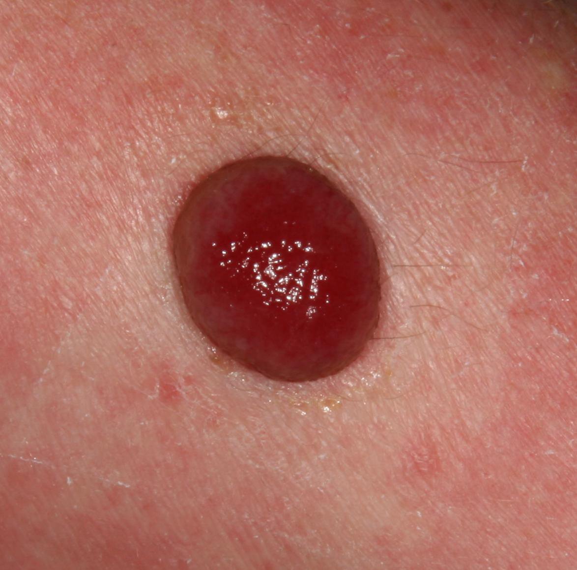
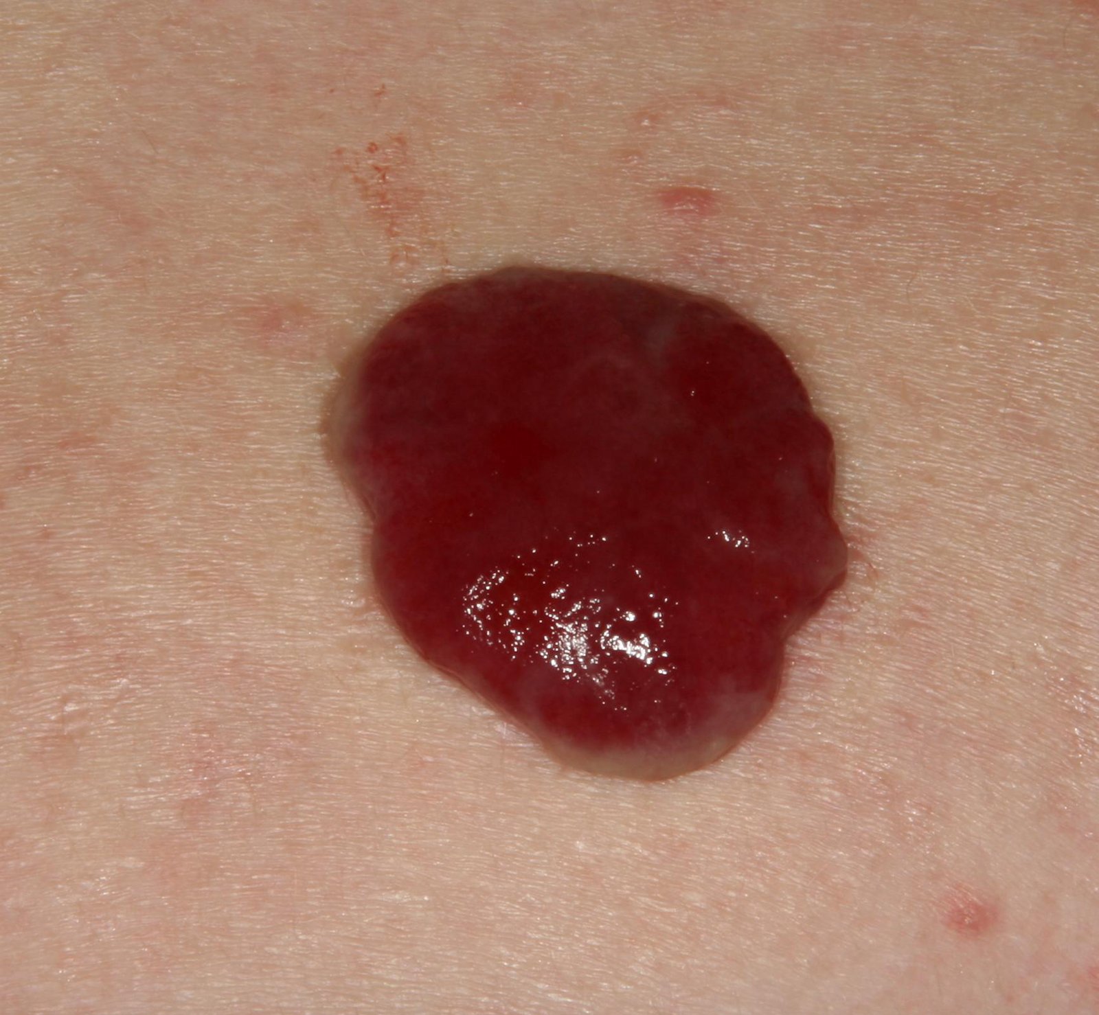
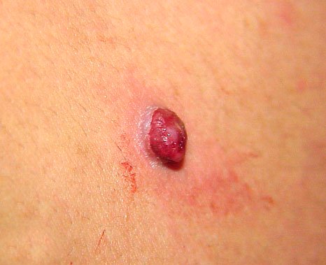
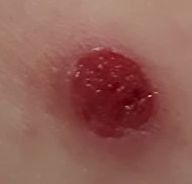
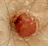
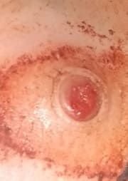
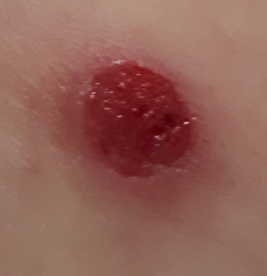
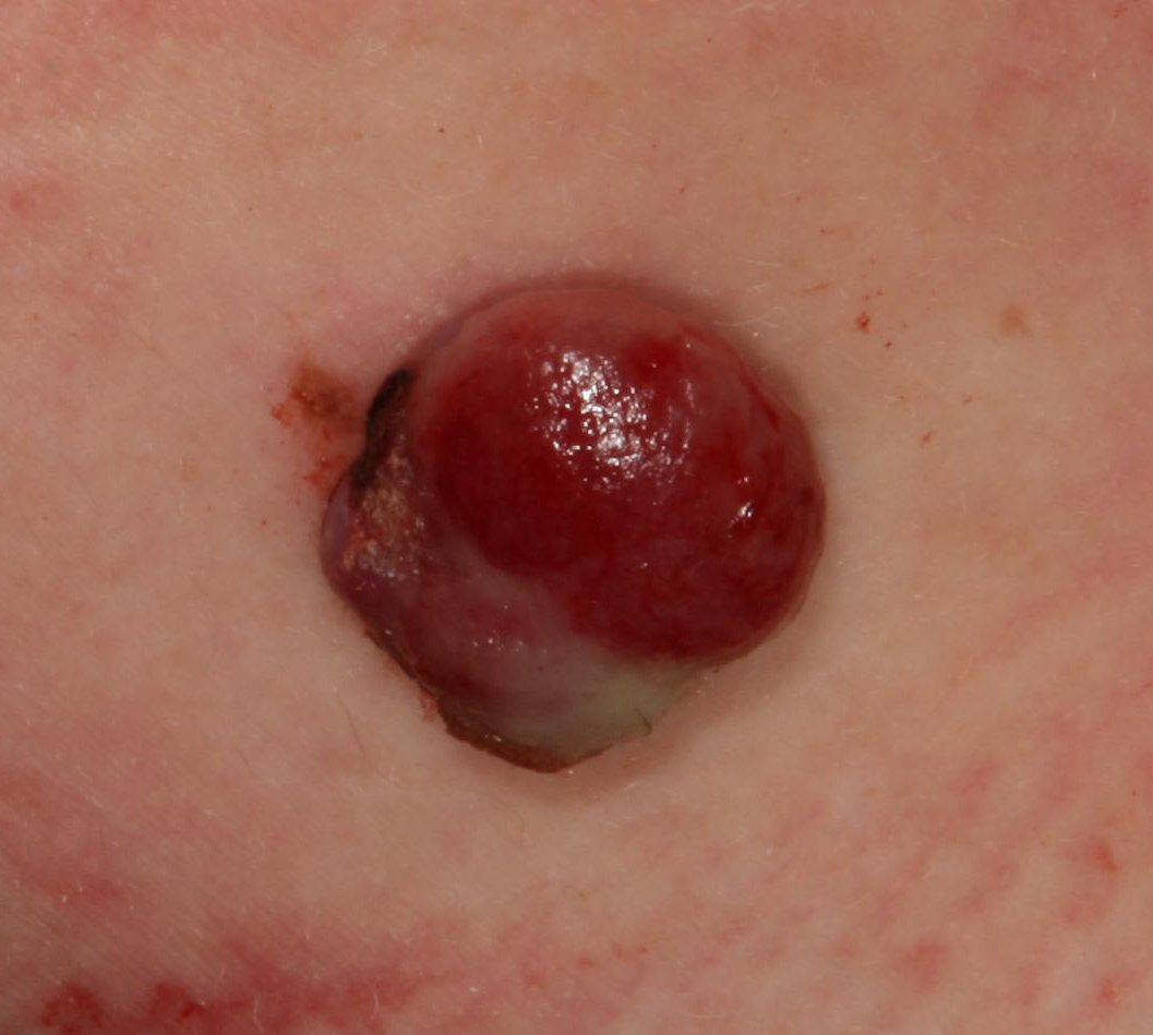
Upon visual examination, pyogenic granulomas present as a hemispherical or dome-shaped growth that rises above the skin on a short, wide stalk (pedicle). Most often, these growths are symmetrical, appearing oval or round. The surface texture of the granuloma can differ from the surrounding skin, sometimes appearing smooth or lobed, resembling raspberries. The granuloma may have a shiny or “wet” appearance due to the presence of minor erosion or crusts. If the granuloma is injured, it may bleed easily, and larger granulomas that become infected can form a purulent plaque with areas of necrosis.
The boundaries of pyogenic granulomas are usually well-defined but can be uneven in larger lesions. The color of the granuloma is often bright red, though it may appear cyanotic (blue or purple) or even yellow or gray in the presence of purulent material or necrotic tissue. When pressure is applied to the granuloma, the red color fades temporarily.
Hair does not grow in the area of a pyogenic granuloma. However, in some cases, coarse hair may grow at the center of the lesion, particularly in congenital forms or large granulomas.
The size of pyogenic granulomas typically ranges from 3 to 15 mm. Lesions larger than 15 mm are rarer and are often associated with underlying systemic diseases or immunodeficiency states. The granuloma grows rapidly, often reaching a diameter of 1-1.5 cm within a short period. Its height typically does not exceed 5 mm. Spontaneous regression is rare but may occur, especially in pregnant women after childbirth.
On palpation, pyogenic granulomas feel soft and elastic, without tenderness. After infection, the granuloma may become painful. There are typically no subjective symptoms unless the granuloma becomes injured or infected.
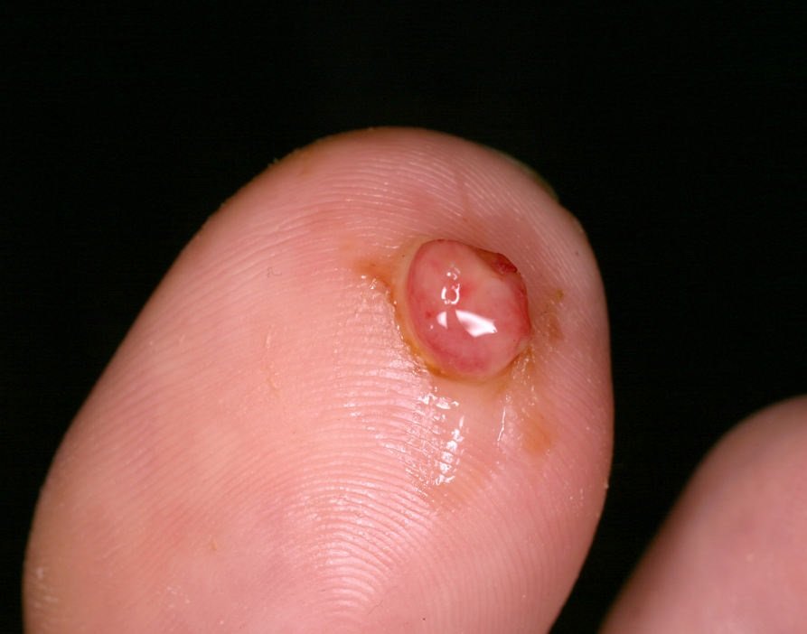
These lesions are most commonly found on the hands and feet, particularly on the palmar and plantar surfaces of the fingers, where injuries and contact with foreign bodies are more likely. Pyogenic granulomas can also appear near the nail ridges (such as from ingrown nails) or on the face. Less commonly, they may appear on mucous membranes, depending on predisposing factors like burns, pressure sores, or physical trauma.
Dermatoscopic examination of a pyogenic granuloma reveals the following characteristics:
Pyogenic granulomas must be differentiated from other skin lesions with similar characteristics, including:
From an oncological perspective, pyogenic granulomas are safe and do not carry an increased risk of malignant transformation. In the absence of external factors such as trauma, UV radiation, or ionizing radiation, the risk of malignancy remains comparable to that of healthy skin. However, if there is a noticeable change in the appearance of the granuloma, such as rapid growth, increased density, or the appearance of symptoms like itching or pain, a dermatologist or oncologist should be consulted.
One of the primary risks associated with pyogenic granulomas is their susceptibility to injury due to their raised and delicate nature. This can lead to bleeding, soreness, and infection, which can further complicate the condition. In large granulomas, bleeding can be difficult to manage without medical intervention. Furthermore, because some malignant tumors can resemble pyogenic granulomas or develop next to them, it is important to undergo a timely differential diagnosis to ensure that no malignant growth is overlooked.
If a pyogenic granuloma is detected, a consultation with a dermatologist or oncologist is recommended. After confirming that the lesion is benign, the possibility of conservative management or the need for treatment can be evaluated. Small granulomas, especially in pregnant women, may resolve spontaneously, and observation may be sufficient. In all other cases, treatment is typically recommended.
For patients who refuse treatment, active monitoring is essential. This includes photo documentation of the lesion to track any changes in appearance. Patients with multiple pyogenic granulomas should see a dermatologist or oncologist in the spring and autumn (before and after the summer season). Regular mapping of skin neoplasms can also help with observation and tracking any new or altered lesions.
The most common treatment for pyogenic granulomas is surgical excision, which involves removing the lesion along with its surrounding skin. This can be done using a classic excision method or with an electric or radio scalpel. After removal, a histological examination is mandatory to confirm that the granuloma is benign.
If the nature of the lesion is confirmed and there are no doubts, less invasive treatments such as laser coagulation, cryodestruction (liquid nitrogen), or electrocoagulation (using an electrical current) may be used for smaller lesions.
Given the vascular nature of pyogenic granulomas, bleeding during removal is common. Therefore, proper hemostasis (control of bleeding) is critical after excision.
Preventing the appearance of pyogenic granulomas involves a careful approach to skin care and proper management of injuries:
In addition, regular examination of the skin, timely consultation with a healthcare specialist in the event of changes in skin lesions, and the removal of potentially dangerous neoplasms are key to maintaining good skin health.