Understanding Linear Skin Eruptions Along Blaschko’s Lines: Diagnosis and Care
Why this skin pattern matters
Some skin rashes follow very specific lines on the body called Blaschko’s lines. These are invisible lines of skin cell development that show up in certain skin conditions. When a rash follows those lines, the list of possible causes is small, but the differences between them are important. A wrong diagnosis can mean months or years of the wrong treatment.
A teen’s long road to the right diagnosis
A 15-year-old boy had a growing, itchy, red, scaly band of skin along his right arm and hand for two years. It began about a month after a routine vaccination, starting at the injection site and slowly spreading down the arm and onto the hand along a clear linear path. He tried several creams without improvement and was misdiagnosed multiple times before reaching a dermatology clinic.
What the doctor found on exam
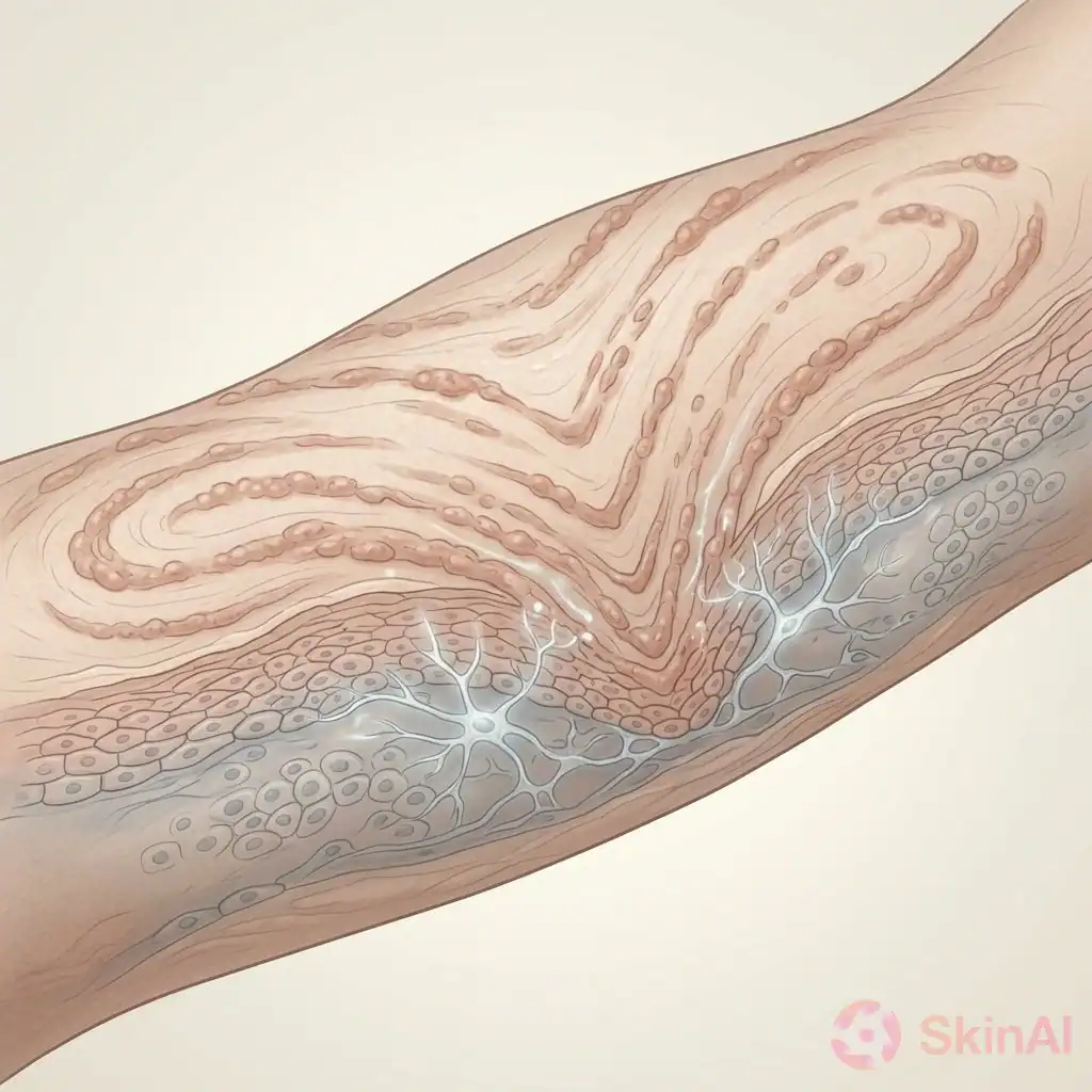
- Unilateral, linear patches that followed Blaschko’s lines on the right arm and hand
- Red, scaly plaques with areas of skin thinning (atrophy)
- Changes in skin color (dyspigmentation)
- Local hair loss in the affected skin
- No nail changes, no mouth sores, and normal sweating
- No signs of systemic illness (no fever, joint pain, muscle weakness, or other symptoms)
The timing around the vaccine was noted, but whether the shot caused the rash is unclear. The diagnostic steps below would be the same regardless of the trigger.
How doctors narrowed the diagnosis
When a rash is strictly along Blaschko’s lines, doctors use that distribution as the first clue. That pattern makes some conditions much more likely than others. From there, two more steps are important: a skin biopsy and checking for any sign of systemic disease.
Step 1: The distribution
The fact the rash was unilateral and strictly linear greatly narrowed the list of likely causes. Other helpful clues were the thinning of the skin, hair loss in the area, and the scaly red appearance. The rash didn’t look like acne-type lesions, it wasn’t a purple hue typical of lichen planus in every case, and it didn’t show an Auspitz sign (tiny pinpoint bleeding when scales are scraped off, which can happen in psoriasis).
Step 2: The biopsy
A small punch biopsy of the skin was taken and examined under a microscope. The lab report described several specific features:
- Lichenoid interface dermatitis — inflammation where the top layer of skin meets the deeper layer.
- Epidermal acanthosis — thickening of the outer skin layer.
- Focal parakeratosis — small areas where the outer skin cells kept their nuclei instead of fully maturing.
- Follicular plugging — openings of hair follicles filled with debris.
- Basal vacuolar degeneration with Civatte bodies — damage to the bottom layer of the epidermis with small dead skin cells visible.
- Vacuolar changes of follicular structures — damage around hair follicle structures.
- Deep periadnexal lymphoplasmacytic infiltrate — immune cells clustered around deeper skin structures like hair follicles.
- Prominent dermal mucin deposition — an increased amount of mucous-like material in the deeper layers of the skin.
Special stains for fungus or mycobacteria were not done because the clinical picture did not suggest those infections. A lupus band test (a direct immunofluorescence test that can support lupus diagnosis) was not available at that center.
Why this matters: of all those findings, the presence of dermal mucin is the most important clue pointing toward a form of cutaneous lupus. Other look-alike conditions that can follow Blaschko’s lines — such as linear lichen planus, blaschkitis, linear psoriasis, lichen striatus, or inflammatory linear verrucous epidermal nevus (ILVEN) — typically do not show this mucin deposit. The combination of mucin with the lichenoid pattern, follicular plugging, and Civatte bodies strongly pointed to a diagnosis called linear cutaneous lupus erythematosus (LCDLE).
Step 3: Making sure the disease is limited to the skin
Even when cutaneous lupus looks likely, doctors check whether lupus is affecting the rest of the body. This patient had no systemic symptoms: no joint pain, no mouth ulcers, no unusual fatigue, and no sensitivity to sunlight. That supported a skin-limited diagnosis.
What this diagnosis means and how it’s handled
LCDLE is a rare, localized form of cutaneous lupus that presents in a linear pattern. It usually stays confined to the skin and does not typically develop into full systemic lupus, but doctors still perform tests to be sure there is no internal involvement.
Treatment decisions are individualized and should be discussed with a dermatologist. Doctors may use oral antimalarial medications, such as hydroxychloroquine, together with topical corticosteroid creams applied to the affected skin. These treatments often help, but responses vary.
Important safety note: because hydroxychloroquine can cause problems in people with glucose-6-phosphate dehydrogenase deficiency, screening for G6PD deficiency is recommended before starting this medicine.
Practical lessons from this case
- The skin distribution is the first and most powerful clue. A unilateral linear rash that follows Blaschko’s lines narrows the likely causes.
- A biopsy is often required to tell similar-looking conditions apart.
- Dermal mucin on biopsy is a key feature that points toward cutaneous lupus rather than other linear skin conditions.
- After biopsy confirms LCDLE, doctors still check for systemic lupus even though progression is uncommon.
- G6PD testing should be done before starting hydroxychloroquine.
When to see a doctor
If you have a new, changing, or spreading rash, or a rash that is painful, bleeding, or causing hair loss in the area, see a dermatologist. Also seek care if you have skin changes plus systemic symptoms such as fever, unexplained joint pain, muscle weakness, mouth sores, or marked sensitivity to sunlight.
A short note about tracking skin changes
Keeping photos or a brief diary of how a rash looks and changes over time can be helpful for your doctor visit. Note when it first appeared, what treatments you tried, and any other symptoms that came with it.
Disclaimer
This article is for general information and does not replace medical advice. Treatment decisions should be made together with your healthcare provider.
Sources
- Jackson R. The lines of Blaschko: a review and reconsideration: Observations of the cause of certain unusual linear conditions of the skin. doi:10.1111/j.1365-2133.1976.tb00835.x
- Saberi F, Ghanadan A, Razavi Z, Azhari VS, Akhdar M, Al-Zahawi S. The first case of linear cutaneous lupus erythematosus following covid-19 vaccination: a case report. Published 2026 May 11. doi:10.1002/ccr3.72703