
New Study Reveals Hidden Scalp Inflammation in Dissecting Cellulitis
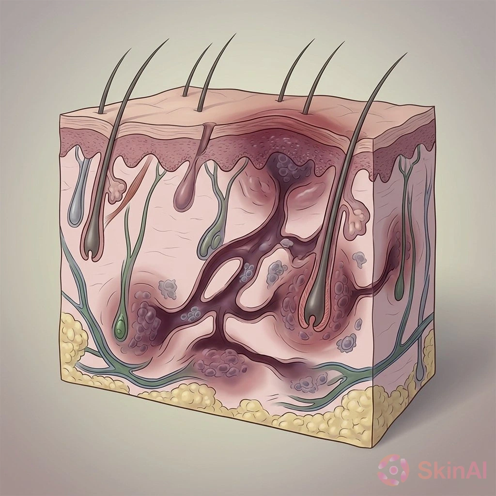
Hidden inflammation may help explain why dissecting cellulitis of the scalp comes back
If you or someone you know has dissecting cellulitis of the scalp, the experience can be painful and frustrating. This condition causes painful bumps on the scalp that can drain, form tunnels under the skin, and lead to scarring and hair loss. A small new study suggests that there may be a quieter, wider inflammatory problem under the skin that isn’t always visible — and that could help explain why the condition often returns or spreads.
Quick summary
Researchers looked at scalp biopsies from 12 men with confirmed dissecting cellulitis of the scalp (often shortened to DCS). They found a consistent pattern of inflammation and scarring around the upper part of hair follicles in areas of scalp that looked normal to the eye. The active, painful lesions had extra signs of destructive inflammation. These findings are early and don’t prove cause and effect, but they suggest a “field” of low-level damage that might lie under visible disease (Source: PR Newswire; Umar et al., Clin Cosmet Investig Dermatol, 2026).
What is dissecting cellulitis of the scalp?
DCS is a chronic inflammatory condition of the scalp that mostly affects young men. It usually shows up as painful lumps or nodules that can become filled with pus, drain, or form tunneling tracks under the skin. Over time these lesions can damage hair follicles and cause scarring hair loss. Because flares can be obvious and painful, people typically notice the disease only when it is active.
What did the study look at?
The researchers did a small retrospective study at a single dermatology clinic. They included 12 men, ages 21 to 46, who had biopsy-confirmed DCS (Source: Umar et al., 2026).
Each person had a 6-mm punch biopsy guided by trichoscopy (a close-up scalp exam). One biopsy came from an area of scalp that looked normal (usually 2 to 3 cm away from an obvious lesion). A second biopsy came from either an active lesion or, if no active lesions were present, a clinically inactive nodule.
The samples were read by two dermatopathologists who did not know which biopsy was which (they were blinded). The team looked at both vertical and cross-section slices of the tissue.
What did they find?
The main finding was a pattern the authors call perifollicular infundibulo-isthmic lymphocytic inflammation and fibrosis (PIILIF). That phrase describes lymphocytic (white blood cell) inflammation and scarring around the upper part of the hair follicle (the infundibulum and isthmus).
PIILIF was present in all 12 biopsies from clinically normal-appearing scalp. It was also seen in all three clinically inactive nodules. In the active, painful lesions the same upper-follicle changes were present, but those lesions also showed extra features of destructive inflammation.
- All 9 active lesions (from 9 patients) had neutrophilic inflammation (neutrophils are another type of white blood cell tied to active, pus-forming infection or inflammation). None of the matched normal-appearing scalp biopsies showed that neutrophil pattern.
- Five of 9 active lesions had granulation tissue (a sign of ongoing tissue repair).
- Six of 9 active lesions showed complete loss of sebaceous glands (oil glands that sit next to hair follicles).
- Follicle rupture or destruction and extravasated hair shafts (hairs pushed out into surrounding tissue) were each seen in 6 of 9 active lesions.
When the team used immunohistochemistry on 8 matched pairs, the inflammatory cells were mostly CD4-predominant (a type of immune cell), and CD117-positive mast cells were common at both sites.
The study also found histologic signs of acne keloidalis nuchae (another scarring follicle problem) in 3 of the 12 people, and PIILIF showed up in one sideburn biopsy from a participant. These findings hint that related follicular inflammatory conditions may overlap in some people.
Why this could matter
Right now DCS is often recognized once lesions are painful, draining, or tunneling. These new results suggest that those dramatic flares may sit on top of a wider, quieter inflammatory process that affects apparently normal scalp skin (Source: Umar et al., 2026).
If a low-grade inflammation and scarring process is present across a broader area, it could help explain why lesions come back, why disease seems to spread into nearby scalp, or why the scalp can flare again even after looking quiet for a while. The authors call this a “field-effect” idea — a background problem that makes new lesions more likely to appear in the same area.
Importantly, the study does not prove that PIILIF causes active disease, predicts recurrence, or needs to be treated directly. It shows a consistent pattern that deserves further study. The authors suggest future research should test whether treating this broader inflammation alongside obvious disease improves long-term control.
Limitations — why we should be cautious
This was a small, retrospective study at a single center with 12 people. Patients had different prior treatments and there were no healthy control biopsies for comparison. Because of these limits, the findings are preliminary. Larger, prospective, controlled studies are needed to know whether PIILIF is specific to DCS and whether it predicts new lesions, relapse, progression, or response to treatment (Source: Umar et al., 2026).
When to see a doctor
If you notice painful bumps on your scalp, draining sores, growing or changing nodules, bleeding, signs of infection, or new areas of hair loss, see a dermatologist. These are the kinds of signs that usually require medical assessment. A dermatologist can help figure out whether a biopsy, medical treatments, or monitoring are needed. Treatment decisions should always be made with your doctor.
If you want to track changes in your scalp between visits, taking occasional photos or noting when new symptoms start can help you and your clinician see whether the condition is changing over time.
Disclaimer
This article summarizes early research and is for informational purposes only. It does not provide medical advice, diagnosis, or treatment. For personalized medical guidance, talk with a dermatologist or your health care provider.
Sources
- New Study Finds Hidden Scalp Inflammation That May Help Explain Why Dissecting Cellulitis Returns and Spreads. PR Newswire. Published August 4, 2026. Accessed August 13, 2026. https://www.prnewswire.com/news-releases/new-study-finds-hidden-scalp-inflammation-that-may-help-explain-why-dissecting-cellulitis-returns-and-spreads-302842501.html (Source: PR Newswire press release)
- Umar S, Ogah O, Yang J, Tan BH, Aiead N, Shitabata PK. Perifollicular Lymphocytic Inflammation and Fibrosis in Dissecting Cellulitis: Evidence of a Consistent Histopathologic Pattern. Clinical, Cosmetic and Investigational Dermatology. Published July 17, 2026. doi:10.2147/CCID.S614816 (Source: Umar et al., Clin Cosmet Investig Dermatol, 2026)