Dermatofibroma (ICD-10: D23) 💚
Dermatofibroma
Dermatofibroma (also known as skin fibroma or benign fibrous histiocytoma) is a benign neoplasm originating from connective tissue, and it is characterized by a firm knot-like structure that can either be raised above the skin’s surface or occur as a dense nodule within the dermis. Dermatofibromas are typically acquired skin growths, and they are extremely rare in newborns and children. These lesions most commonly appear in individuals over the age of 25-30 years. The occurrence of multiple dermatofibromas is rare, except in cases of specific systemic diseases, hereditary syndromes, or conditions like HIV infection. Dermatofibromas are more frequently found in women compared to men.
Predisposing Factors
The exact cause of dermatofibromas is not fully understood, but several factors have been identified that may contribute to the development of these benign neoplasms. These factors increase the likelihood of dermatofibroma formation or influence the rate at which they grow:
- Female Gender: Dermatofibromas are more common in women, although they can appear in both sexes.
- Skin Damage: Skin trauma, micro-injuries, or insect bites are common triggers for the formation of dermatofibromas, as they often arise at the site of prior injury.
- Genetic Factors: In some cases, heredity can play a role in the development of dermatofibromas, suggesting a potential genetic predisposition to these benign growths.
Diagnostics
The diagnosis of dermatofibroma is primarily based on clinical examination, which involves a physical examination of the growth followed by dermatoscopy to assess its characteristics. If there is any suspicion that the lesion may be malignant or show signs of rapid growth, a biopsy may be performed to confirm the diagnosis and rule out other possible conditions.
Symptoms
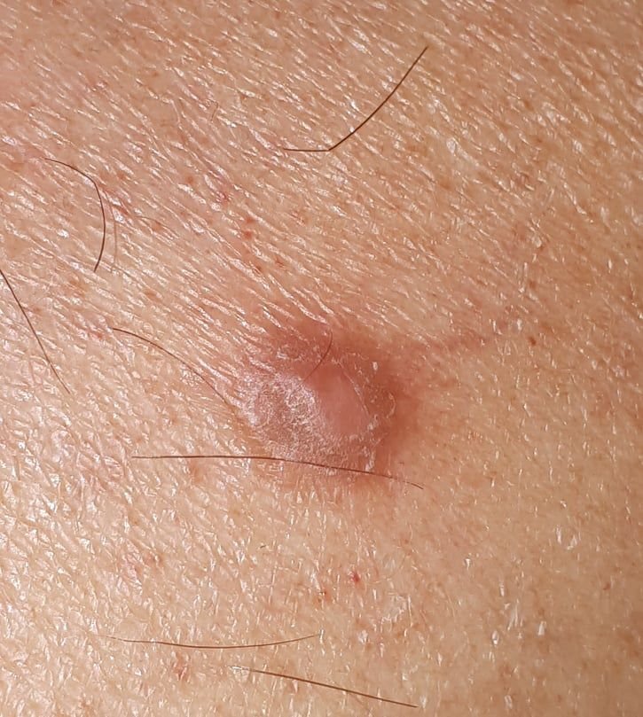
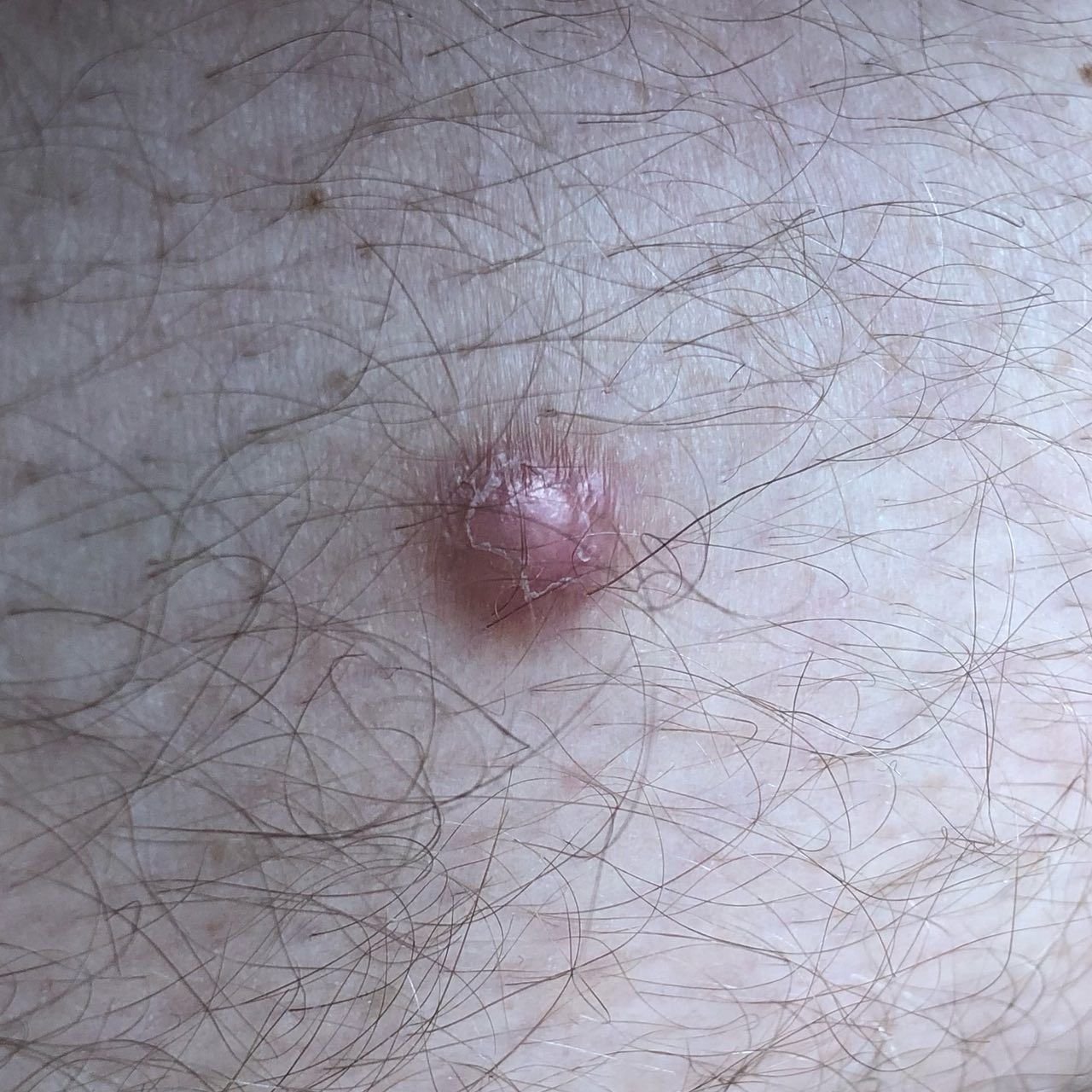
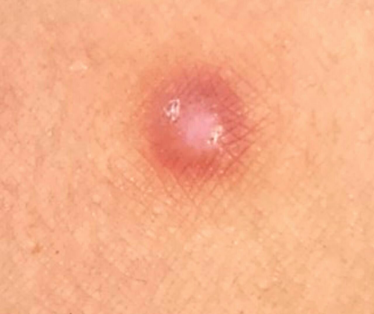
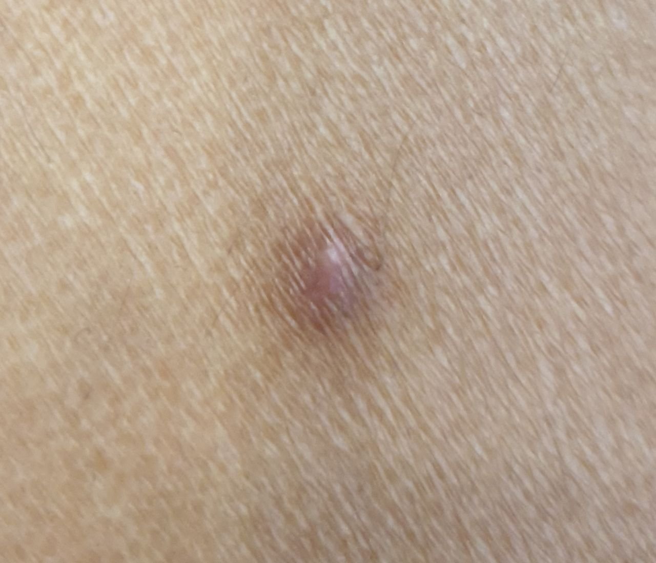
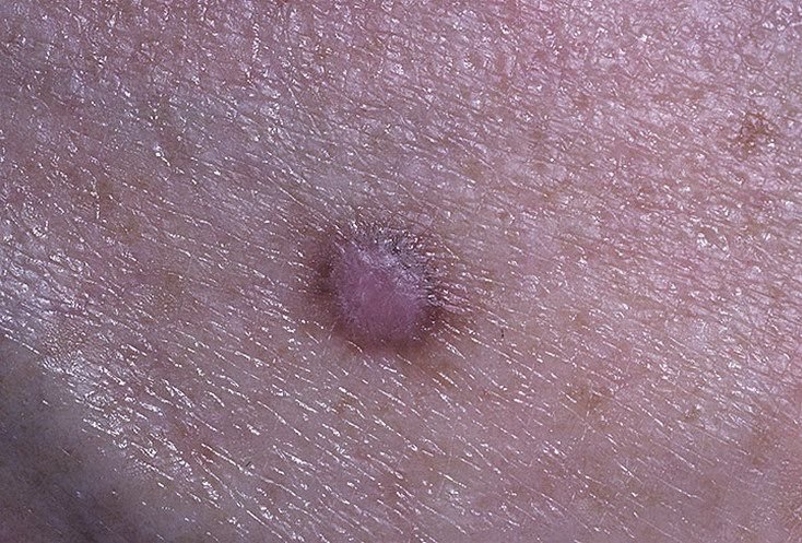
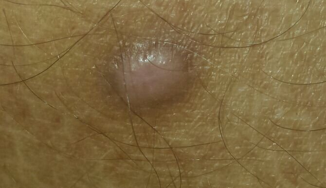
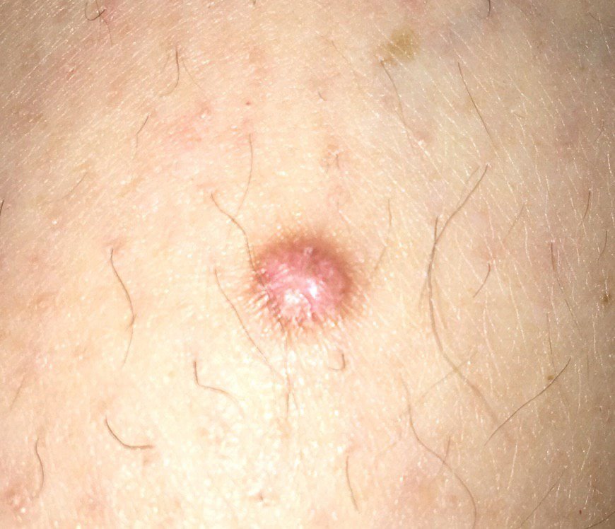
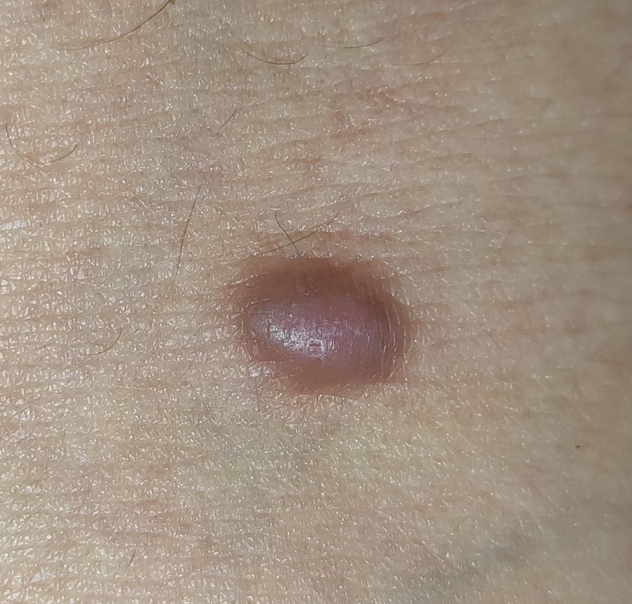
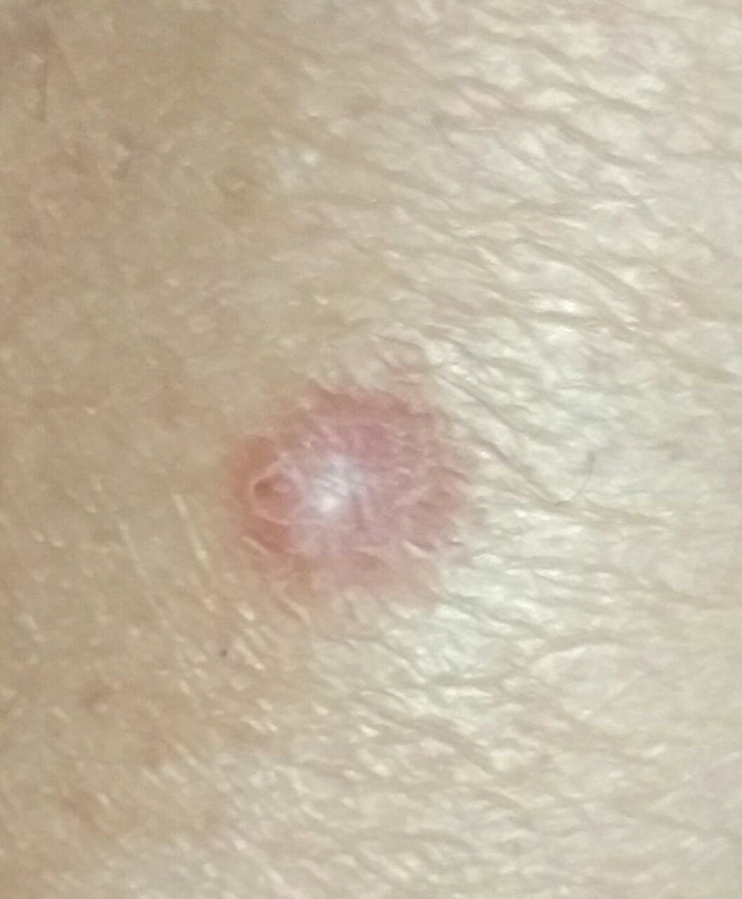
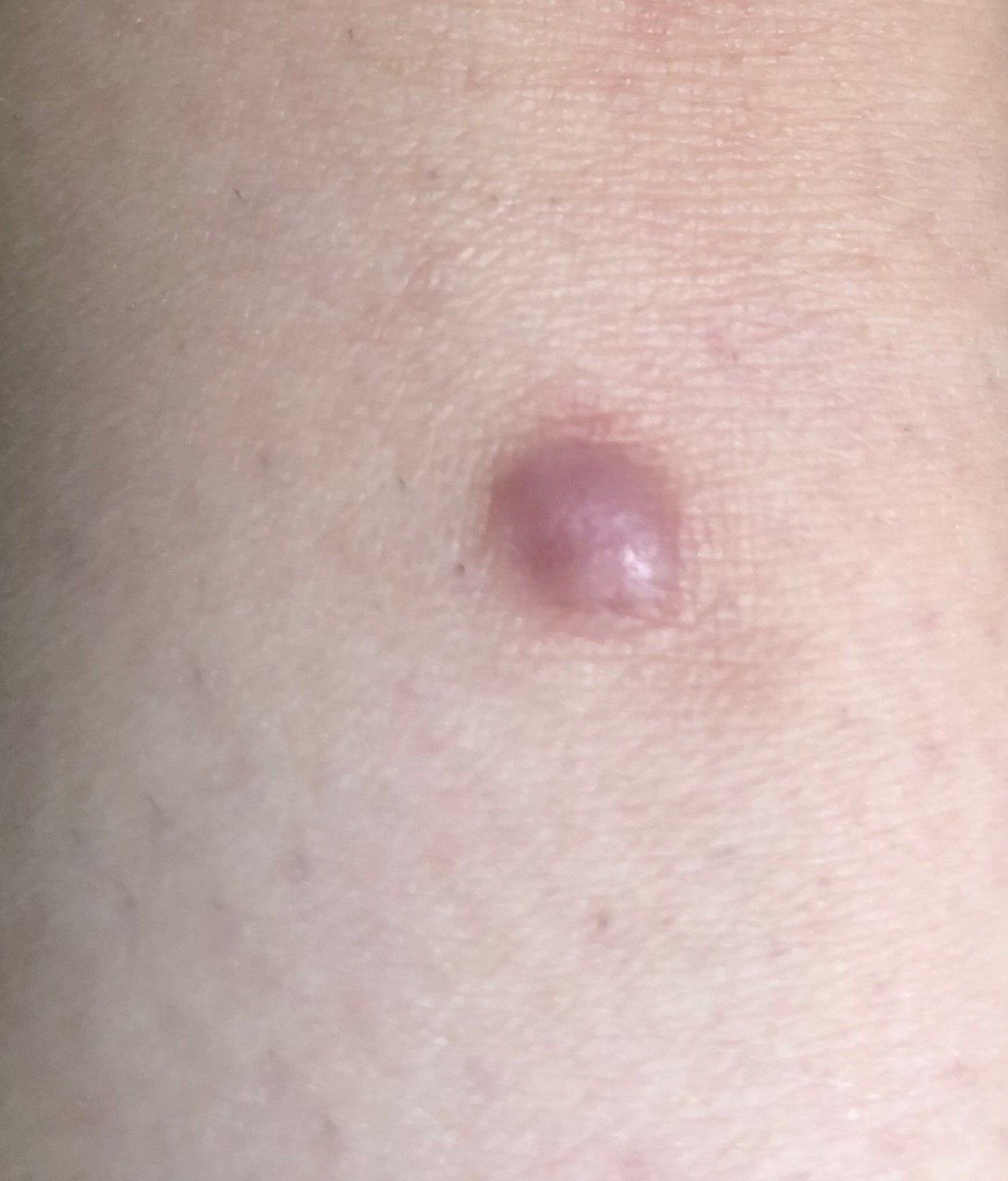
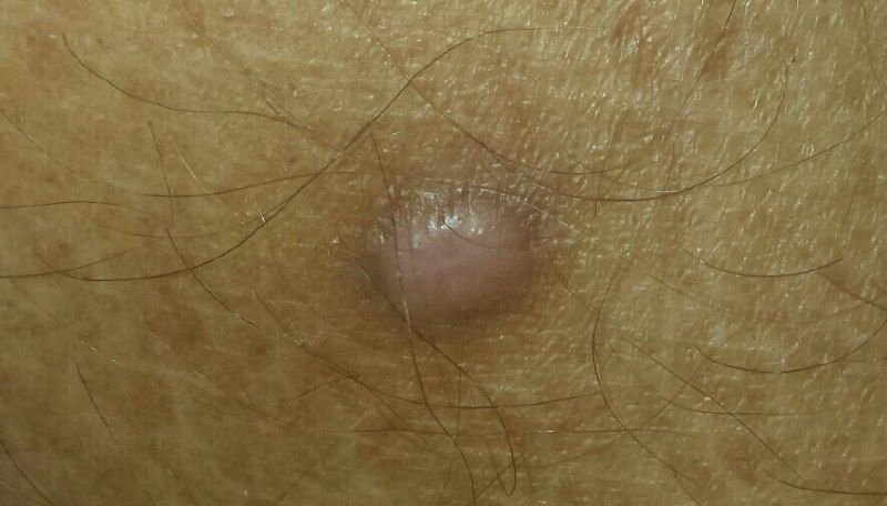
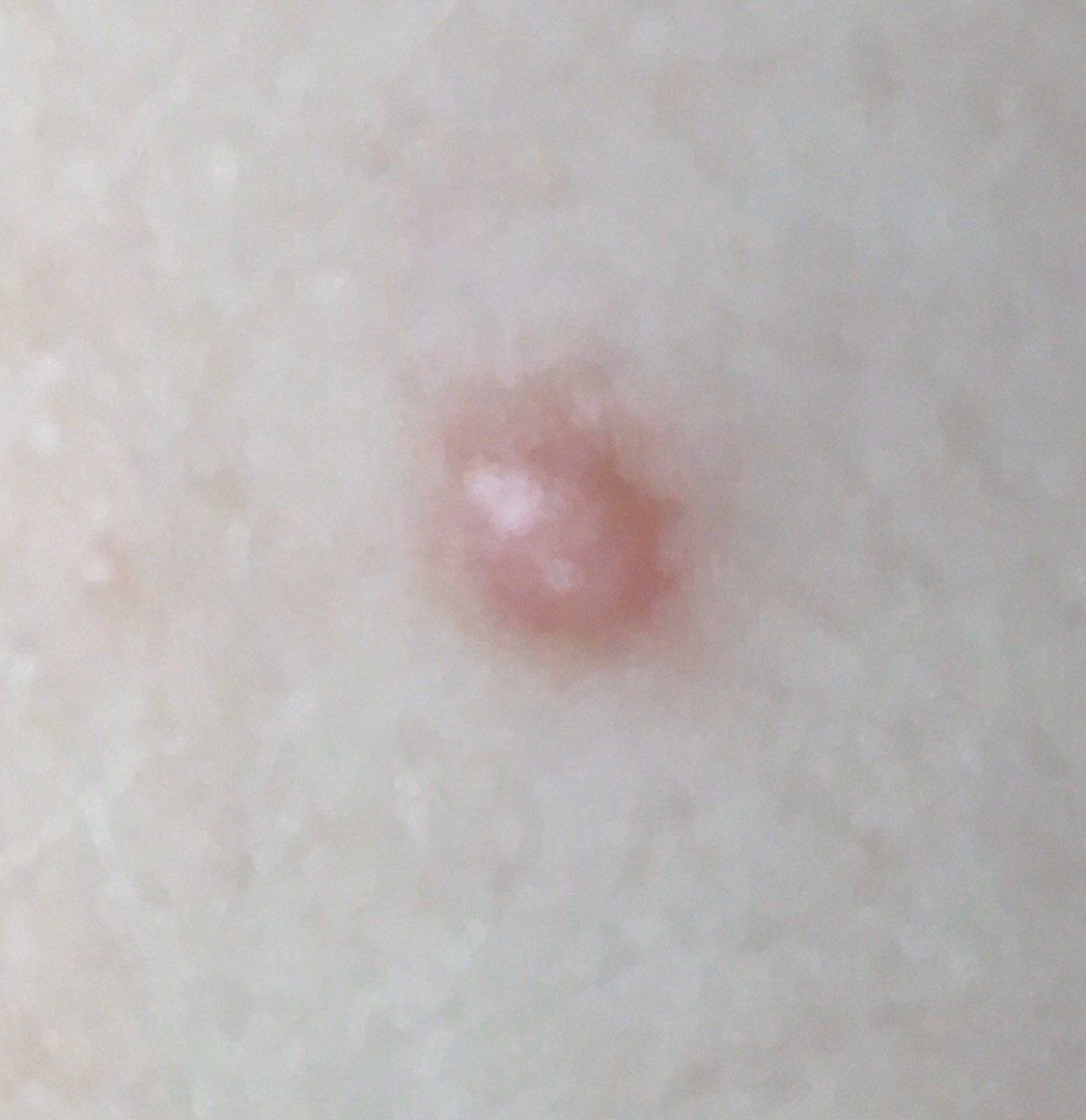
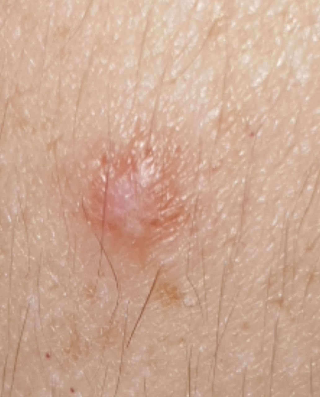
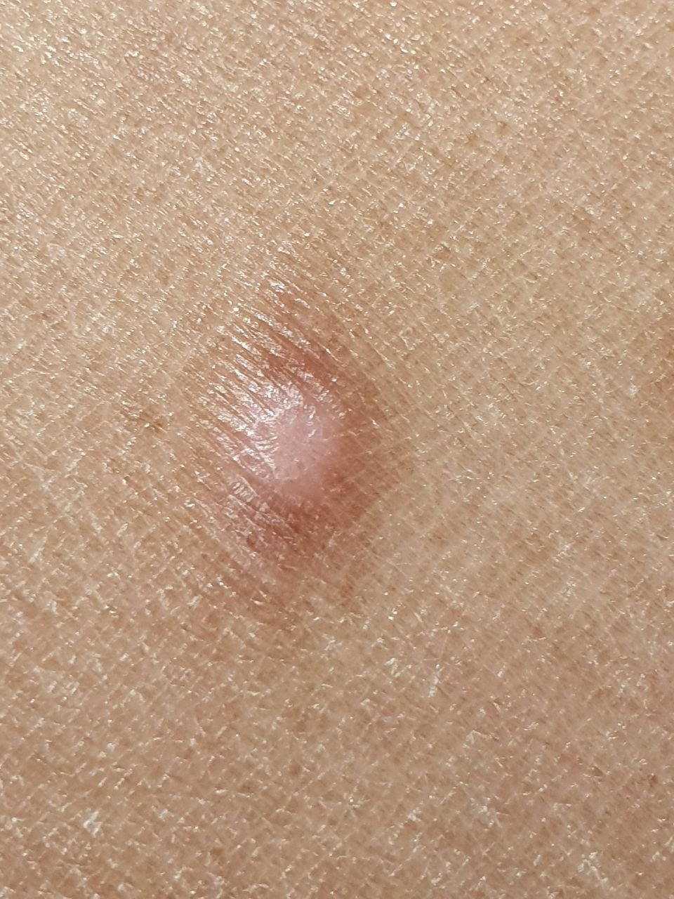
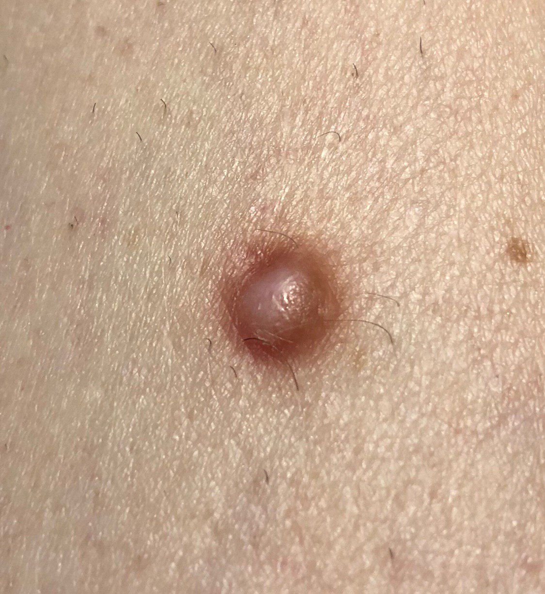
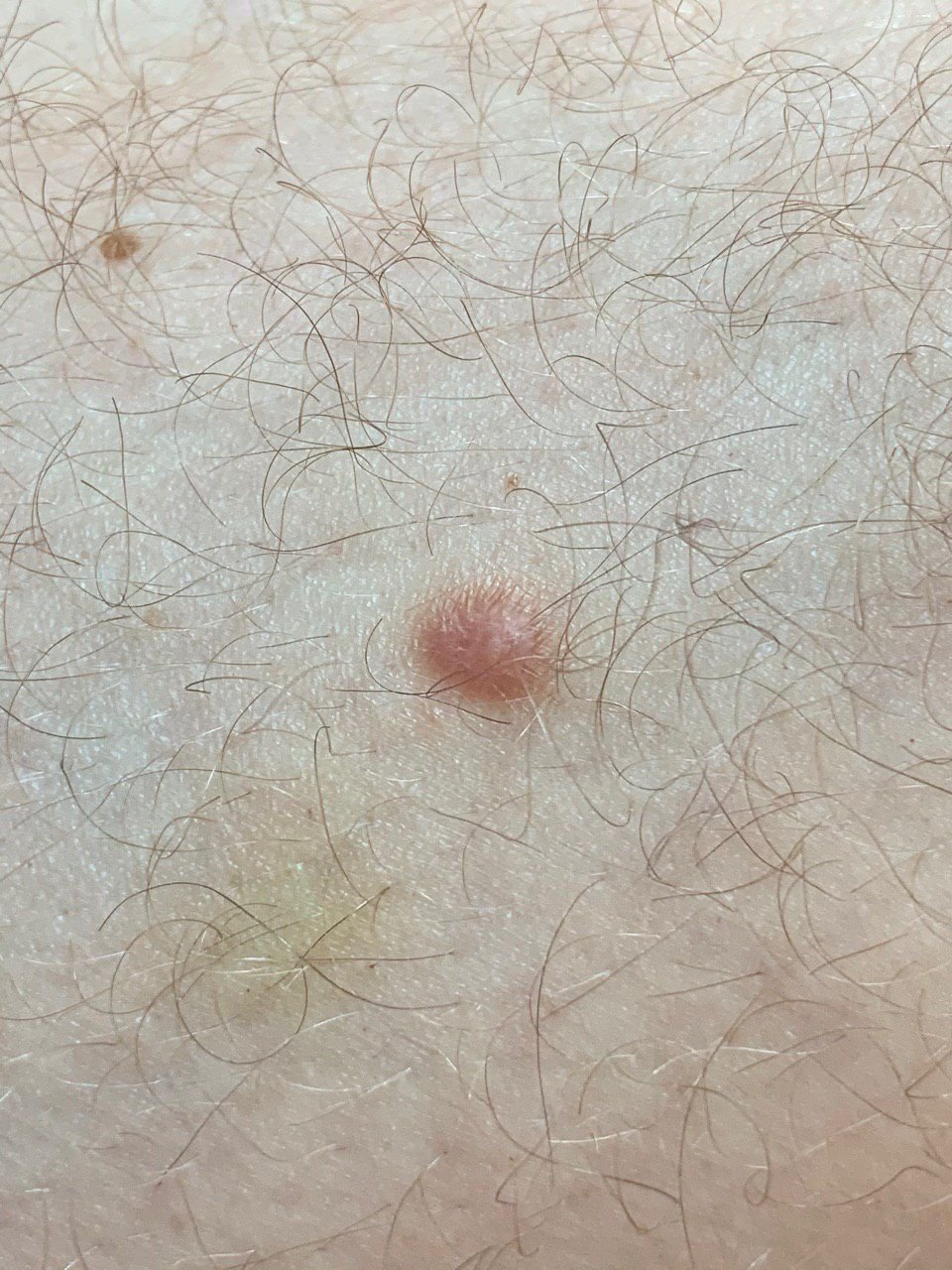
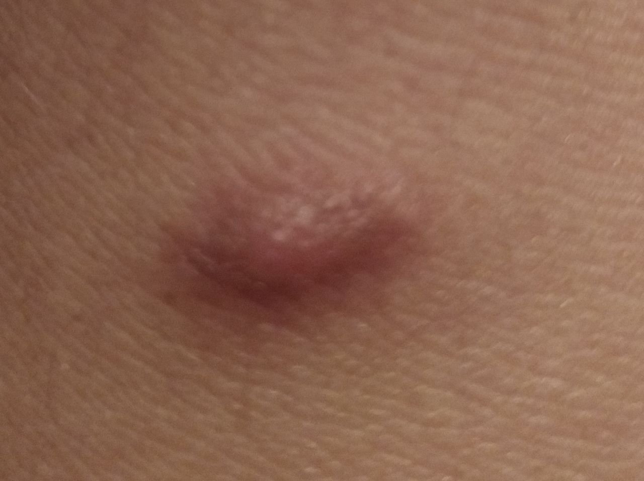
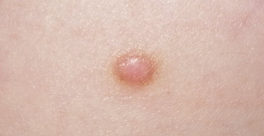
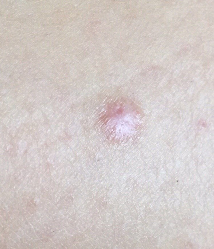
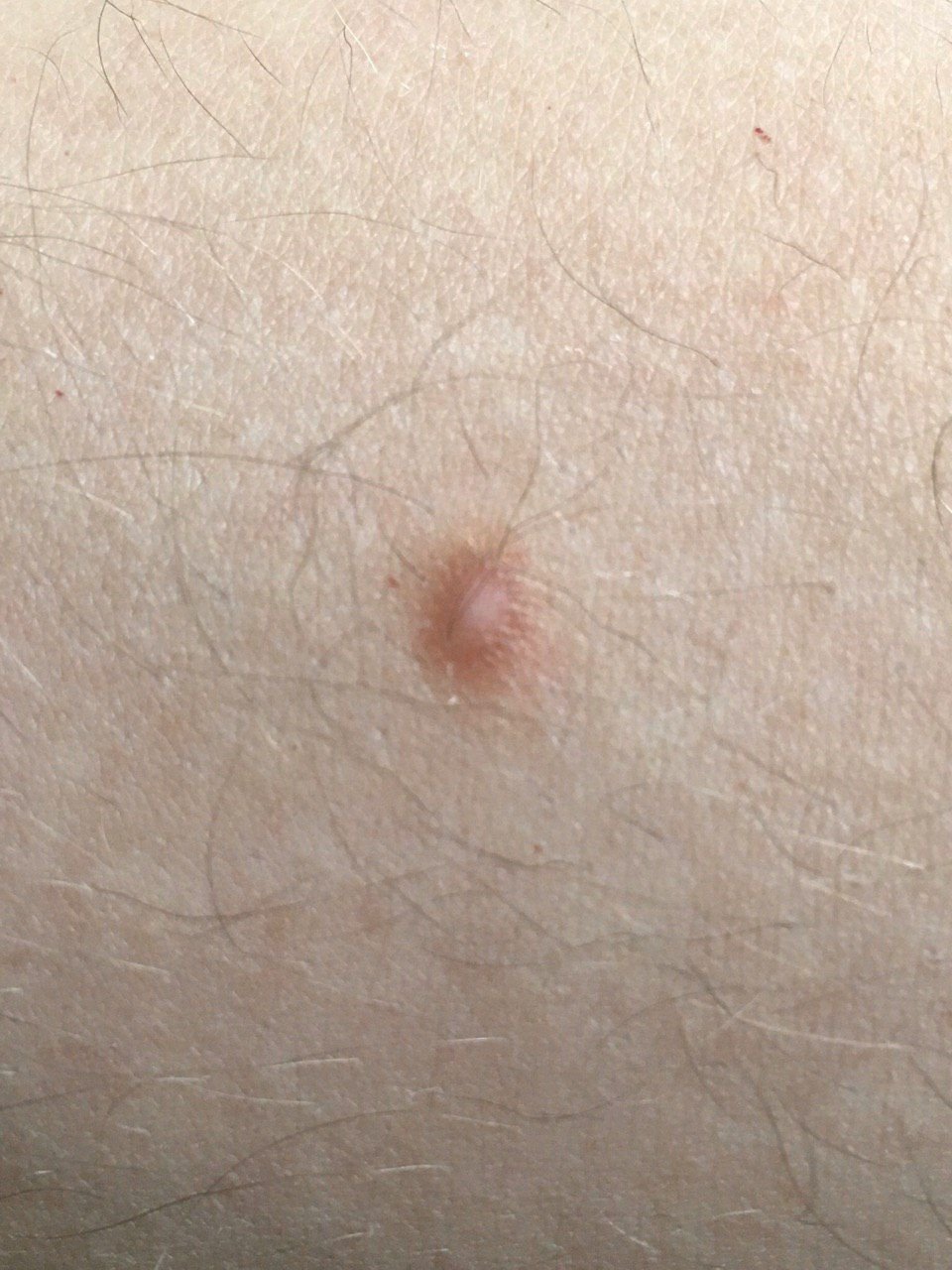
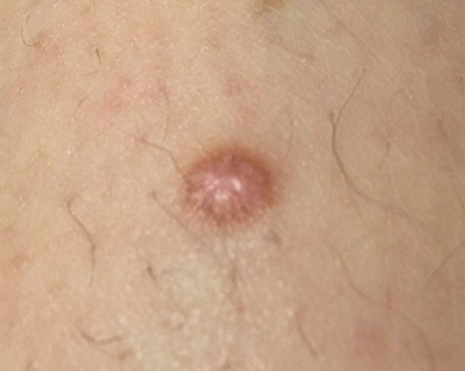
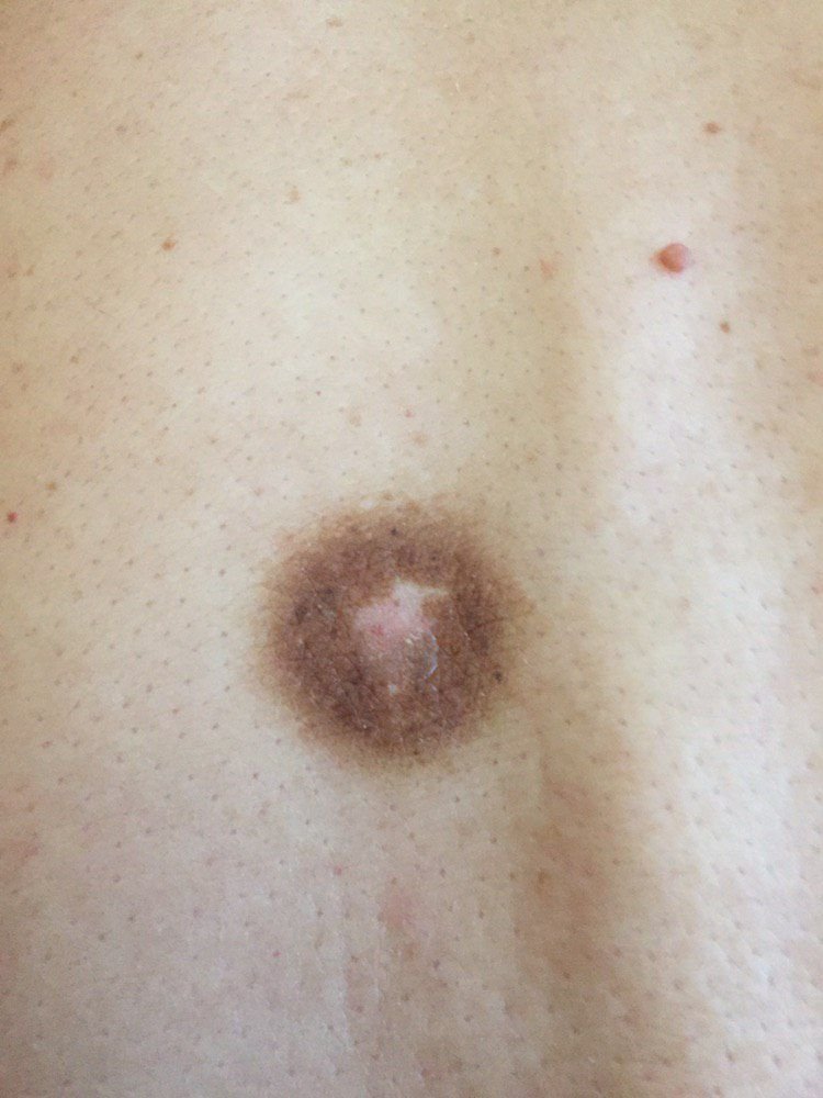
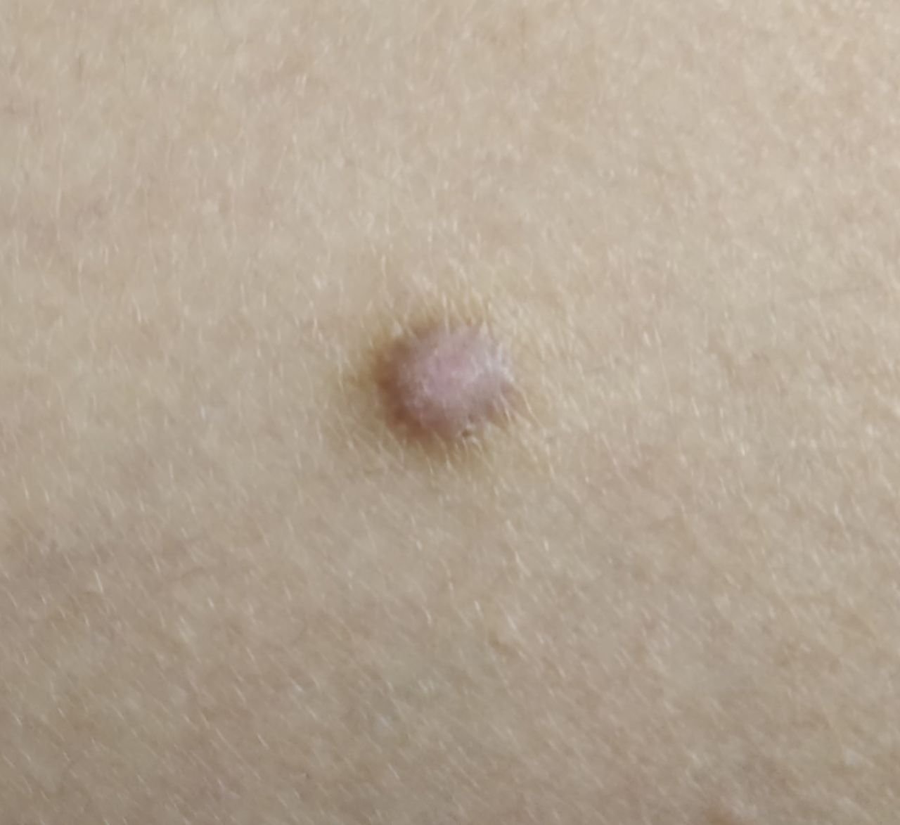
Upon visual inspection, dermatofibromas appear as slightly sunken or hemispherical growths, often symmetrical in shape (commonly oval or round). In some cases, the lesion may be spindle-shaped. The surface of the dermatofibroma is usually smooth, with the skin pattern often absent in the center. Less frequently, a small tuberous appearance or slight peeling may be observed.
The borders of the dermatofibroma can either be clear and well-defined (typical of hemispherical forms) or less distinct (especially when the growth is flat or slightly sunken). The color of the lesion ranges from flesh-colored to gray or brown shades, with the pigment distribution being uneven. The intensity of the color tends to gradually increase from the center to the outer edges, with the center often being lighter in color, and the periphery darker. In larger dermatofibromas, color heterogeneity can be observed across the entire lesion, including areas of polychrome (multiple colors).
Hair is typically absent from the central part of the dermatofibroma, but in some cases, small hairs may grow at the periphery, especially in larger lesions. Dermatofibromas typically do not exceed 10 mm in diameter, with the majority being smaller. Larger lesions are rare and are referred to as “giant dermatofibromas.” The height of hemispherical lesions above the skin is usually between 5-7 mm.
On palpation, dermatofibromas feel firm and dense. A characteristic feature known as the “fossa sign” is present: when the surrounding skin is compressed, a depression forms in the center of the lesion. In general, there are no subjective symptoms associated with dermatofibromas, although mild itching may occur in rare instances.
Dermatofibromas are most commonly located on the lower extremities and the shoulder girdle, with other locations being less frequent. In the case of giant dermatofibromas, they are most often found in the sacro-gluteal region.
Dermatoscopic Description
Dermatoscopic examination of dermatofibromas typically reveals the following features:
- Hypopigmentation in the Central Zone: The central portion of the dermatofibroma often appears lighter in color compared to the outer regions.
- Irregular Contours of the Central Zone: The boundaries of the hypopigmented central area may be unclear or irregular in shape.
- Peripheral Light Brown Zone: A light brown area surrounding the central zone is a common feature.
- Delicate Pigment Network: The peripheral zone often exhibits a fine pigment network with small cells.
- Smooth Transition to Healthy Skin: The peripheral zone blends smoothly into the surrounding healthy skin, with no abrupt borders.
- Fine-Meshed Uniform Structure: At the point where the lesion meets healthy skin, a fine, evenly spaced mesh structure may be visible.
- Occasional Presence of Vessels: Point-like blood vessels may occasionally be seen in the lesion.
Differential Diagnosis
Dermatofibromas must be differentiated from other pigmented or nodular skin lesions, including:
- Simple nevus
- Papillomatous nevus
- Comedones
- Hemangioma
- Blue nevus
- Dysplastic nevus
- Keratoacanthoma
- Basal cell carcinoma
- Nodular melanoma
- Dermatofibrosarcoma protuberans
Risks
Dermatofibromas are typically benign and do not pose a significant risk of becoming malignant. Dermatofibromas are benign and malignant degeneration is not expected. Noticeable changes in appearance, new pain or tenderness, or new symptoms should prompt re-evaluation to exclude another lesion.
Dermatofibromas are benign lesions and malignant transformation is not expected; any concerning changes should be evaluated to rule out another diagnosis. Large dermatofibromas or those that change rapidly in appearance should be monitored closely for any signs of malignancy.
Tactics
If no changes are observed in the dermatofibroma’s appearance, and there are no symptoms such as pain or discomfort, self-monitoring is usually sufficient. This should include annual checks, with assistance from others if the lesion is in a difficult-to-see location. If the dermatofibroma experiences mechanical trauma, exhibits changes in appearance, or if new sensations develop, it is important to consult with a dermatologist or oncologist.
A healthcare professional will determine whether continuous monitoring or surgical removal is required. Dermatofibromas that are subject to chronic irritation, either from clothing, jewelry, or specific occupational activities, should be considered for removal to prevent further trauma.
Photographic documentation of the lesion can provide a useful record, allowing for the identification of any changes over time. Individuals with multiple skin neoplasms, including dermatofibromas, should also undergo an examination by a dermatologist or oncologist at least twice a year (typically before and after the summer months). Maintaining a map of skin neoplasms is also recommended to simplify monitoring, track changes, and identify any new lesions.
Treatment
When treatment is needed, options include surgical excision using standard techniques, electrosurgery, or radiofrequency; asymptomatic lesions may be monitored. A histological examination of the excised tissue is essential to confirm the benign nature of the lesion.
Destructive treatments such as laser removal or cryodestruction are not advised for dermatofibromas, as these methods can lead to a high rate of local relapse and do not provide a sufficient histological diagnosis.
Prevention
Preventing the development of dermatofibromas and reducing the risk of malignancy involves careful and gentle skin care:
- Avoiding chronic skin trauma, including repetitive friction and pressure.
- Following safety protocols to minimize exposure to skin-damaging chemicals or radiation.
- Maintaining good personal hygiene and being vigilant for any changes in existing skin lesions.
Regular checks of dermatofibromas, early consultation with a healthcare professional if any changes occur, and timely removal of potentially dangerous lesions are essential for maintaining skin health.