Dermatofibroma (ICD-10: D23) 💚
Dermatofibroma
Dermatofibroma (also known as skin fibroma or benign fibrous histiocytoma) is a benign neoplasm originating from connective tissue, and it is characterized by a firm knot-like structure that can either be raised above the skin’s surface or occur as a dense nodule within the dermis. Dermatofibromas are typically acquired skin growths, and they are extremely rare in newborns and children. These lesions most commonly appear in individuals over the age of 25-30 years. The occurrence of multiple dermatofibromas is rare, except in cases of specific systemic diseases, hereditary syndromes, or conditions like HIV infection. Dermatofibromas are more frequently found in women compared to men.
Predisposing Factors
The exact cause of dermatofibromas is not fully understood, but several factors have been identified that may contribute to the development of these benign neoplasms. These factors increase the likelihood of dermatofibroma formation or influence the rate at which they grow:
- Female Gender: Dermatofibromas are more common in women, although they can appear in both sexes.
- Skin Damage: Trauma, micro-injuries, insect bites and injections have been proposed as triggers, since dermatofibromas are sometimes noticed at the site of a previous injury. This is not established: the cause of dermatofibromas remains uncertain, and most arise without any identifiable injury.
- Genetic Factors: In some cases, heredity can play a role in the development of dermatofibromas, suggesting a potential genetic predisposition to these benign growths.
Diagnostics
The diagnosis of dermatofibroma is primarily based on clinical examination, which involves a physical examination of the growth followed by dermatoscopy to assess its characteristics. For a typical lesion that is usually enough, and no biopsy is needed. Biopsy or excision becomes appropriate where the lesion is atypical, has recently enlarged, has ulcerated or bled, or shows asymmetric dermatoscopic features.
Symptoms
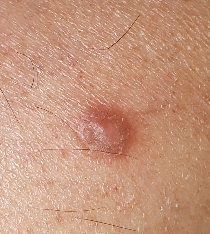
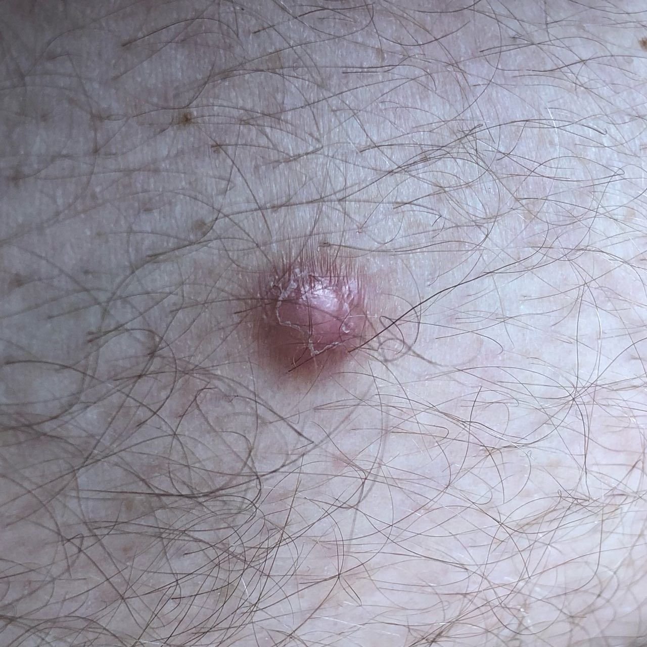
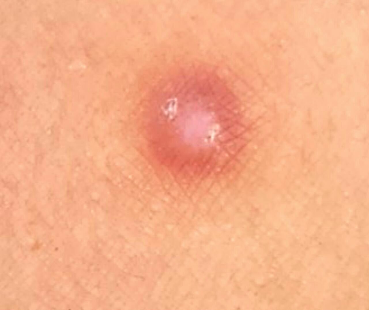
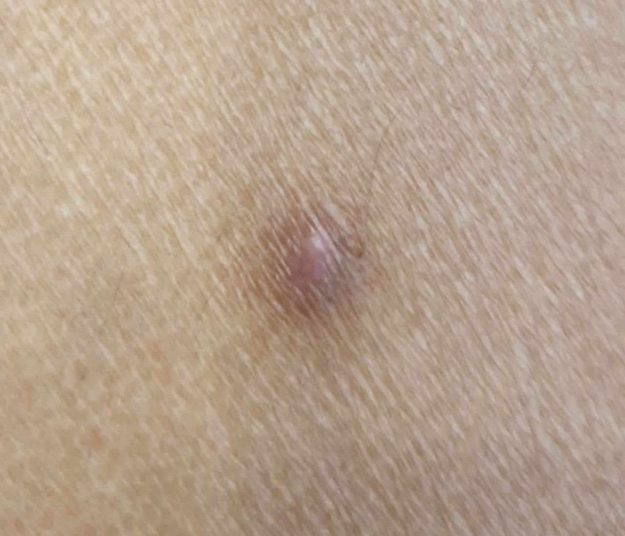
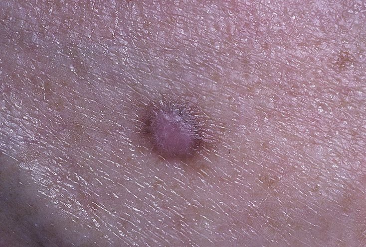
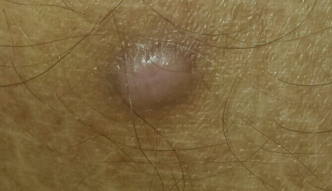
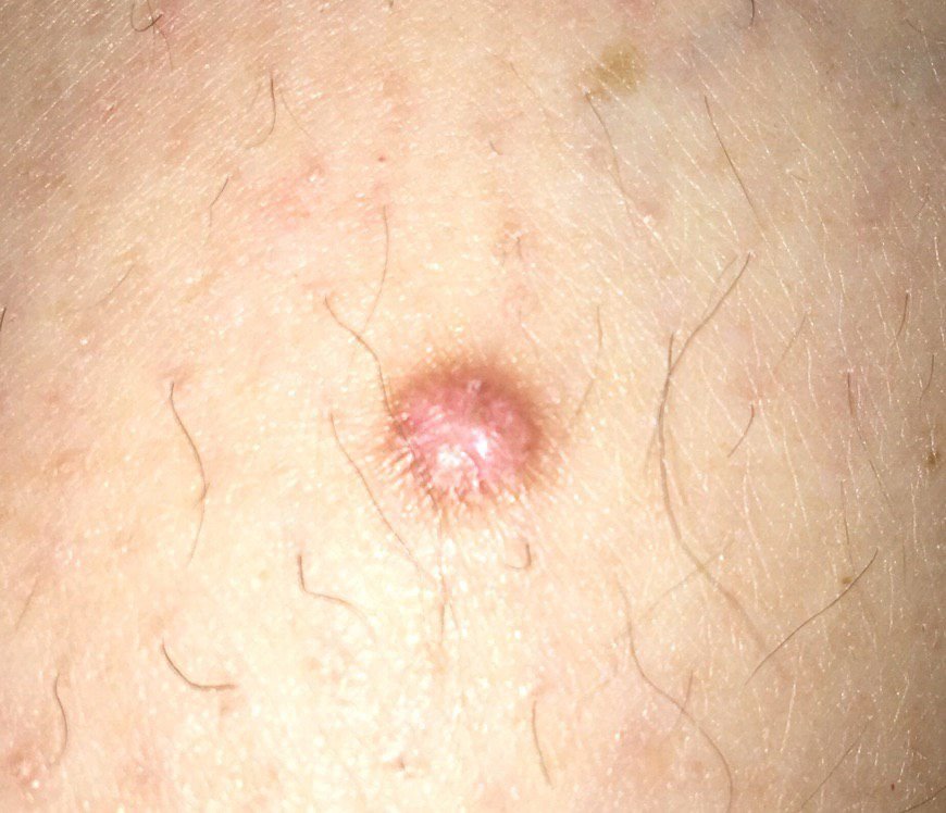
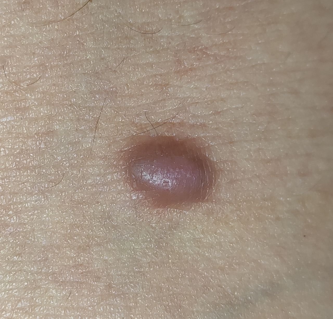
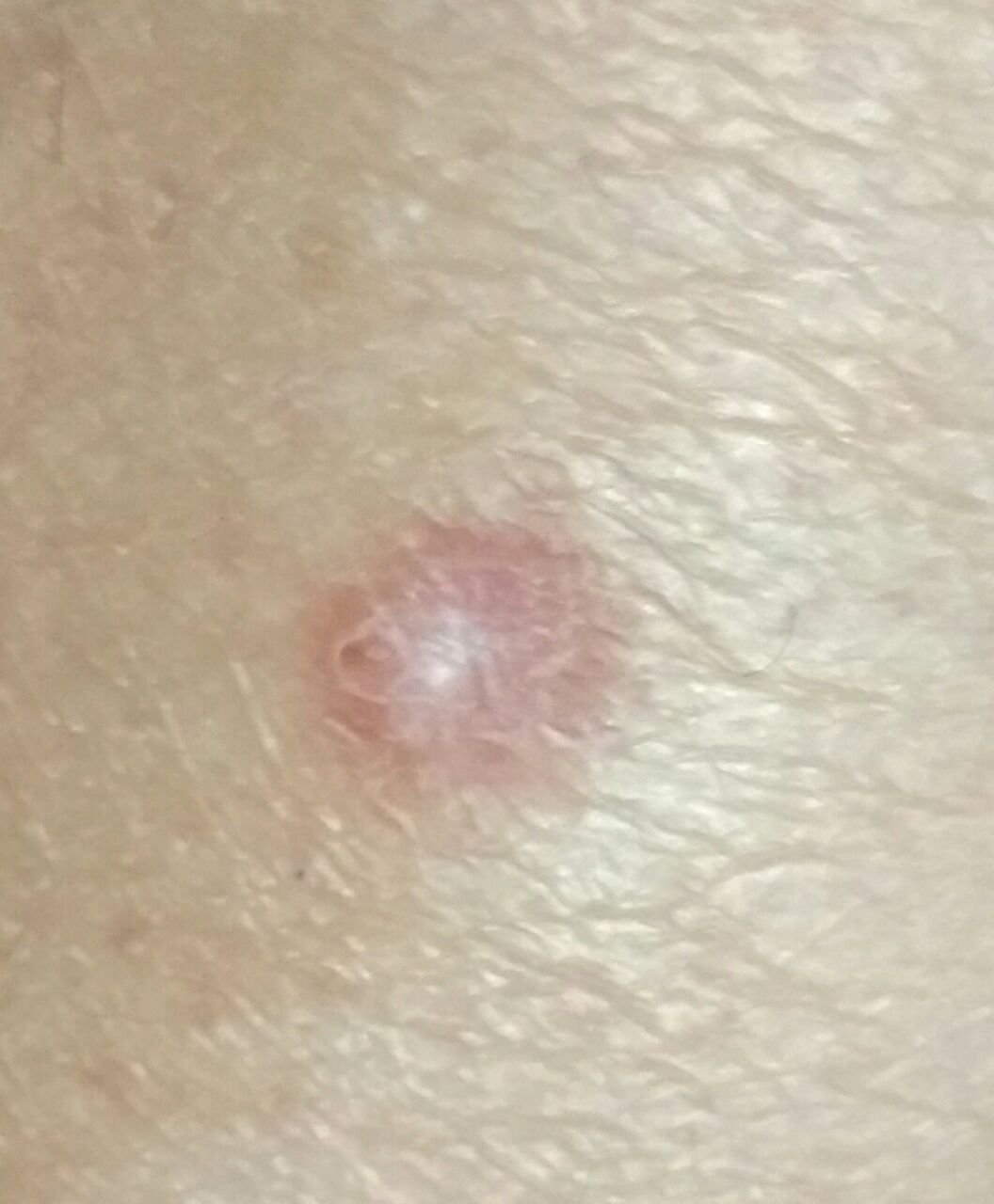
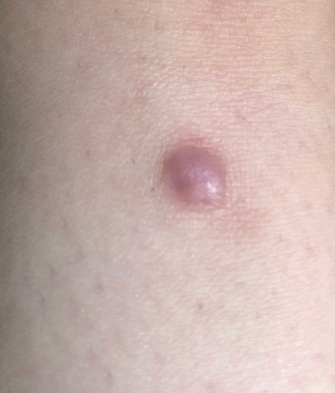
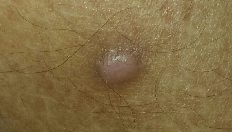
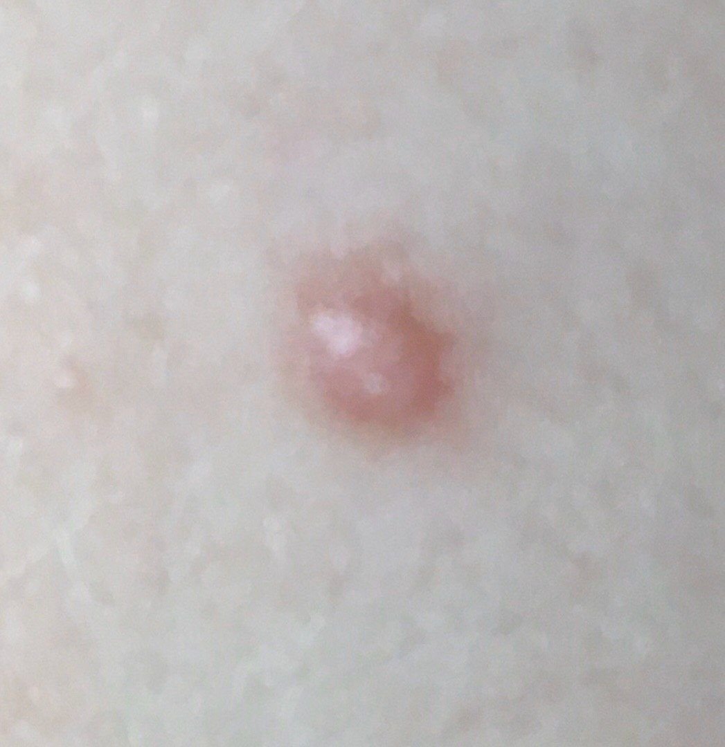
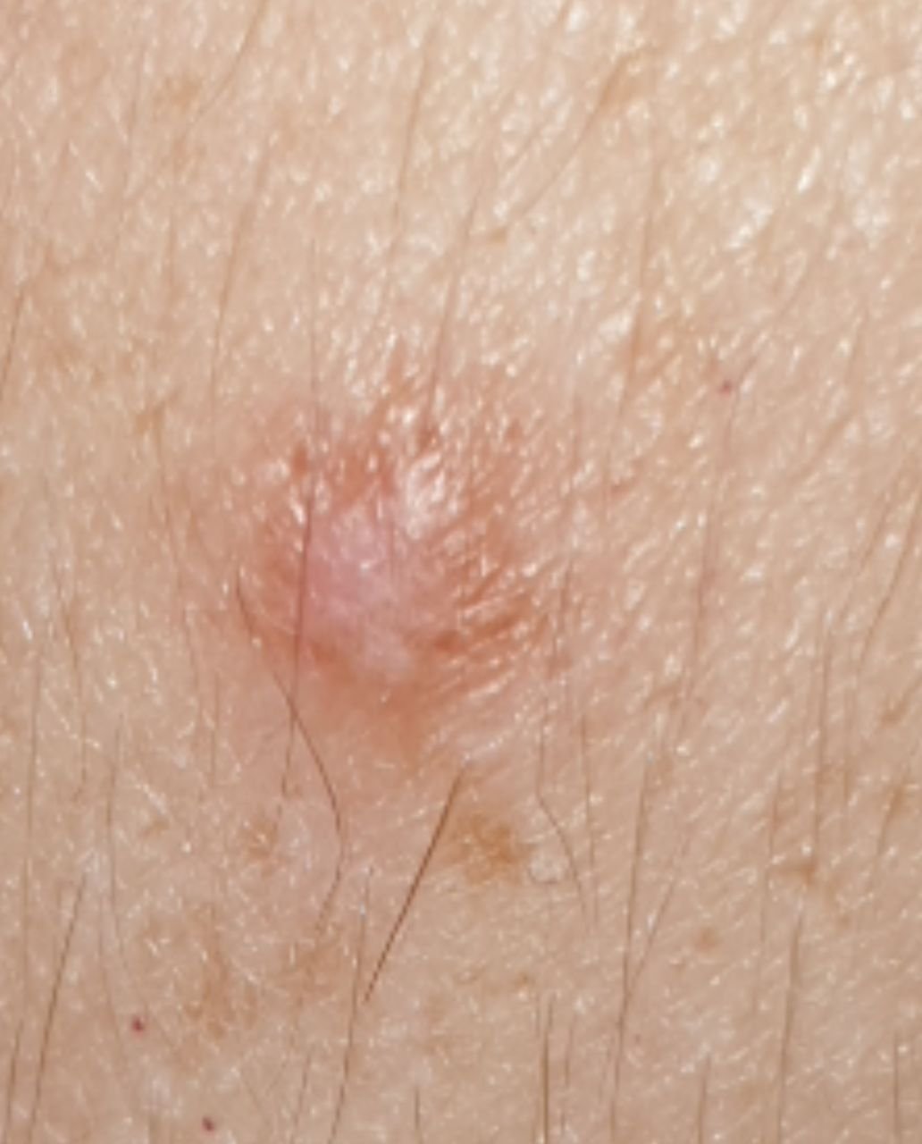
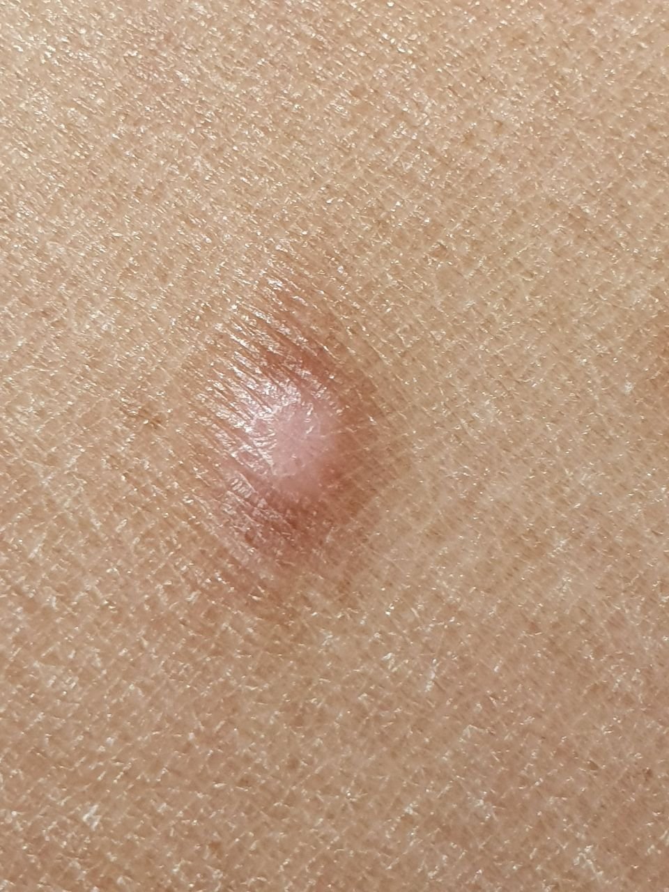
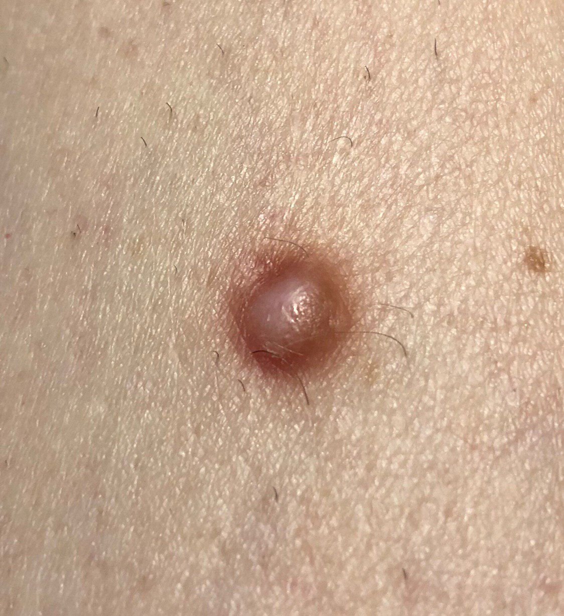
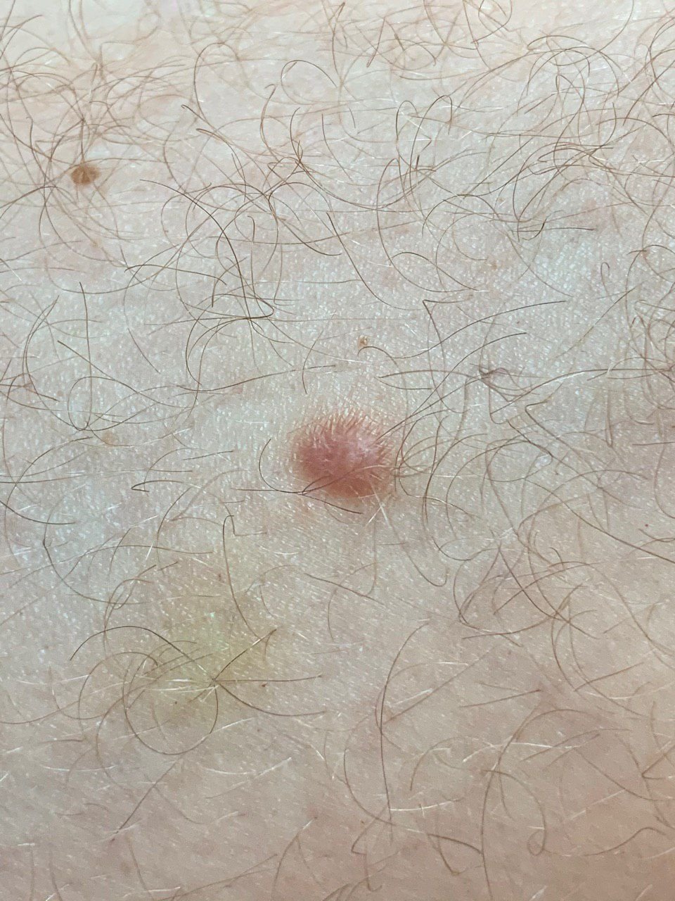
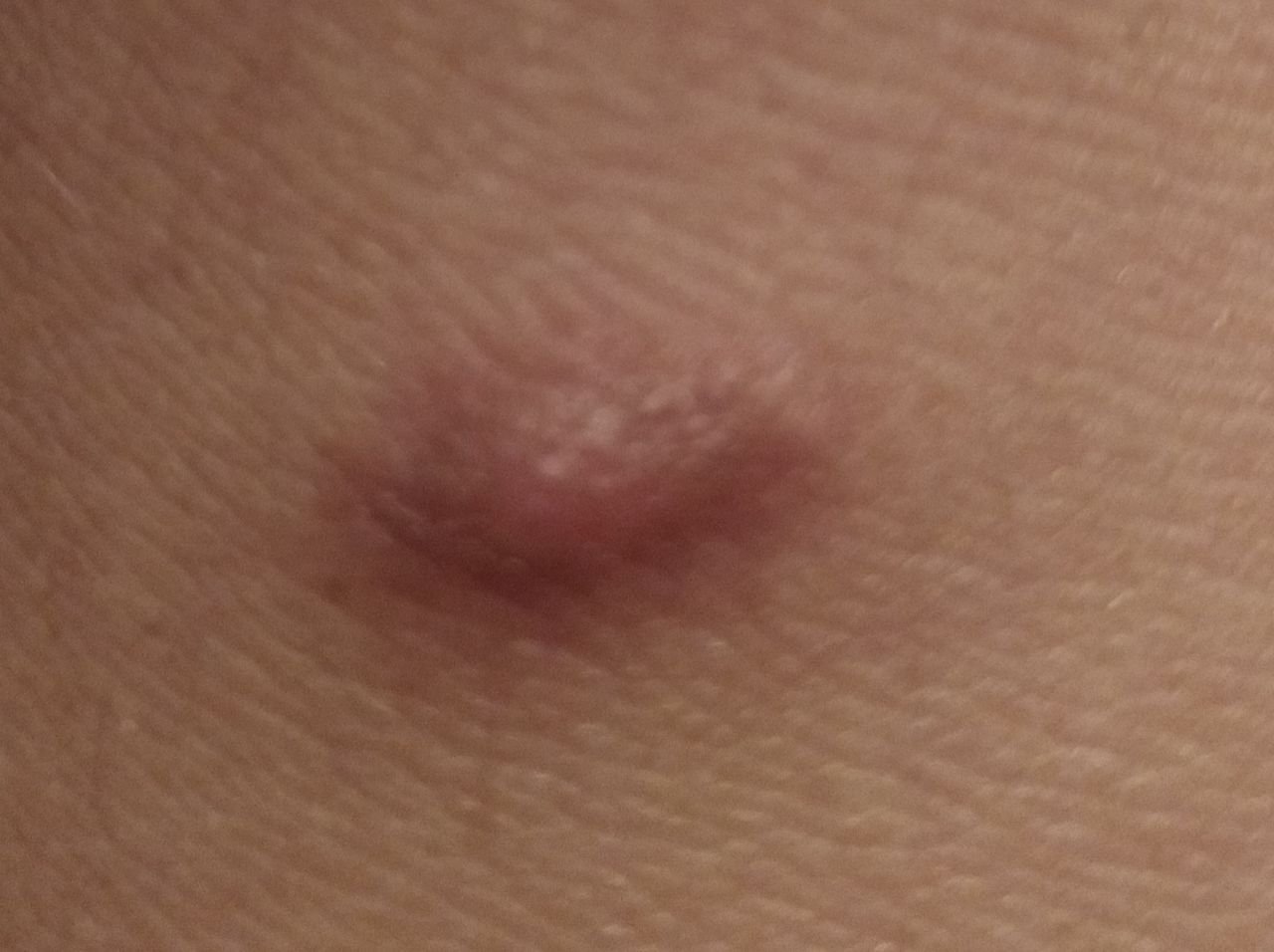
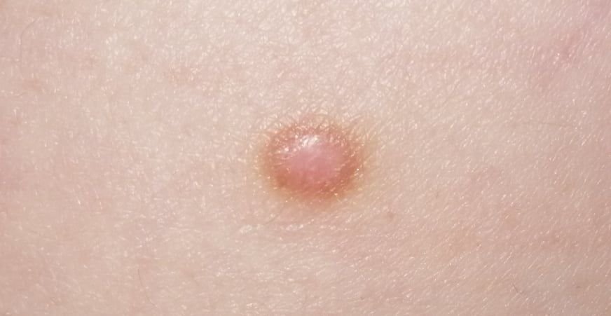
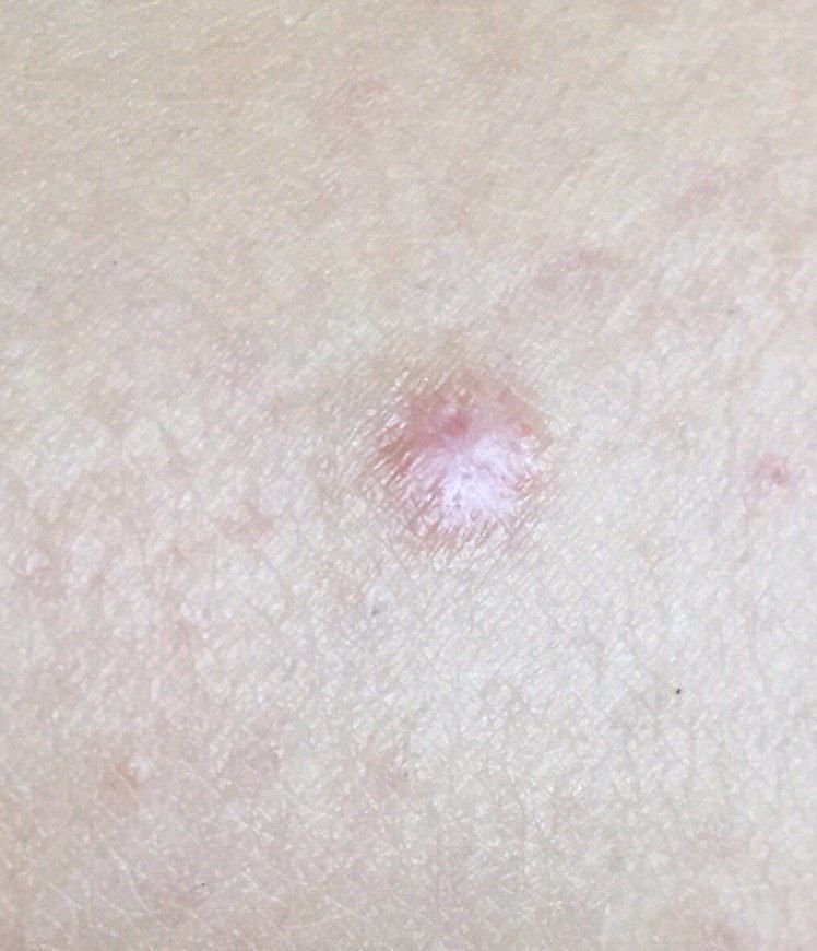
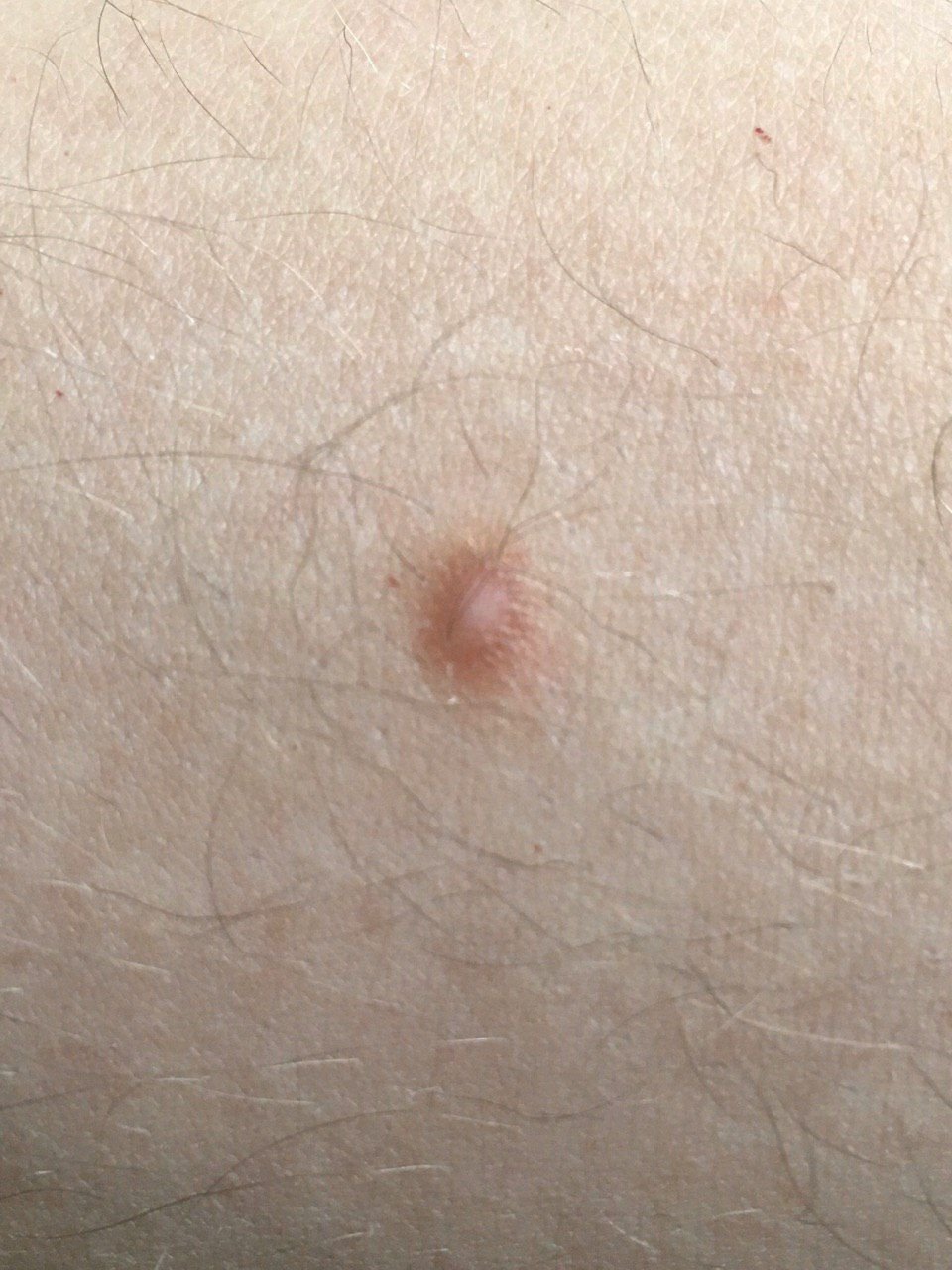
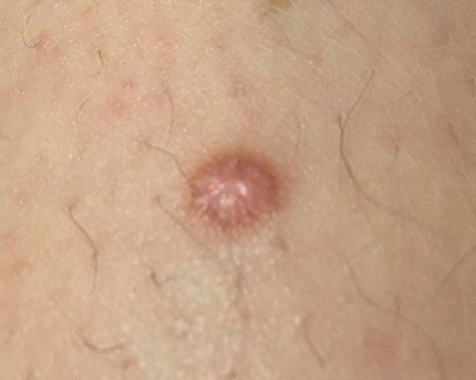
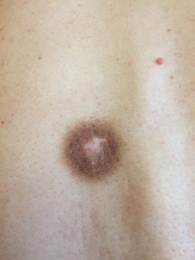
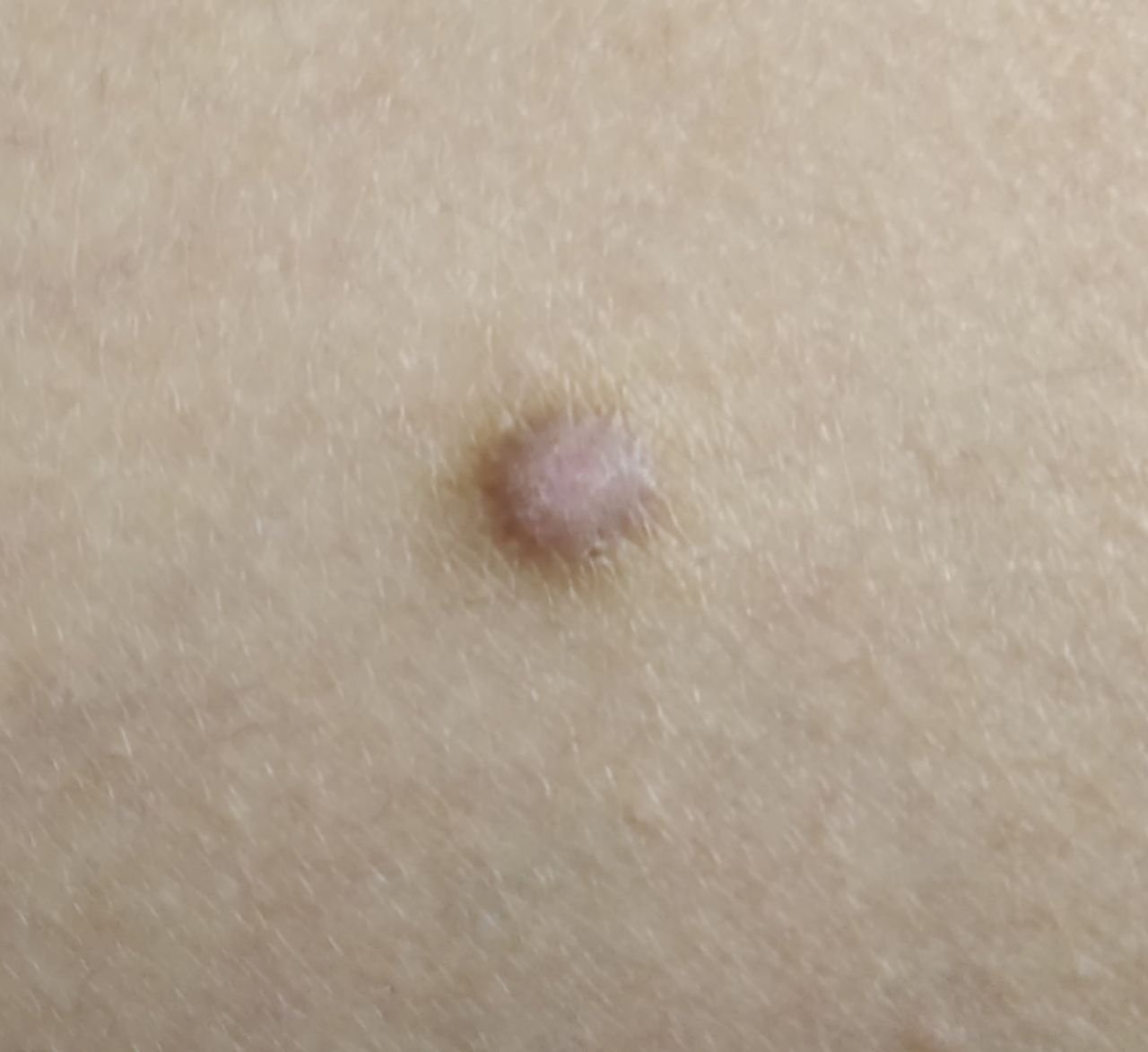
Upon visual inspection, dermatofibromas appear as slightly sunken or hemispherical growths, often symmetrical in shape (commonly oval or round). In some cases, the lesion may be spindle-shaped. The surface of the dermatofibroma is usually smooth, with the skin pattern often absent in the center. Less frequently, a small tuberous appearance or slight peeling may be observed.
The borders of the dermatofibroma can either be clear and well-defined (typical of hemispherical forms) or less distinct (especially when the growth is flat or slightly sunken). The color of the lesion ranges from flesh-colored to gray or brown shades, with the pigment distribution being uneven. The intensity of the color tends to gradually increase from the center to the outer edges, with the center often being lighter in color, and the periphery darker. In larger dermatofibromas, color heterogeneity can be observed across the entire lesion, including areas of polychrome (multiple colors).
Hair is typically absent from the central part of the dermatofibroma, but in some cases, small hairs may grow at the periphery, especially in larger lesions. Dermatofibromas typically do not exceed 10 mm in diameter, with the majority being smaller. Larger lesions are rare and are referred to as “giant dermatofibromas.” The height of hemispherical lesions above the skin is usually between 5-7 mm.
On palpation, dermatofibromas feel firm and dense. A characteristic feature known as the “fossa sign” is present: when the surrounding skin is compressed, a depression forms in the center of the lesion. In general, there are no subjective symptoms associated with dermatofibromas, although mild itching may occur in rare instances.
Dermatofibromas are most commonly located on the lower extremities and the shoulder girdle, with other locations being less frequent. In the case of giant dermatofibromas, they are most often found in the sacro-gluteal region.
Dermatoscopic Description
The classic dermatoscopic pattern of a dermatofibroma is a peripheral faint pigment network surrounding a central white, scar-like area. Dermatoscopic findings are variable, however, and are not diagnostic in isolation:
- Central White, Scar-like Area: The centre appears white or paler than the outer regions, with the look of scar tissue.
- Irregular Contours of the Central Zone: The boundaries of the central area may be unclear or irregular in shape.
- Peripheral Faint Pigment Network: A delicate, fine pigment network in a light brown zone surrounding the centre.
- Smooth Transition to Healthy Skin: The peripheral zone blends smoothly into the surrounding healthy skin, with no abrupt borders.
- Fine-Meshed Uniform Structure: At the point where the lesion meets healthy skin, a fine, evenly spaced mesh structure may be visible.
- Occasional Presence of Vessels: Point-like blood vessels may occasionally be seen in the lesion.
Differential Diagnosis
Dermatofibromas must be differentiated from other pigmented or nodular skin lesions, including:
- Simple nevus
- Papillomatous nevus
- Comedones
- Hemangioma
- Blue nevus
- Dysplastic nevus
- Keratoacanthoma
- Basal cell carcinoma
- Nodular melanoma
- Dermatofibrosarcoma protuberans
Risks
A dermatofibroma is benign, and it is not expected to turn malignant. The clinical issue lies elsewhere: atypical variants, and other lesions that resemble a dermatofibroma, can be something quite different. Melanoma and dermatofibrosarcoma protuberans both enter the differential, which is why a lesion that looks clinically suspicious should be assessed histopathologically rather than watched.
Noticeable change in appearance, new pain or tenderness, rapid enlargement, or a firm nodule that lacks the typical central dimple should prompt re-evaluation — not because the dermatofibroma is degenerating, but because it may not be a dermatofibroma.
Tactics
A lesion you have not had looked at is a different matter from one a clinician has already identified. Monitoring follows assessment, not the other way round: a firm nodule that has not been examined should be shown to a dermatologist. Dermatofibrosarcoma protuberans, melanoma and squamous cell carcinoma can all present as a firm lump, and a lesion lacking the typical central dimple is not reliably a dermatofibroma.
Once a lesion has been assessed as a dermatofibroma and is stable, watching it between reviews is reasonable. Go back sooner for rapid enlargement, marked asymmetry, ulceration, bleeding, persistent pain, change in colour, or a lesion that becomes fixed to deeper tissue.
A healthcare professional will determine whether continuous monitoring or surgical removal is required. Dermatofibromas that are subject to chronic irritation, either from clothing, jewelry, or specific occupational activities, should be considered for removal to prevent further trauma.
Photographic documentation of the lesion can provide a useful record, allowing for the identification of any changes over time. How often someone with multiple skin lesions needs to be examined depends on those lesions rather than on the time of year. Maintaining a map of skin neoplasms is also recommended to simplify monitoring, track changes, and identify any new lesions.
Treatment
An uncomplicated dermatofibroma generally requires no treatment at all. Excision is undertaken mainly where the lesion is symptomatic or cosmetically bothersome, or where the diagnosis needs to be established. It can be done with standard surgical technique, electrosurgery or radiofrequency, and tissue from an excised lesion should be examined histologically.
Cryotherapy and shave procedures carry a higher rate of recurrence and incomplete removal, and destructive methods such as laser do not yield tissue for a histological diagnosis — which is the reason to avoid them on any lesion whose nature has not been settled.
Prevention
Preventing the development of dermatofibromas and reducing the risk of malignancy involves careful and gentle skin care:
- Avoiding chronic skin trauma, including repetitive friction and pressure.
- Following safety protocols to minimize exposure to skin-damaging chemicals or radiation.
- Maintaining good personal hygiene and being vigilant for any changes in existing skin lesions.
Regular checks of dermatofibromas, early consultation with a healthcare professional if any changes occur, and timely removal of potentially dangerous lesions are essential for maintaining skin health.