
Hemangioma (ICD-10: D18) 💚
Hemangioma (Angioma, Vascular Nevus)
Hemangioma is a benign growth made up of small blood vessels, usually capillaries, which appears as a flat or slightly raised red lesion. Several distinct things are grouped under the name, and they behave differently, so it is worth separating them.
Infantile hemangioma is the common one. It is usually not fully formed at birth: it appears during the first weeks of life, grows during a proliferative phase over the following months, and then involutes spontaneously over a period of years. Congenital hemangioma is present and fully formed at birth and is biologically a different lesion, with its own course. A vascular malformation — a port-wine stain, for instance — is not a hemangioma at all: it is a malformed vessel network present from birth that grows with the child and does not involute. Lesions appearing in adult life, such as cherry angiomas, are again separate.
Multiple lesions can occur, and diffuse multiple forms are referred to as hemangiomatosis. Infantile hemangiomas are more frequent in girls.
Predisposing Factors
The exact cause of hemangiomas remains unclear, but several factors are believed to increase the likelihood of their development. These predisposing factors can influence the formation of hemangiomas and their growth patterns:
- For Congenital Hemangiomas:
- Female Gender: Female infants are more likely to develop congenital hemangiomas.
- Prematurity: Premature infants are at higher risk of developing congenital hemangiomas.
- Maternal Viral Infections and Intoxications: Exposure to certain viral infections or toxic substances during pregnancy may contribute to the development of congenital hemangiomas.
- Mother’s Age Over 40: Older maternal age may increase the risk of hemangioma formation in the infant.
- Intrauterine Hypoxia: A lack of oxygen during fetal development can lead to an increased risk of hemangiomas.
- Multiple Pregnancies: Multiple gestations (e.g., twins or more) are associated with a higher occurrence of hemangiomas.
- Genetic Factors: Family history and genetic predisposition may play a role in the formation of congenital hemangiomas.
- For Acquired Hemangiomas:
- Vascular Wall Pathology: Abnormalities in the structure of the blood vessel walls may contribute to the development of hemangiomas.
- Liver Dysfunction: Impaired liver function can increase the likelihood of hemangioma formation.
- Endocrine Disorders: Hormonal imbalances and endocrine pathologies are associated with a higher risk of hemangiomas.
- Metabolic Disorders: Disorders in metabolism can contribute to the development of these lesions.
- Environmental Factors: Environmental influences, such as toxins or pollutants, may trigger hemangioma formation.
- Genetic Factors: A family history of hemangiomas or other vascular anomalies may increase the likelihood of developing these growths.
Diagnostics
A typical infantile hemangioma is diagnosed clinically, by examination and dermatoscopy. Neither biopsy nor laboratory testing is part of the routine work-up. Imaging — ultrasound, or MRI — is reserved for the cases where the diagnosis is genuinely uncertain, or where the extent of a lesion and any associated anomalies need to be assessed.
Lesions that are deep, cover large areas, or lie near vital organs and vascular structures are where that imaging is warranted. A large or segmental lesion may need evaluation by several specialists together, to establish its extent and to look for any associated anomalies.
Symptoms
Hemangiomas can present with various visual characteristics, depending on their type (congenital or acquired).
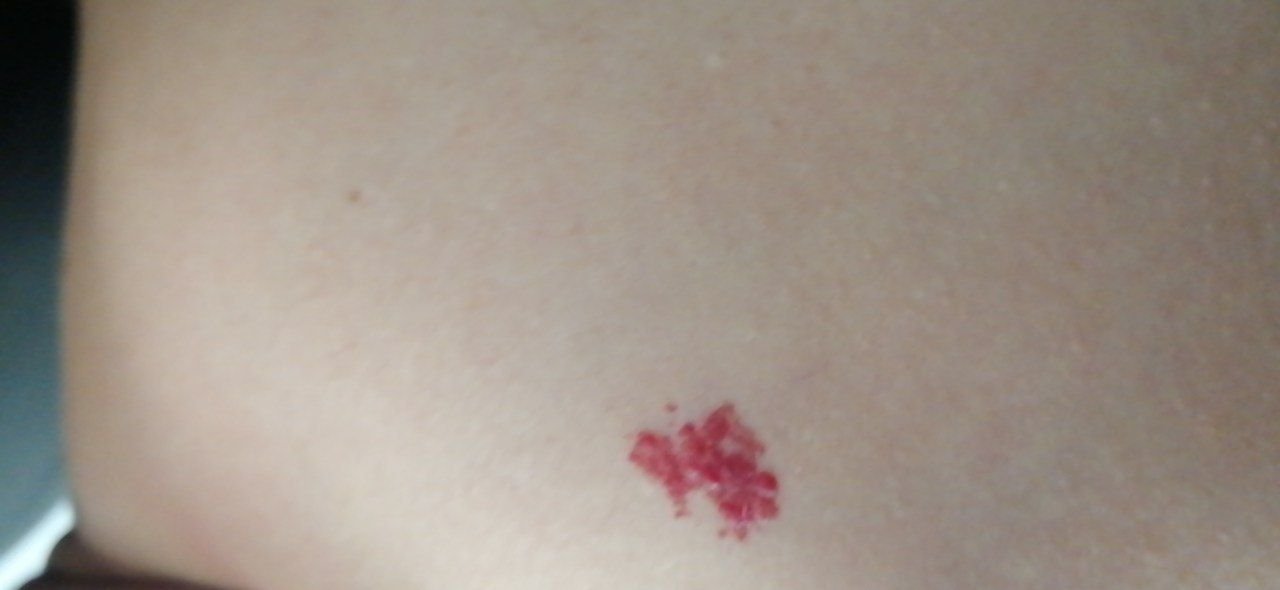
Congenital Hemangiomas: These lesions can take on many different shapes, including oval, asymmetric, or large masses that occupy multiple anatomical regions. The surface of the hemangioma may be smooth, especially in flat forms, or slightly bumpy, which is typical of more prominent hemangiomas that rise above the skin. The color can vary from pink to bright red, raspberry, crimson, or even cyanotic shades. The color is usually uniform across the lesion, although it may sometimes exhibit a spotted or variegated pattern. In general, congenital hemangiomas do not affect hair growth, although a few coarse or fluffy hairs may appear in the central area of the lesion.
The size of congenital hemangiomas can range widely, from small lesions just a few millimeters in diameter to large ones that occupy several anatomical regions (20-30 cm in some cases). On palpation, these hemangiomas are typically soft and more delicate than the surrounding skin. When pressed, they may briefly lose their color and pale. There are no associated sensations like pain or itching. Congenital hemangiomas are most commonly located on the head, face, and neck, although they can appear in other areas as well.
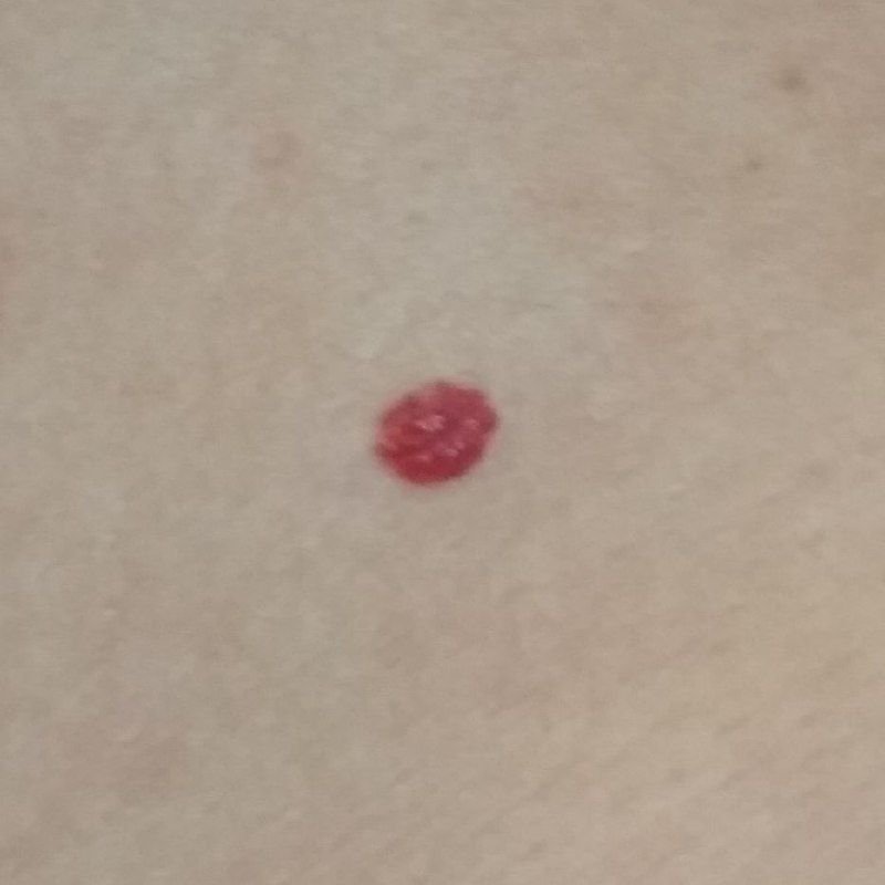
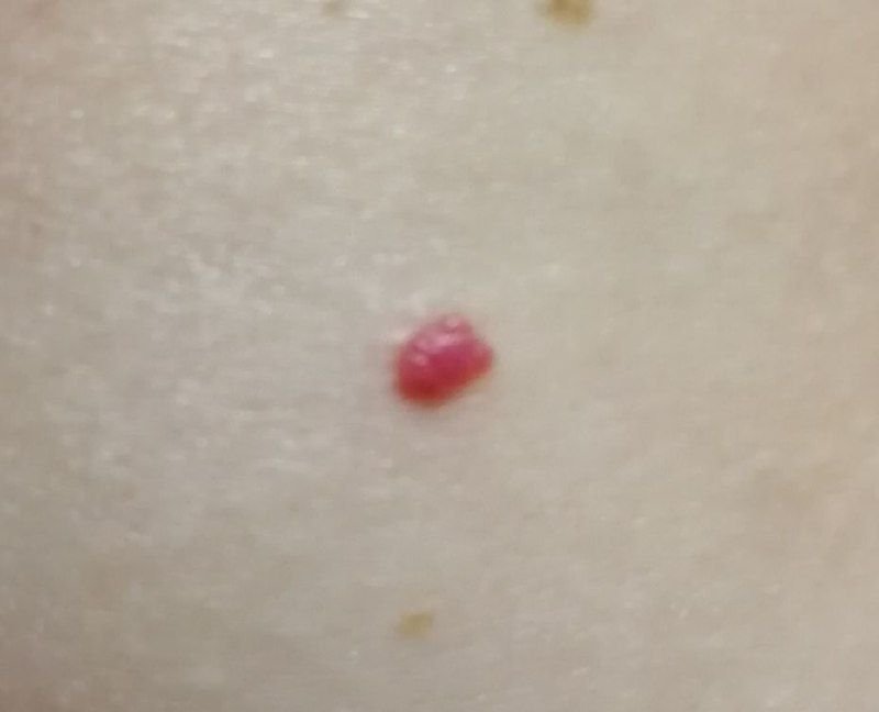
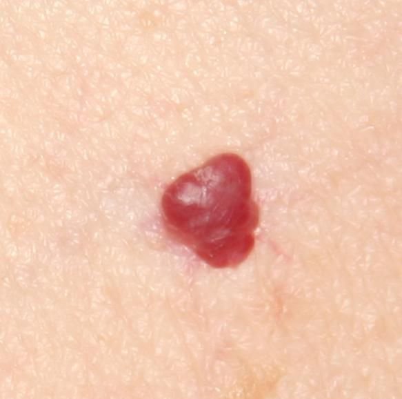
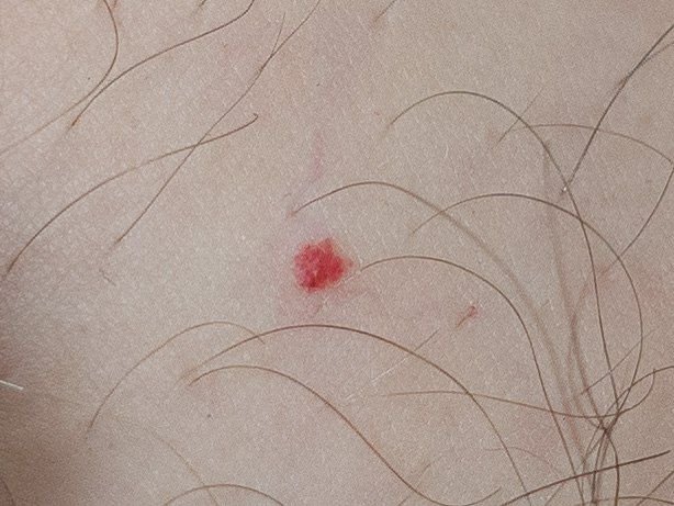
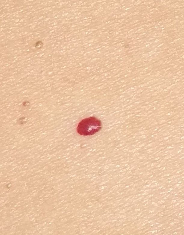
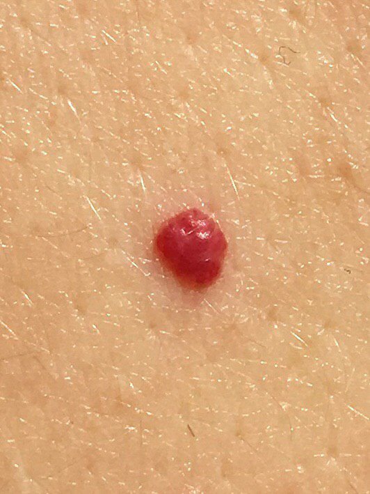
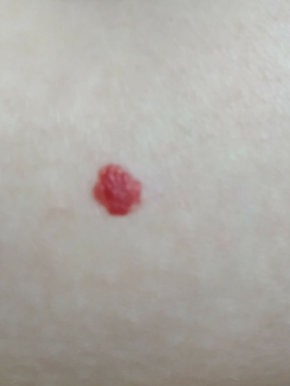
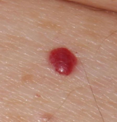
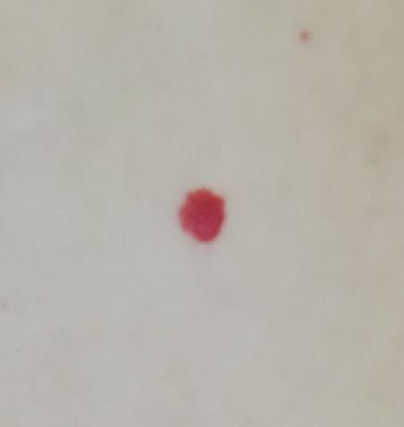
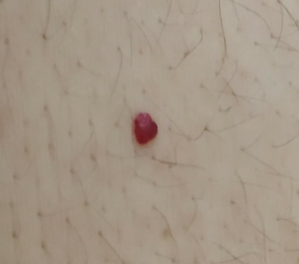
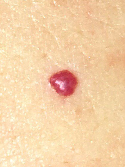
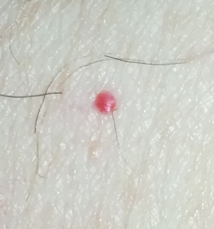
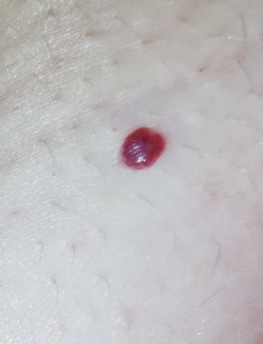
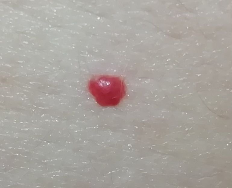
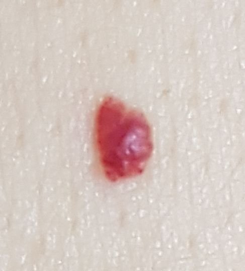
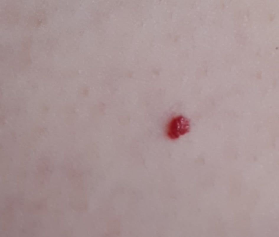
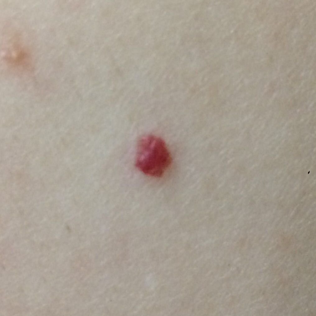
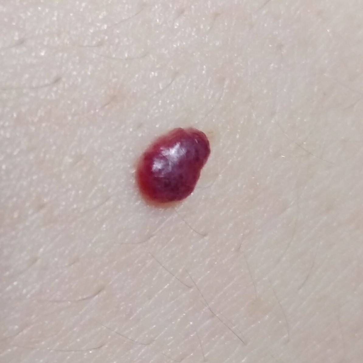
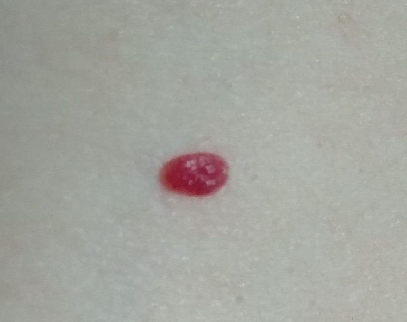
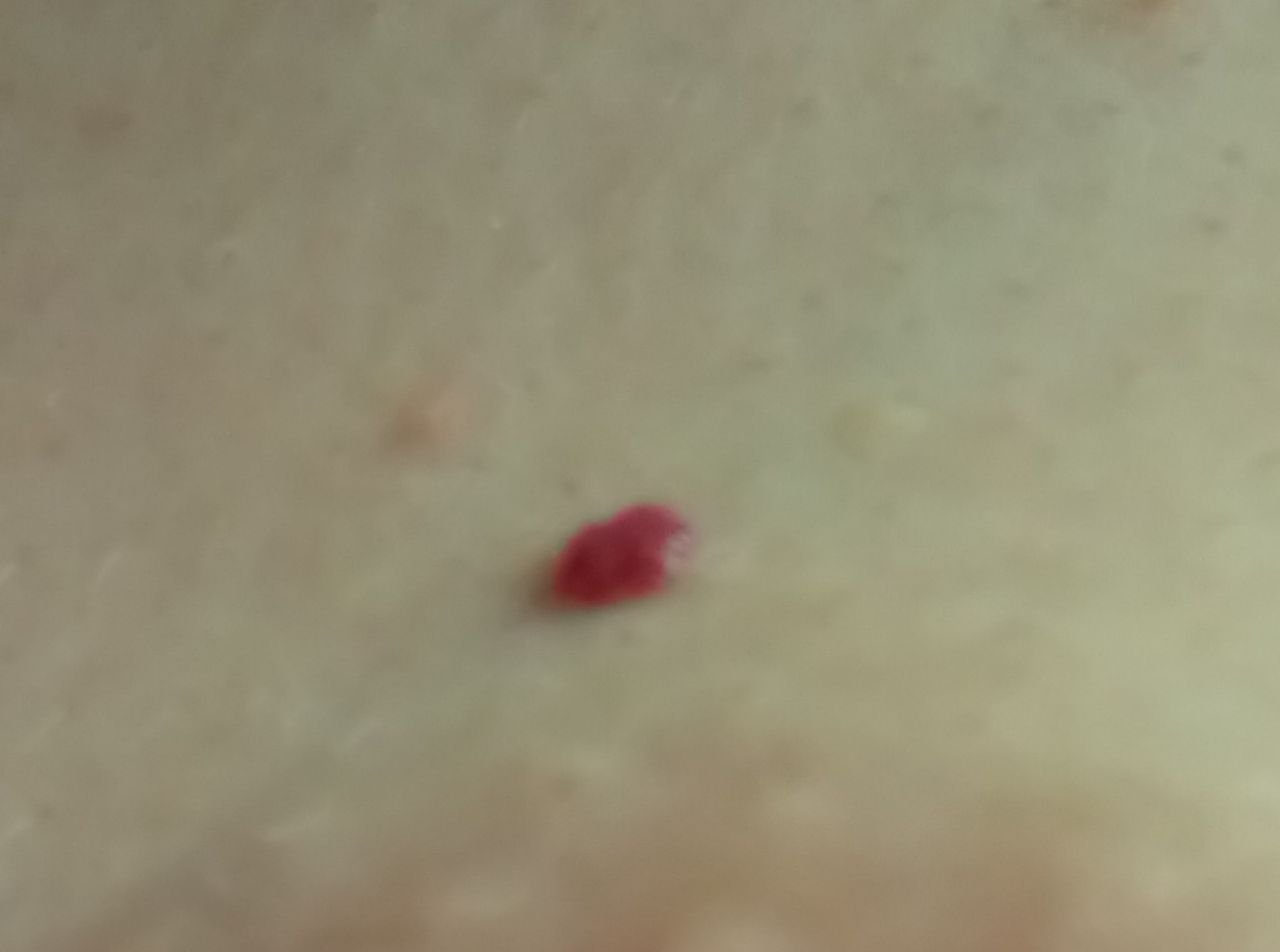
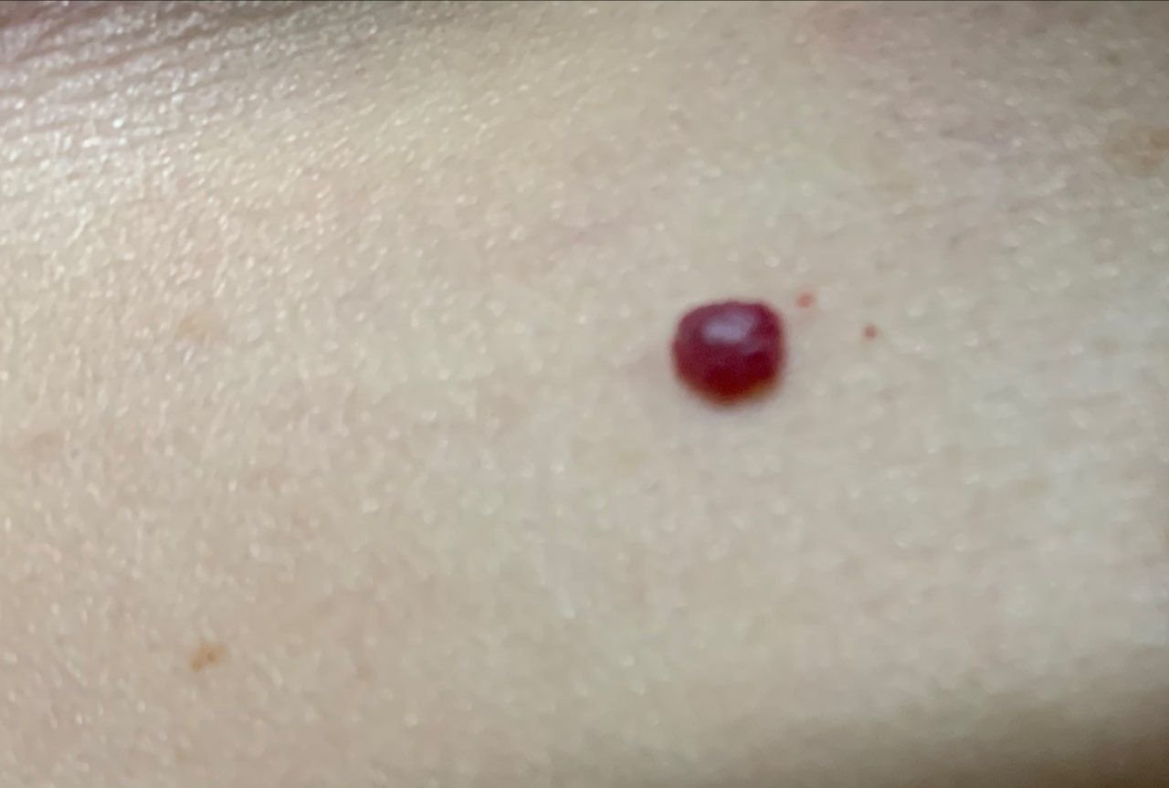
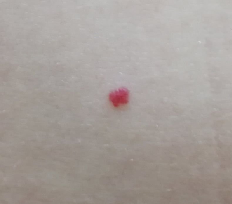
Acquired Hemangiomas: These lesions usually appear as small, hemispherical (sometimes on a narrow stalk), symmetrical nodules that protrude slightly above the skin. The surface may be slightly different from the normal skin texture, sometimes appearing smooth or even glossy. The color is generally bright red, but other shades of red may occasionally be seen. Like congenital hemangiomas, acquired hemangiomas do not affect hair growth. The size of acquired hemangiomas is typically small, usually up to 5-7 mm in diameter, although larger nodules may occasionally form. These hemangiomas are most commonly found on the trunk or upper limbs, though they can also develop on other parts of the body.
Dermatoscopic Description
On dermatoscopy, the following features are typically observed in hemangiomas:
- Elasticity and Filling: The tumor exhibits elasticity and can change size when compressed, turning pale temporarily.
- Small Vascular Gaps: A large number of small red gaps, separated by thin blue partitions, may be visible.
- Large Venous Gaps: The presence of large blue-violet gaps indicates that the hemangioma is deep and may involve venous blood vessels.
- Thrombosed Lacunae: Hemangiomas that are injured may show blue-black or black-red lacunae with a yellowish rim, which indicate thrombosis (clotting) in the vascular spaces.
Differential Diagnosis
Hemangiomas should be differentiated from other neoplasms or lesions, including:
- Various types of hemangiomas (congenital, acquired, deep, superficial, etc.)
- Pyogenic granuloma
- Blue nevus (particularly deep, venous hemangiomas)
- Angiosarcoma
- Kaposi’s sarcoma
- Mycosis fungoides (T-cell lymphoma of the skin)
Risks
Hemangiomas are benign and do not typically undergo malignant transformation. However, signs that suggest possible malignancy include changes in the appearance of the hemangioma or the development of new symptoms such as pain or itching.
Large hemangiomas can cause significant bleeding if injured. Additionally, large hemangiomas can ulcerate and become infected if they are subject to chronic trauma.
Some infantile hemangiomas need prompt specialist assessment rather than watchful waiting, because of where they are or what they are doing:
- Around the eye: A periocular lesion can interfere with vision as it grows.
- Airway: A lesion involving the airway can cause obstruction.
- Ulceration: An ulcerating lesion is painful and can scar.
- Large or segmental lesions: These may be associated with underlying structural anomalies.
- Five or more skin lesions: Multiple infantile hemangiomas warrant evaluation for liver involvement.
A lesion that is proliferating rapidly, or one that is interfering with feeding, breathing or vision, should be assessed without waiting.
Tactics
The management of hemangiomas depends on the size, location, and type (congenital or acquired). For large congenital hemangiomas, decisions about treatment should involve a team of specialists, including pediatricians, dermatologists, and surgeons, among others. The timing of treatment interventions is determined on a case-by-case basis, considering the hemangioma’s impact on vital functions and the presence of other associated health issues.
A lesion you have not had looked at is a different matter from one a clinician has already identified. Monitoring follows assessment, not the other way round: a vascular lesion that has not been examined should be shown to a dermatologist, since not every red lesion is a hemangioma. Once a small lesion has been assessed and is stable, no treatment is required and watching it between reviews is reasonable. Go back sooner if it is injured, changes in appearance, ulcerates, or develops new symptoms.
For dynamic monitoring, it is helpful to take photographs of the hemangioma to track any changes over time. How often someone with multiple skin lesions needs to be examined depends on those lesions rather than on the season. A skin neoplasm map can be beneficial for ongoing monitoring and the identification of new or altered lesions.
Treatment
Most uncomplicated infantile hemangiomas need no treatment at all. They involute on their own, and observation — with photographs to track the course — is the appropriate course. Laser, cryotherapy, electrocoagulation and surgery are not routine treatments for them.
Where treatment is needed, it is medical first:
- Oral propranolol: First-line therapy for infantile hemangiomas that are high-risk — because of their site, their size, ulceration, or a threat to function. It is started and supervised by a specialist.
- Topical timolol: An option for selected small, superficial lesions.
Laser has a place for residual changes after involution, such as persistent surface vessels, and surgery for a lesion that is bleeding dangerously or has left tissue that needs reconstruction. Doses and the choice of agent are set by the treating specialist.
Some lesions require staged treatment or follow-up over time.
Prevention
Preventing the appearance of hemangiomas involves proper prenatal care, including minimizing stress, avoiding harmful drugs during pregnancy, and addressing infections promptly. To prevent further lesions or malignancy:
- Avoid chronic skin trauma.
- Observe safety measures when working with skin-damaging agents.
- Address concomitant health conditions promptly.
- Maintain good personal hygiene and awareness of skin changes.
Regular examination of hemangiomas, prompt consultation with a specialist if any changes are observed, and removal of potentially harmful neoplasms are essential for managing skin health.


Medical review
Board-Certified Dermatologist · Fellow of the American Academy of Dermatology (FAAD)