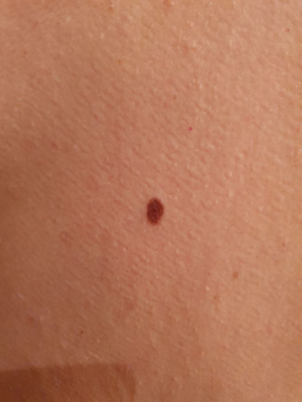
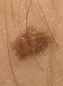
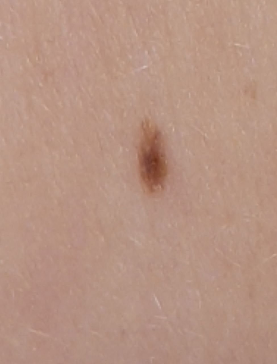
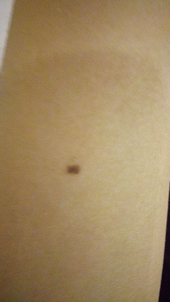
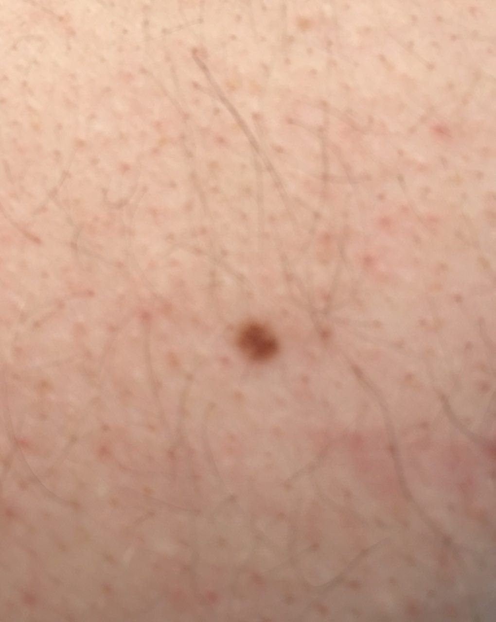
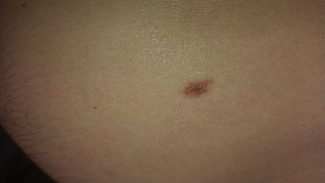
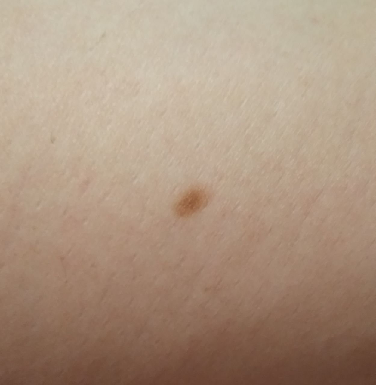
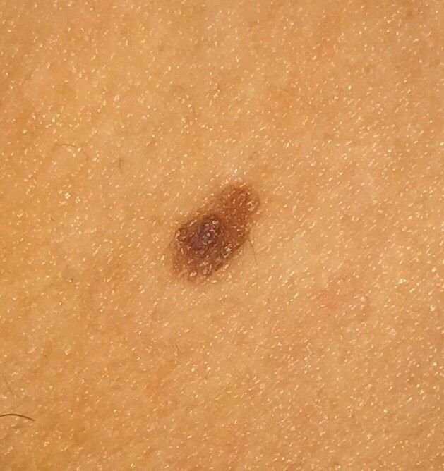
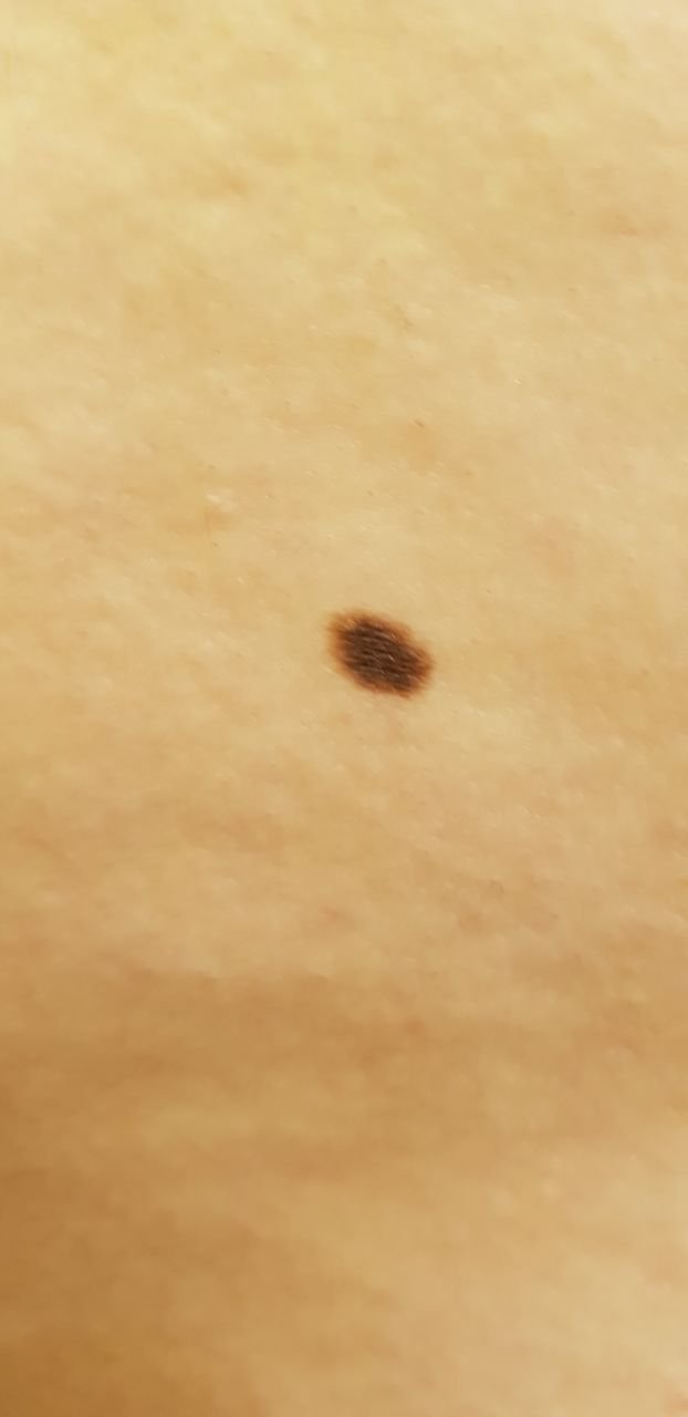
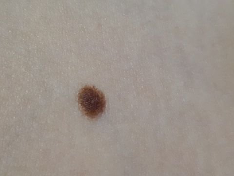
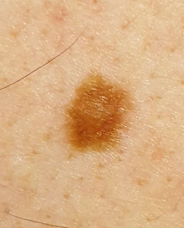
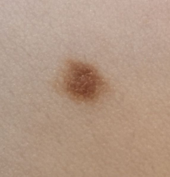
Benign Nevus (ICD-10: D22) 💚
Benign Melanocytic Nevus (Simple Nevus, Common Mole)
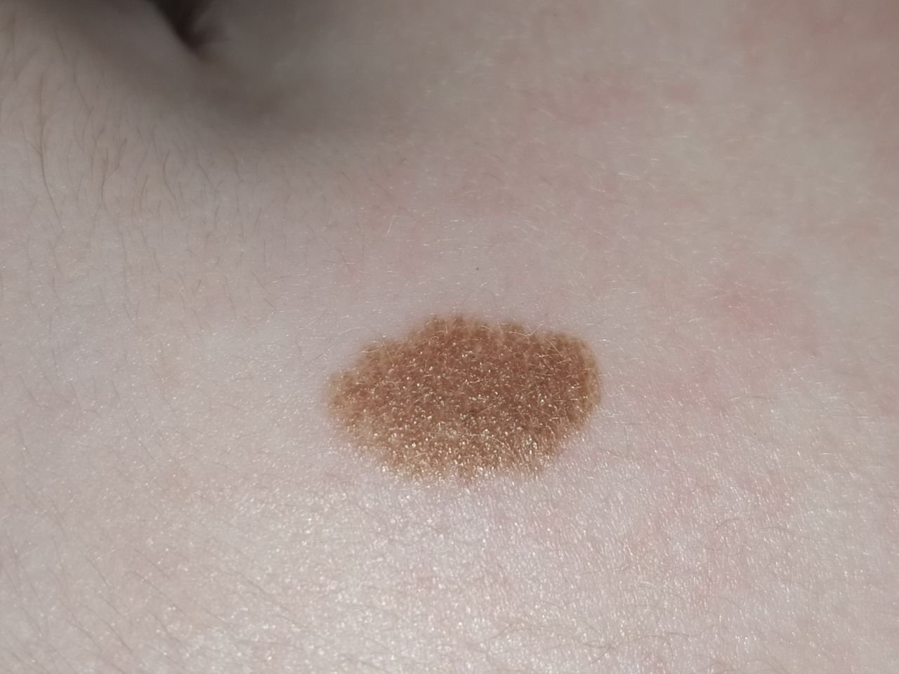
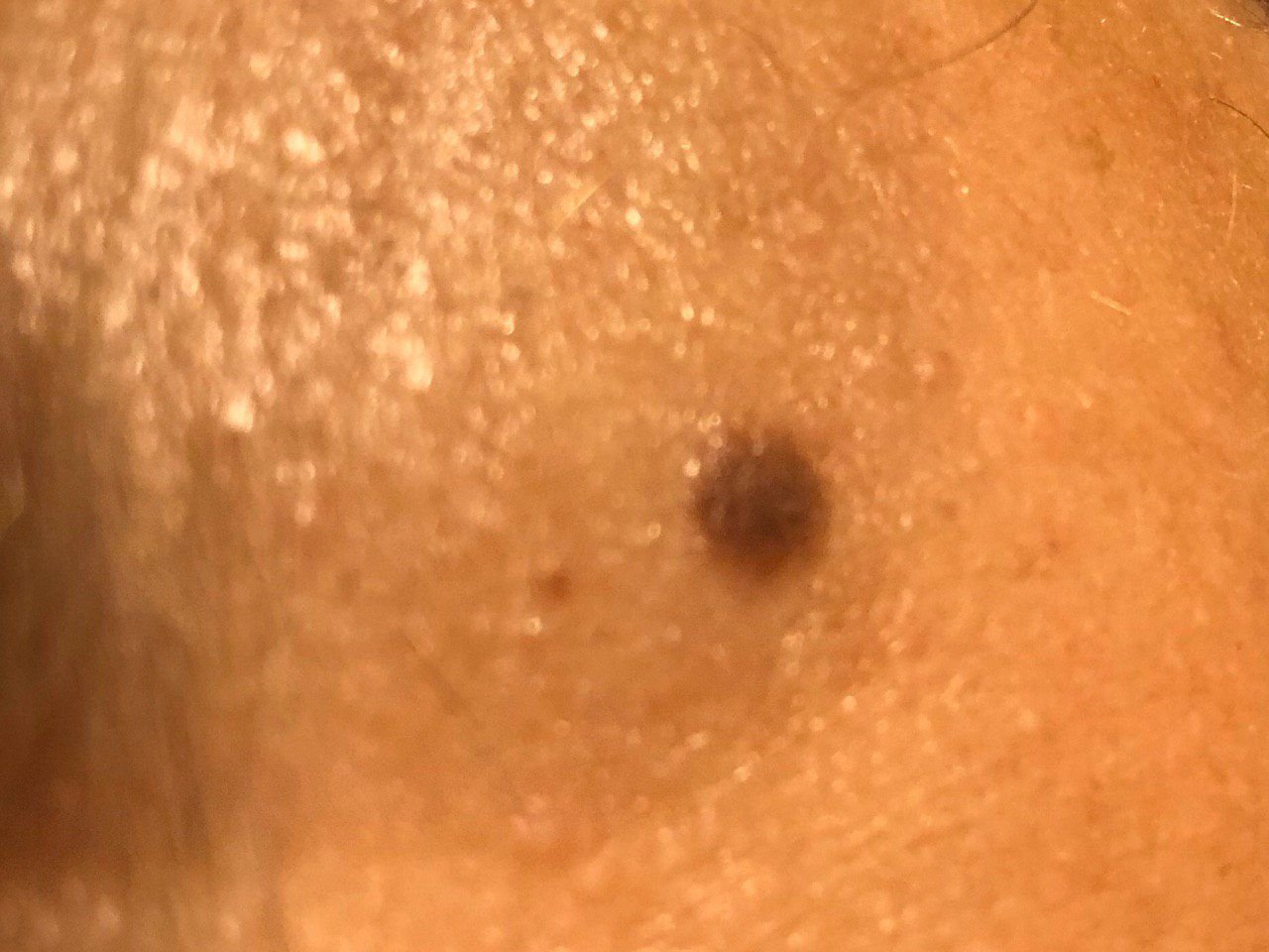
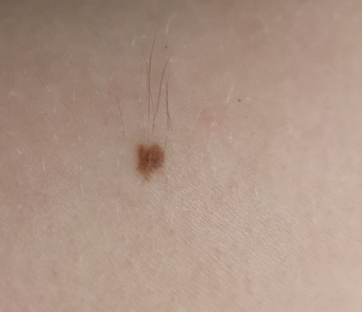
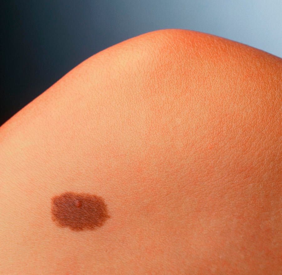
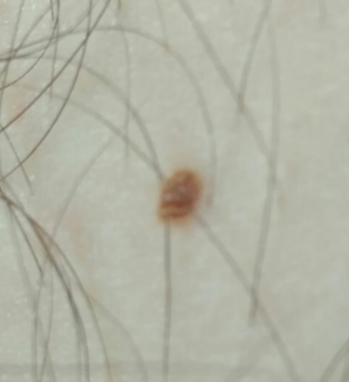
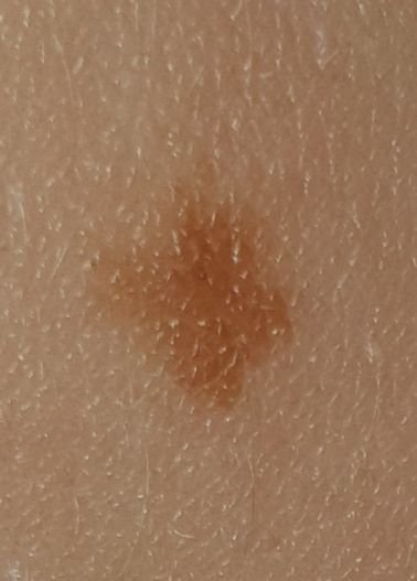
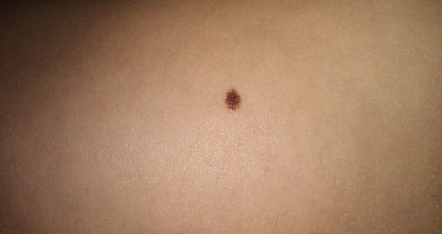
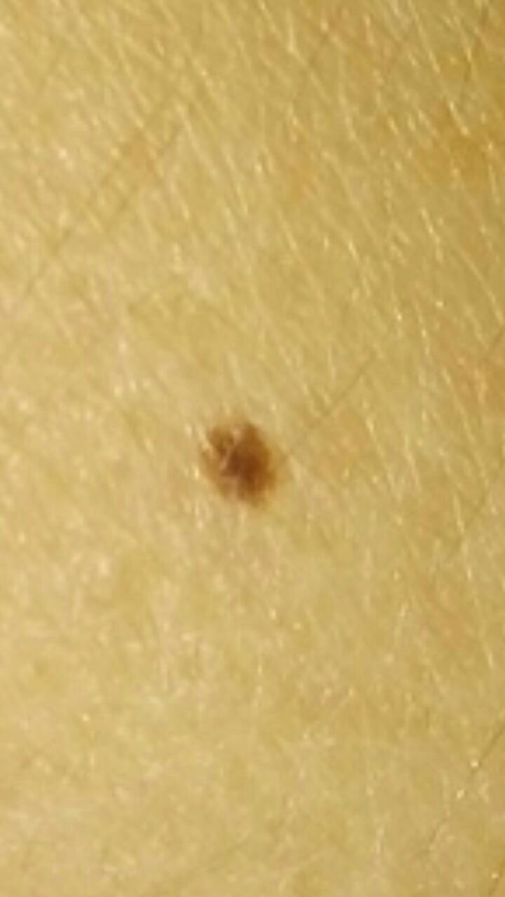
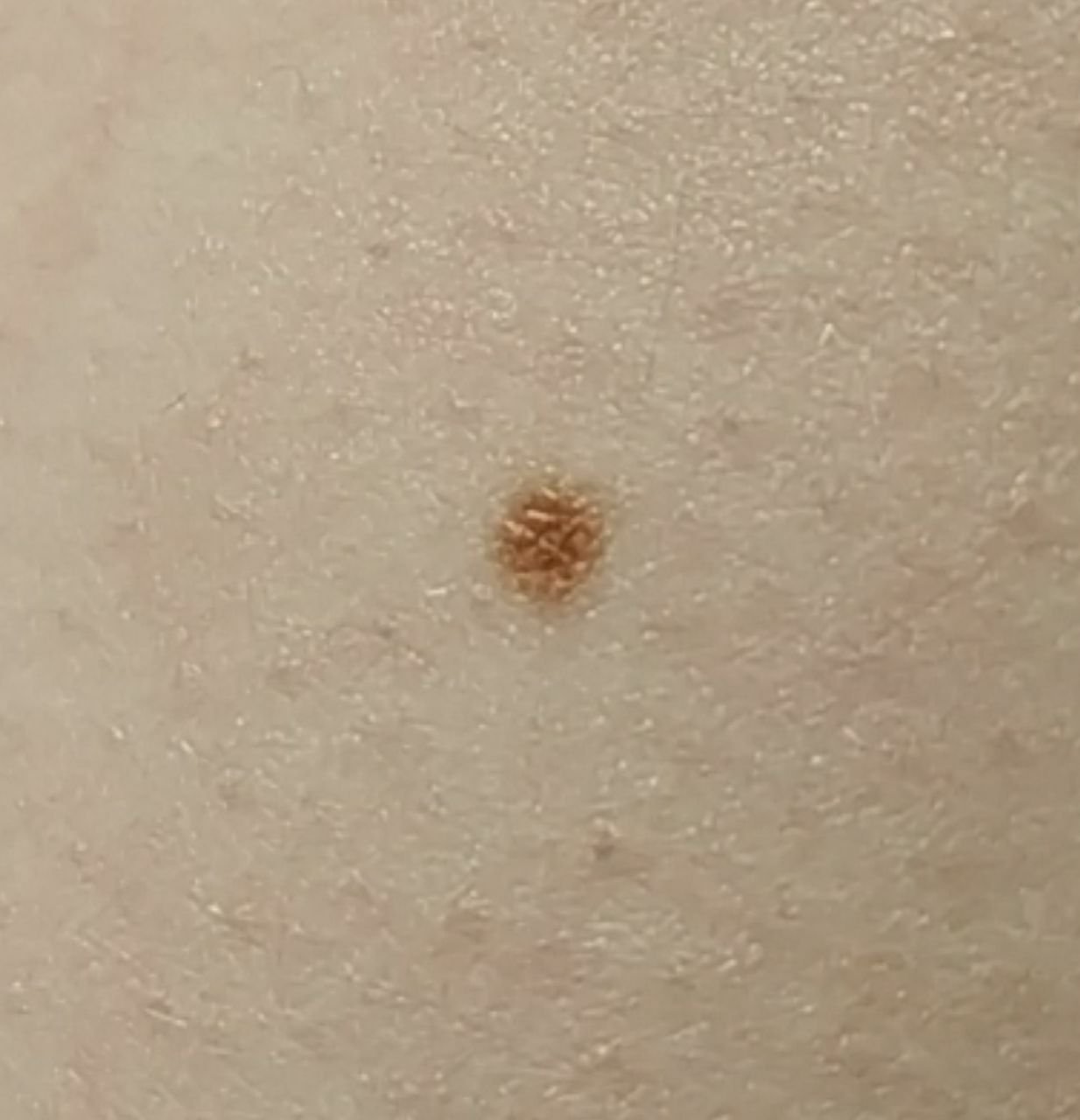
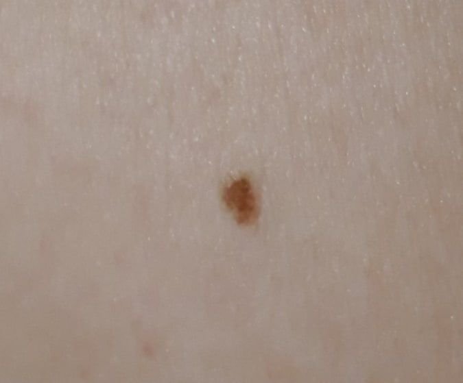
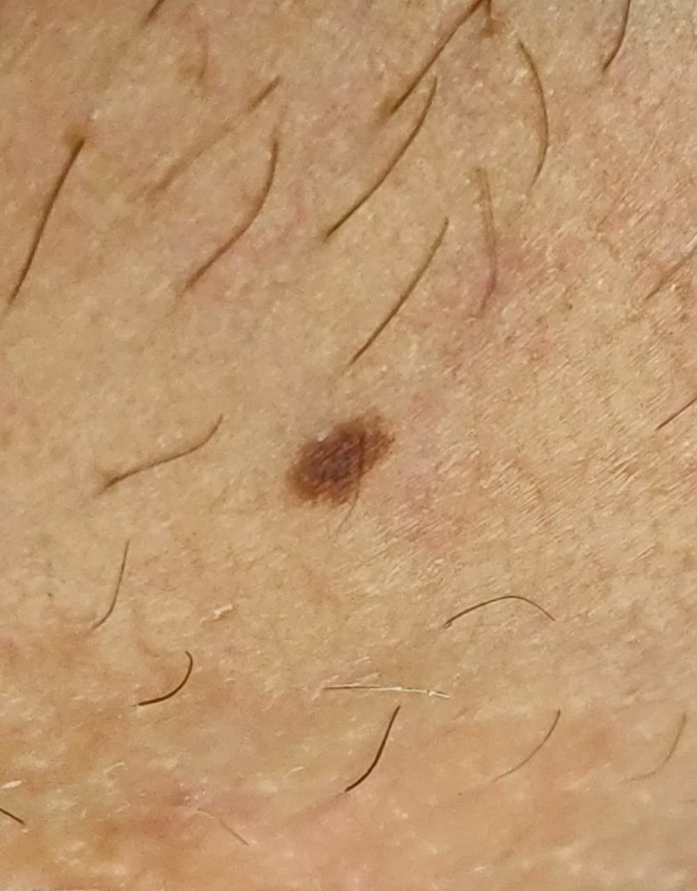
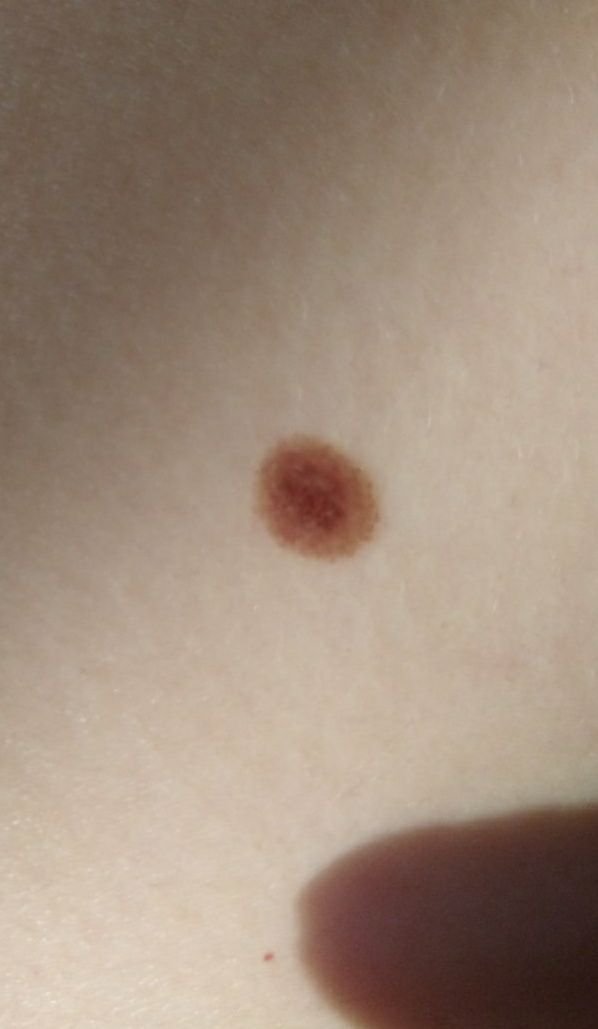
Benign melanocytic nevus — a simple nevus, or in everyday language a mole — is the broad category that benign pigmented lesions of melanocytic origin belong to. It usually appears as a small spot or a slightly raised nodule. Particular kinds within this category have their own pages in this atlas, among them the papillomatous nevus, the blue nevus, the halo nevus and the acral nevus. Simple nevus can be either congenital (present at birth) or acquired at any age. Around 3% of newborns have multiple simple nevi, with the frequency increasing with age. Simple nevi are slightly more common in women than in men, with a ratio of 3:2, respectively.
Predisposing Factors
There is no definitive cause for the formation of simple nevi. However, several predisposing factors may increase the risk of their appearance:
- Genetic Factors: The appearance of pigmented nevi may be influenced by a person’s genetic makeup.
- Ultraviolet Radiation: Both artificial and natural ultraviolet light can stimulate the rapid growth of nevus cells and cause excessive melanin production, leading to pigmentation in the nevus.
- Hormonal Changes: Nevi may become more noticeable during periods of hormonal change, such as puberty or pregnancy. Thyroid and adrenal hormones, viral infections and injuries are sometimes listed as causes of nevus formation; they are not established as such.
Diagnostics
The diagnosis of simple nevi is based on clinical examination, which includes routine visual inspection and dermatoscopy. If there are concerns about malignant growth, a biopsy may be performed for further examination.
Symptoms
A simple nevus typically appears as a small spot or slightly raised nodule. It is usually symmetrical (oval or round), though large congenital nevi may be irregularly shaped. The surface texture of the nevus is similar to that of normal skin, but it may sometimes differ slightly from the surrounding skin pattern.
The edges of benign nevi are well-defined and smooth. Large congenital nevi, however, may have uneven borders. The color of a simple nevus ranges from light brown to dark brown, with uniform pigment distribution. In some cases, the color may gradually fade from the center to the outer edges. The color of congenital nevi may change during the first few years of life.
Simple nevi do not typically affect hair growth. However, some congenital nevi may show increased growth of coarse, dark hair, often accompanied by noticeable pigmentation.
Simple nevi can vary greatly in size, but most commonly they measure up to 10 mm. Nevi larger than 10 mm are usually congenital and are rare, though they can grow to be as large as 20 cm or more (giant congenital nevi).
When palpated, simple nevi feel like regular skin and do not cause any discomfort.
Nevi are mostly found on the trunk (~38%) or limbs (~48%), with fewer located on the head and neck (~14%).
Acral nevi (on the palms and soles) differ slightly in shape, border, and pigment distribution due to the characteristic skin pattern of these areas (“fingerprints”). These nevi are often elongated, with irregular borders, darker pigmentation, and pigment distribution in parallel stripes.
Dermatoscopic Description
On dermatoscopy, a simple nevus exhibits the following characteristics:
- Pigment Network: A pattern of light to dark brown hypopigmented holes and lines, with the lines thinning out towards the periphery of the nevus.
- Dots: Small, round hyperpigmented structures located at the center or along the pigmented lines of the network.
- Globules: Larger, hyperpigmented ring-shaped structures, evenly distributed throughout the nevus or located centrally, rarely found at the periphery.
- Spots: Hyperpigmented, structureless areas in the center of the nevus.
- Uniform Diffuse Staining: The entire nevus shows a uniform pigmentation.
None of these structures, on its own, makes a nevus benign — each of them occurs in melanoma as well. What matters clinically is the arrangement: symmetry, and an organised, homogeneous pattern across the whole lesion. A peripheral starburst of radial streaks, in particular, is not a routine feature of a common nevus; it is associated with Spitz and Reed nevi and, in the wrong context, with melanoma, so it calls for assessment rather than reassurance.
Acral nevi have their own dermatoscopic patterns. In a benign acral nevus the pigment follows the furrows of the skin, in one of three recognised arrangements — the parallel furrow pattern, the lattice-like pattern, or the fibrillar pattern, the last of these especially on the weight-bearing sole. Pigment running along the ridges instead is the parallel ridge pattern, which is a warning sign for acral melanoma and not a feature of a benign nevus.
Differential Diagnosis
Simple nevi should be differentiated from other pigmented lesions, such as:
- Post-inflammatory hyperpigmentation
- Congenital dermal melanocytosis
- Sebaceous gland nevus
- Halo nevus
- Spitz nevus
- Blue nevus
- Lentigo
- Dysplastic nevus
- Lentigo melanoma
- Melanoma
Risks
Any one typical nevus has a very low chance of becoming a melanoma. That is a different question from a person’s overall melanoma risk: having a large number of moles, or having clinically atypical ones, is itself a recognised marker of increased risk. A person with many moles is therefore not in the same position as a person with one, even though each individual lesion is unlikely to cause trouble. Changes in appearance, or new sensations in a nevus, should be assessed by a dermatologist.
Melanoma risk in a congenital melanocytic nevus depends on several things together — its size, its appearance, where it is, whether satellite nevi are present — and it is not settled by a single measurement. A fixed cut-off, with one risk figure above it and another below, states the matter more precisely than the evidence allows.
Large and multiple congenital nevi may be associated with certain genetic syndromes or diseases, so individuals with such nevi require careful observation and medical evaluation.
Tactics
A lesion you have not had looked at is a different matter from one a clinician has already identified. Monitoring follows assessment, not the other way round: a mole you have not had examined should be shown to a dermatologist, and this applies with more force to a new one, or to one that looks unlike the rest of your moles.
Once a lesion has been assessed as a typical benign nevus, watching it between reviews is reasonable, with help from someone else for areas you cannot see. Go back sooner for rapid growth, asymmetry, more than one colour, bleeding, or any new sensation in the lesion.
The doctor will determine if dynamic monitoring is necessary, or if the nevus should be removed. Nevi subject to constant irritation from clothing, jewelry, or occupation should also be removed.
For dynamic monitoring, it is useful to take photographs of the skin neoplasm, which will help detect even minor changes in appearance over time.
How often someone should be examined is decided individually, taking account of what their nevi look like, how many they have, the size of any congenital lesion, and their personal and family history of melanoma — together with any change that has been noticed. It is not set by the calendar. Mapping of skin lesions is not a routine recommendation. Mapping remains a reasonable option for someone at high risk — a patient with many atypical or dysplastic nevi, or with a previous melanoma — where it is decided individually.
Treatment
A typical benign nevus does not need to be removed. Where removal is indicated — to establish a diagnosis, because the lesion is symptomatic, or for cosmetic reasons — the technique depends on the lesion.
A pigmented lesion that looks suspicious is a different matter: it should be sampled or excised so that the tissue can be examined histologically. Destructive methods such as laser removal and cryodestruction leave nothing for the pathologist, so they should not be used on a pigmented lesion whose nature has not been established.
Prevention
Preventing the appearance of nevi and their malignant transformation involves gentle skin care:
- Avoid excessive UV exposure (e.g., tanning beds, sunbathing).
- Use protective sunscreen during peak sunlight hours.
- Avoid chronic skin trauma.
- Follow safety measures when handling skin-damaging agents.
- Maintain personal hygiene and be aware of skin changes.
Regularly examine pigmented nevi, seek timely consultation with a specialist if any changes occur, and remove potentially dangerous nevi when necessary.