
Lentigo (ICD-10: L81) 💚
Lentigo
Lentigo is a benign, flat brown spot, and the word covers more than one condition. Solar lentigo, also called actinic lentigo or a sunspot, follows years of sun exposure: it appears on chronically sun-exposed skin, typically after the age of 35, and becomes common with age — around 90% of people over 60 have at least one. Lentigo simplex is a different thing. It commonly appears in childhood, is not necessarily caused by sun exposure, and can occur on skin that is rarely exposed. Because the two have different contexts and different causes, not every lentigo should be read as sun damage.
Predisposing Factors
The exact cause of lentigo is not fully understood, but several predisposing factors have been identified that may increase the likelihood of developing hyperpigmentation. These factors can influence the onset and growth of lentigo:
- Ultraviolet Radiation: Both natural sunlight and artificial UV radiation contribute to excessive melanin production in the skin, which leads to the formation of lentigo.
- Hormonal Changes: Hormonal fluctuations, particularly those involving sex hormones, thyroid hormones, and adrenal hormones, can influence the development of lentigo.
- What is not established: Ionizing radiation, viral diseases and injuries are sometimes listed as causes of lentigo. None of them is established as a general predisposing factor.
- Genetic Factors: There may be a genetic predisposition to developing lentigo in certain individuals.
- Light Skin: Individuals with a Fitzpatrick skin type I-II are more prone to developing lentigo due to their skin’s sensitivity to UV radiation.
- Age Over 35 Years: The likelihood of developing lentigo increases with age, especially after 35 years.
Diagnostics
The diagnosis of lentigo is based on a clinical examination, which includes visual inspection of the lesion and dermatoscopy to evaluate the characteristics of the pigmentation. Dermatoscopy is an aid here, not a verdict: it supports the diagnosis of a typical lesion but cannot settle an atypical or changing one. Atypical and changing lentigines can look like melanoma, lentigo maligna in particular, and where malignancy cannot be excluded a diagnostic biopsy is the appropriate step.
Symptoms
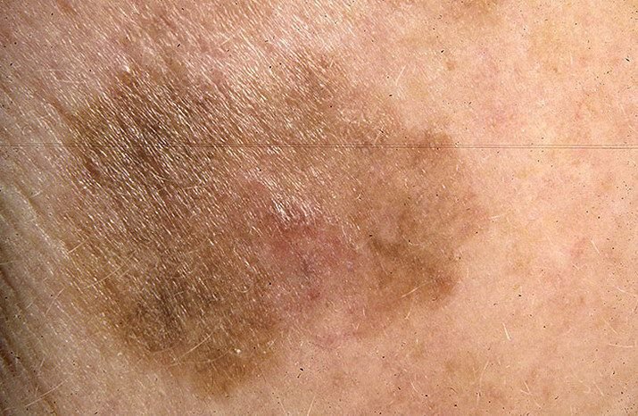
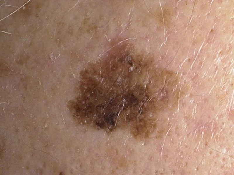
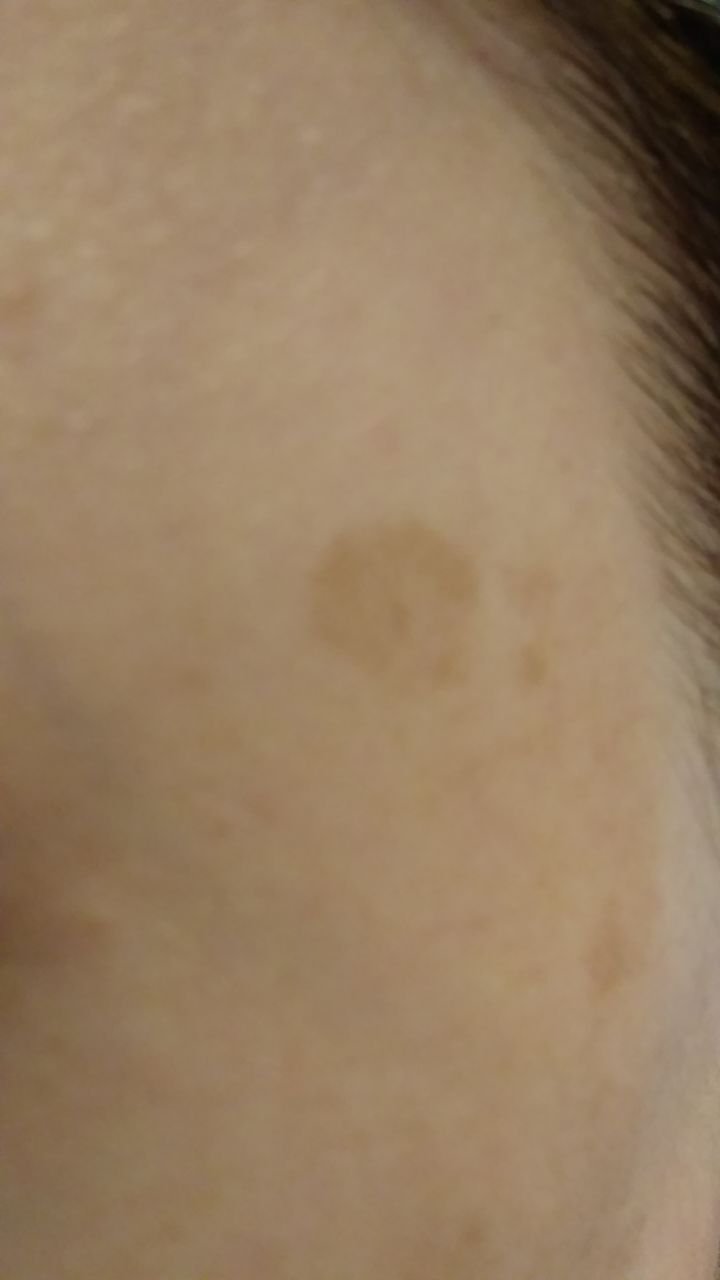
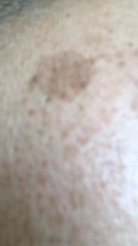
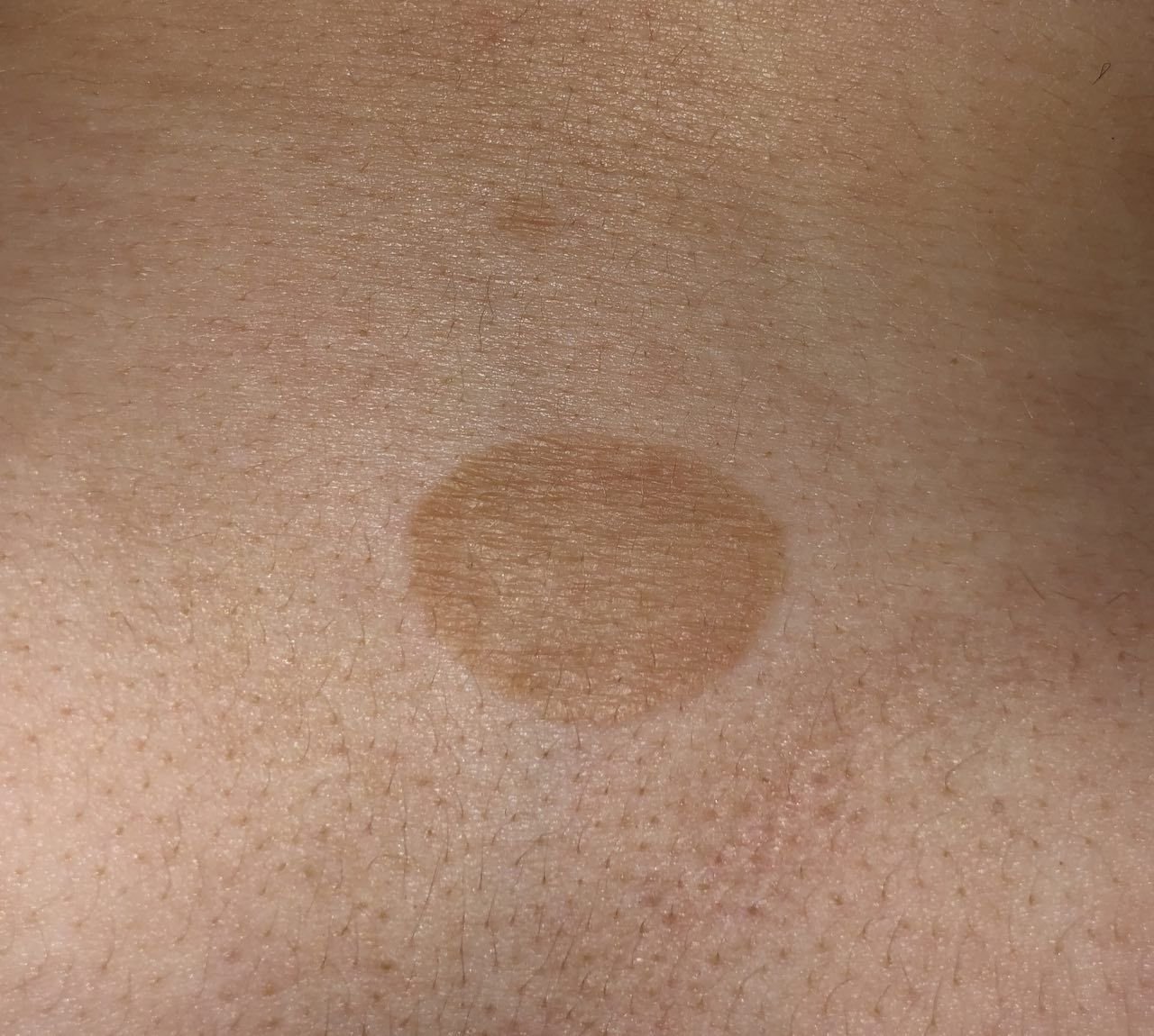
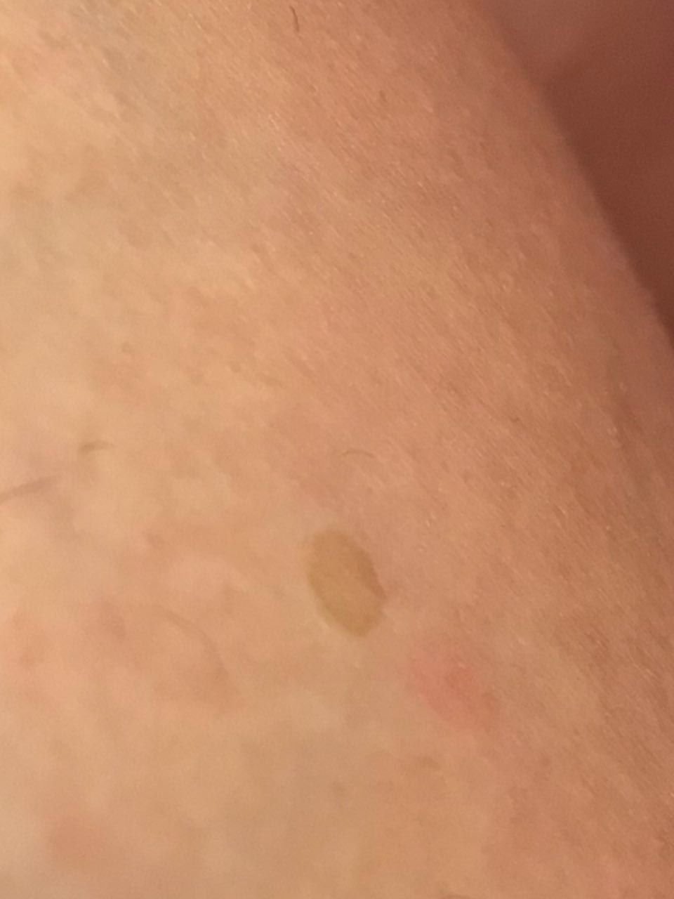
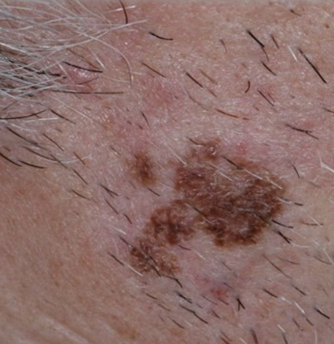
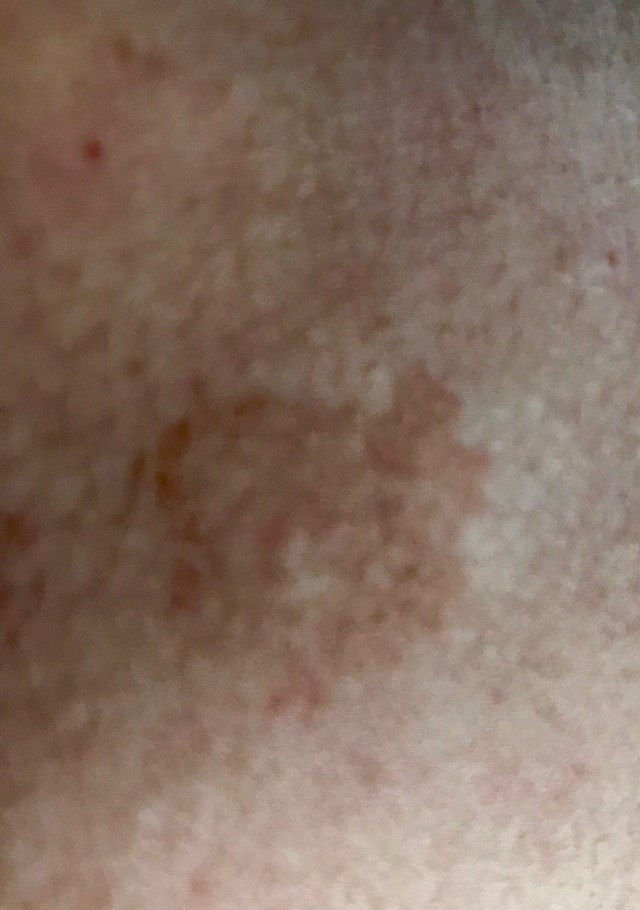
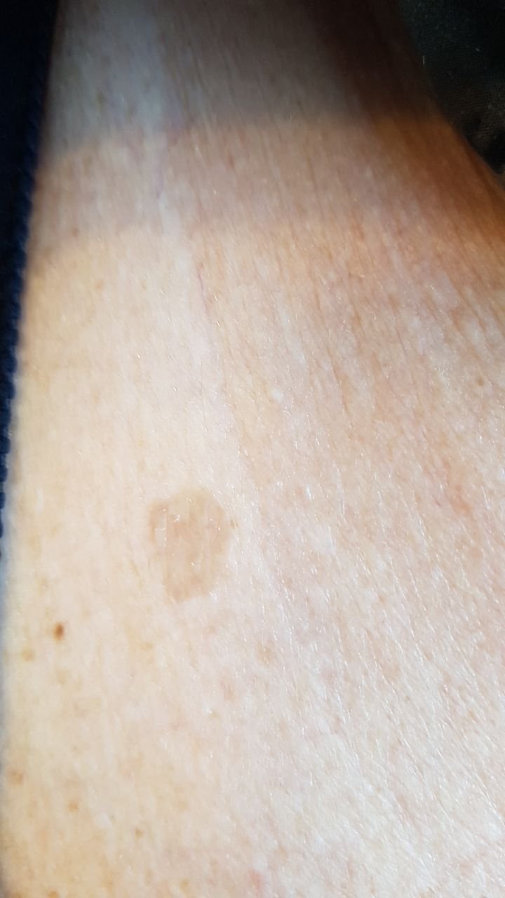
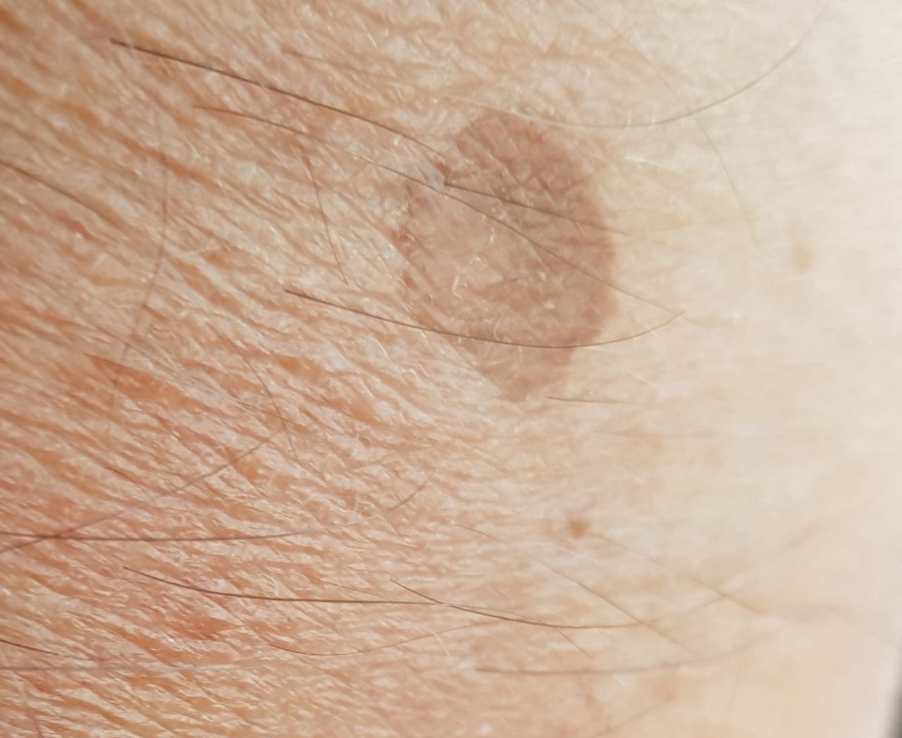
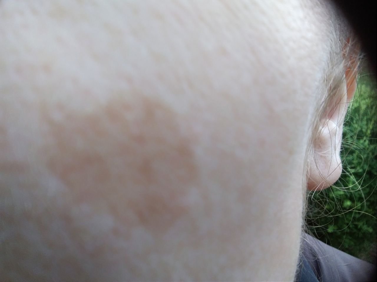
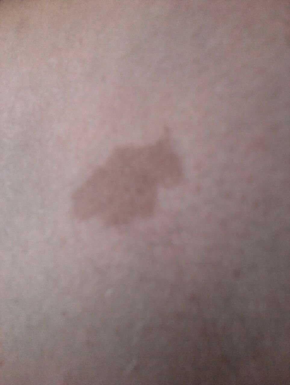
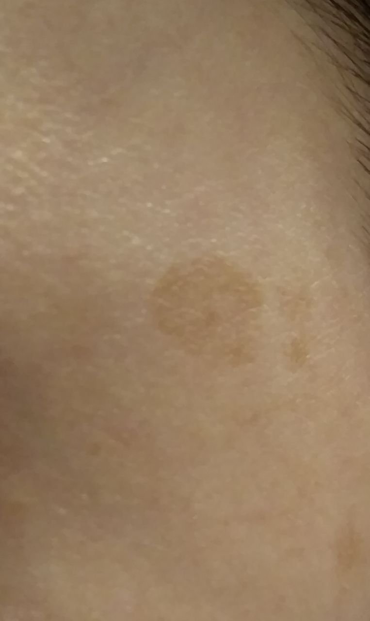
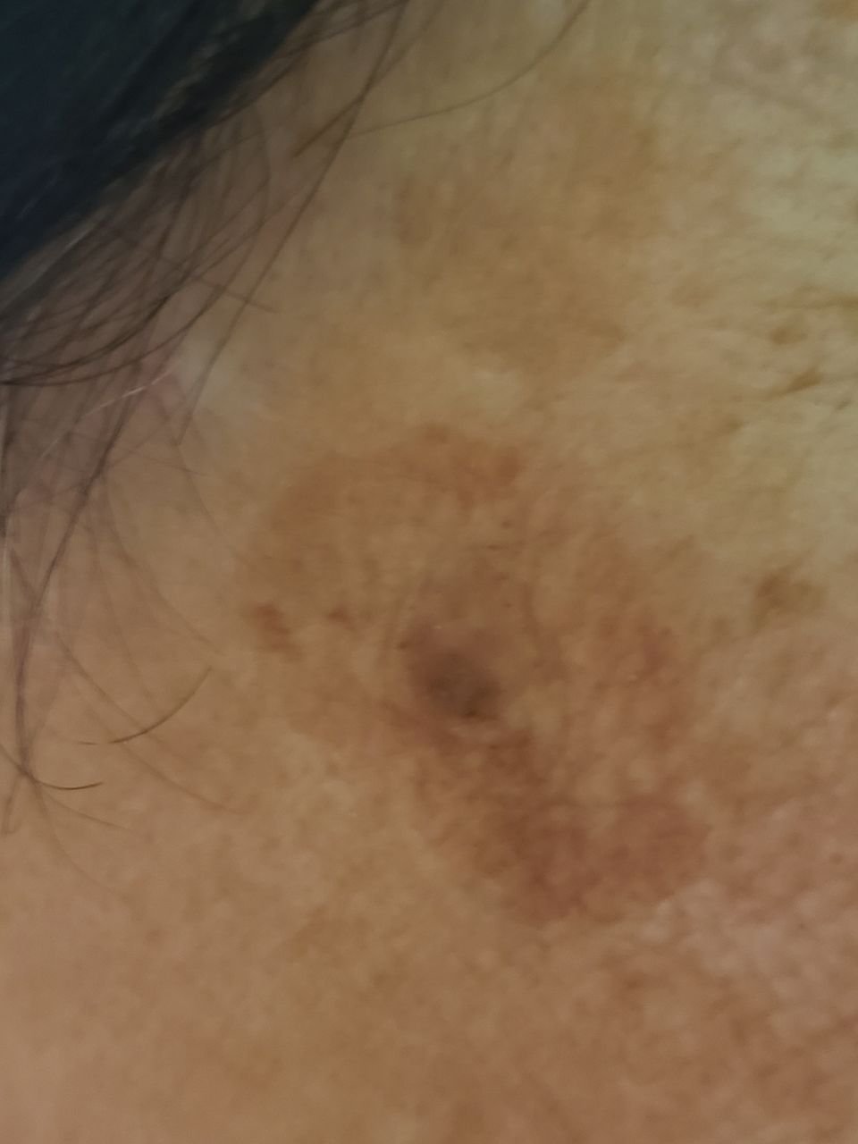
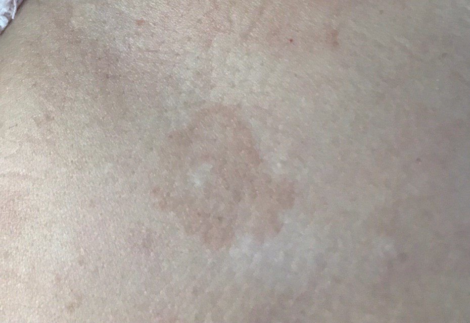
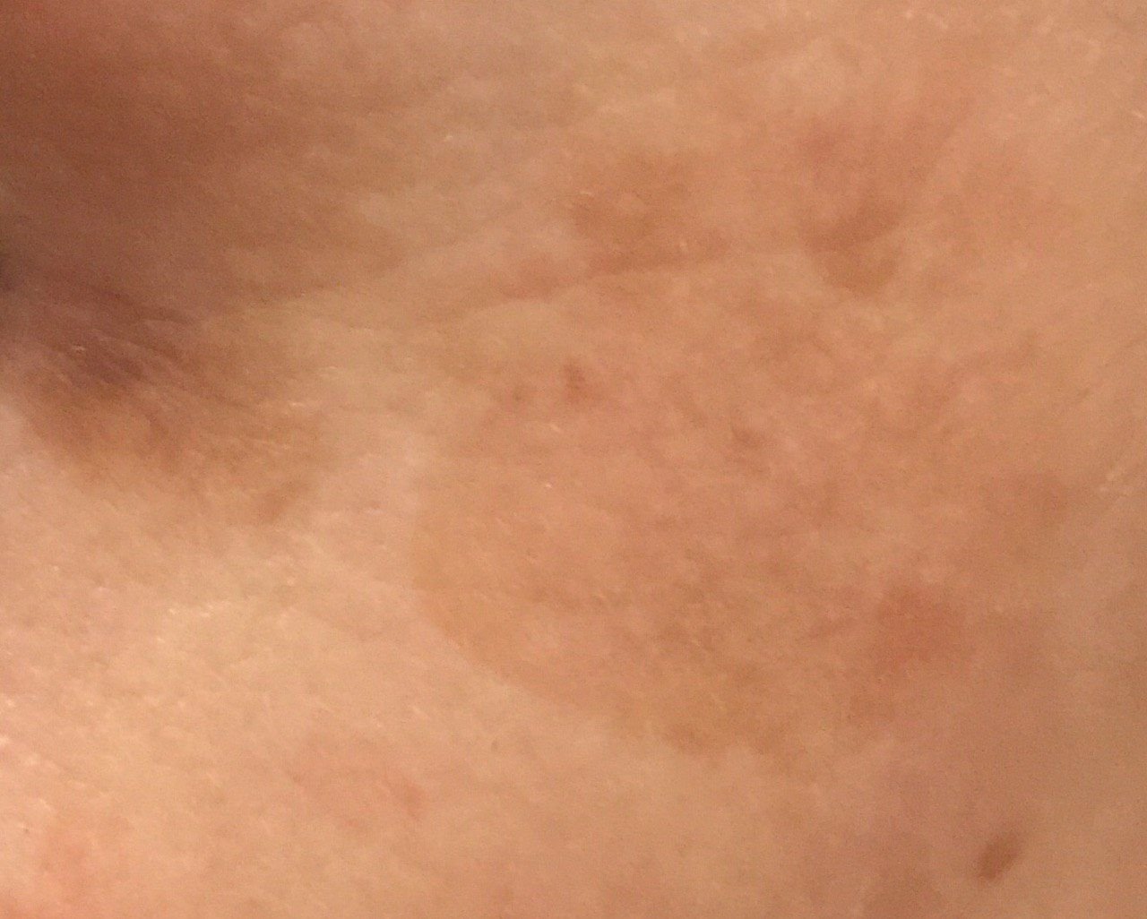
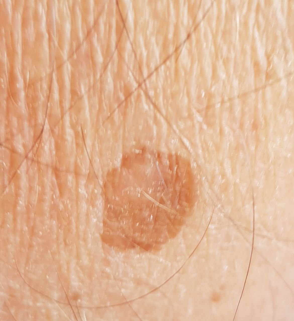
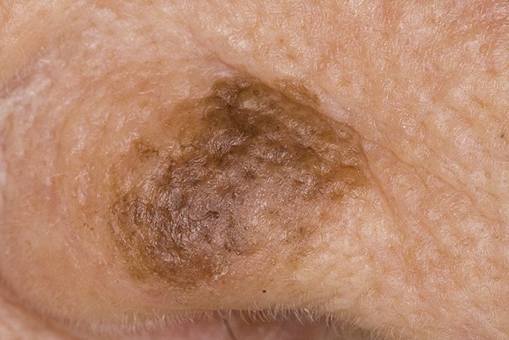
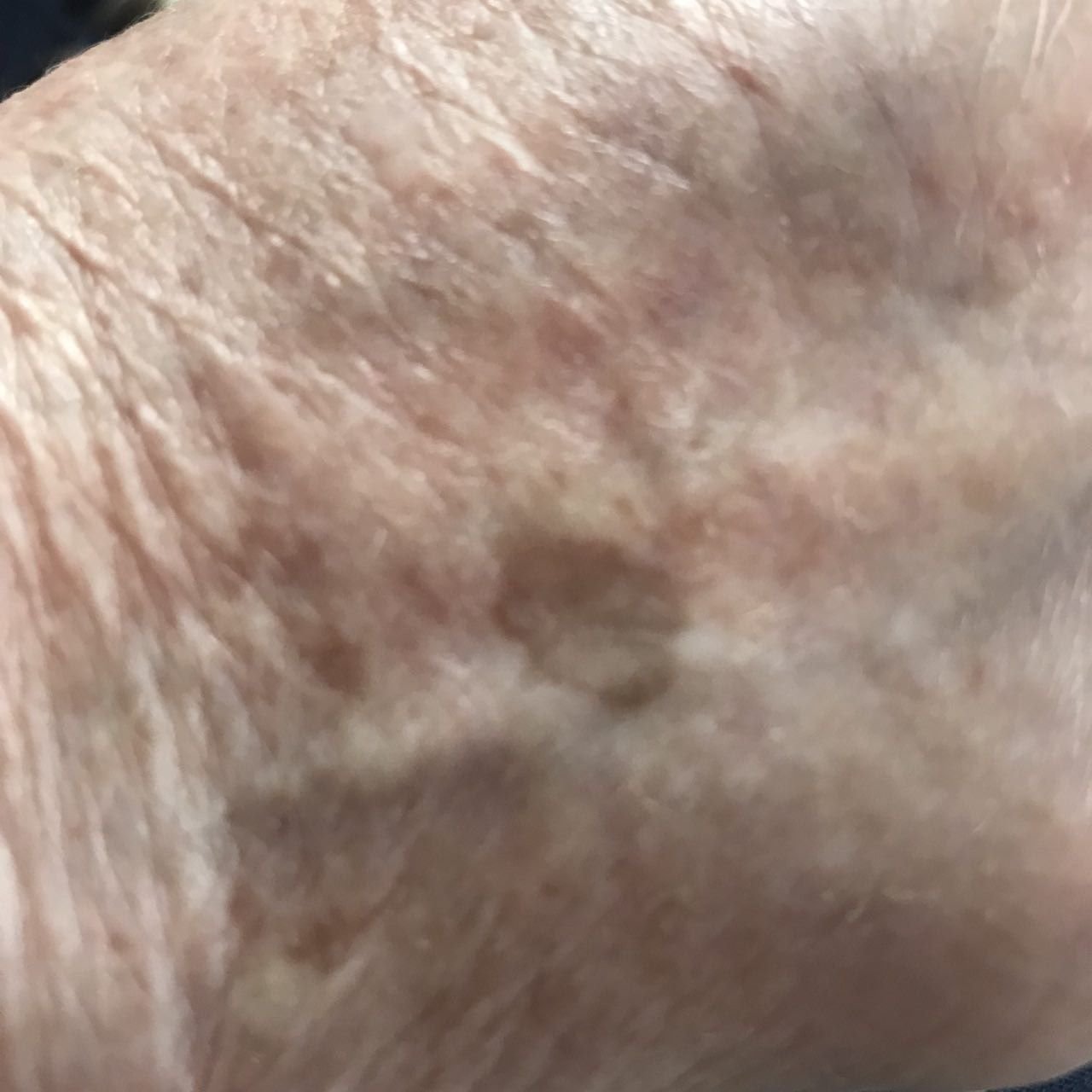
During a visual examination, lentigo appears as a flat spot or a group of spots that are slightly elevated above the skin’s surface (usually no more than 1 mm). The lesions may be symmetrical or irregular in shape, such as when several spots merge together or form a group. Lentigo can appear as a collection of multiple spots, sometimes covering entire anatomical areas. The surface of lentigo typically resembles the texture of normal skin, though small rough areas or peeling may sometimes be observed.
The boundaries of lentigo are usually clear but can often be uneven, especially with larger or multifocal spots. The coloration ranges from light brown to dark brown, and the pigment is usually evenly distributed throughout the lesion. Occasionally, there is a gradual decrease in color intensity from the center towards the edges, or an irregular change in shade within the area of pigmentation. Over time, the color of lentigo may become more intense.
Lentigo does not typically affect hair growth. However, in some cases, coarse or fluffy hair may grow in the central area of the lesion.
The size of lentigo can vary widely. Individual spots can range from 2-3 mm in diameter to much larger spots, up to 3-4 cm. When lesions are grouped together, they may cover several areas, sometimes extending to tens of centimeters. On palpation, lentigo feels like normal skin, though in older lesions, roughness may be noted, and mild itching can occasionally occur.
Lentigo is most commonly found on sun-exposed areas, such as the face, neck, and hands. In elderly individuals, lentigo may also appear on other areas of the body.
Dermatoscopic Description
Dermatoscopic examination of lentigo reveals the following features:
- Faint Pigment Network (solar lentigo): A delicate, pale network rather than a strong one, often against diffuse light brown pigmentation.
- Fingerprint-like Structures (solar lentigo): Fine parallel ridged lines resembling a fingerprint.
- Moth-eaten Borders (solar lentigo): Well-demarcated edges with concave notches, as though bitten out.
- Dark, Relatively Uniform Network (lentigo simplex): A more pronounced and evenly distributed pigment network than in solar lentigo.
- Pseudo-Network: On the face, a reticulate structure formed by follicular and adnexal openings against diffuse brown pigmentation.
Differential Diagnosis
It is important to differentiate lentigo from other pigmented neoplasms or conditions, including:
- Post-inflammatory hyperpigmentation
- Congenital dermal melanocytosis
- Halo nevus
- Spitz nevus
- Dysplastic nevus
- Lentigo maligna melanoma
- Pigmented basal cell carcinoma
- Melanoma
Risks
Lentigo is generally considered safe and does not have a significantly increased risk of malignant transformation. Benign lentigo is not known to undergo malignant transformation on its own; new or changing pigmented lesions should be evaluated to exclude lentigo maligna. However, signs of potential malignancy include changes in the appearance of the lesion, such as increased size, irregular shape, or the appearance of subjective sensations such as itching or bleeding.
Lentigo maligna (also known as Dubreuilh melanosis or melanoma in situ on sun-damaged skin) is a lesion distinct from benign lentigo; it carries a meaningful risk of progression to invasive melanoma and requires specialist evaluation.
The following should prompt dermatologic assessment rather than being put down to an ordinary lentigo: a spot that keeps enlarging; one that is becoming less symmetrical; irregular pigmentation or several shades within one lesion; the appearance of black or blue areas; thickening of a previously flat spot; bleeding; or ulceration.
Tactics
A lesion you have not had looked at is a different matter from one a clinician has already identified. Monitoring follows assessment, not the other way round: a new pigmented macule, particularly on chronically sun-damaged skin of the face, should be examined by a dermatologist rather than assumed to be a lentigo. Lentigo maligna presents as exactly that — a flat brown patch on sun-damaged skin.
Once a lesion has been assessed as a benign lentigo, keeping an eye on it between reviews is reasonable. Go back sooner if it enlarges, becomes less symmetrical, develops several shades or black or blue areas, thickens, bleeds or ulcerates.
A healthcare provider will determine whether further monitoring or removal is required based on the lesion’s characteristics. Lentigo lesions that are subject to chronic trauma (due to clothing, jewelry, or professional activities) should be evaluated by a dermatologist.
For individuals undergoing dynamic observation, it is helpful to photograph the lentigo to document any changes over time. How often someone with multiple lentigines needs to be examined depends on their lesions and on their overall melanoma risk, not on the time of year. Mapping of skin lesions is not needed routinely. Mapping remains a reasonable option for someone at high risk — a patient with many atypical or dysplastic nevi, or with a previous melanoma — where it is decided individually.
Treatment
An uncomplicated lentigo does not need to be treated. It is benign, and leaving it alone and simply keeping an eye on it is a perfectly reasonable course. Where treatment is undertaken — laser, intense pulsed light, cryotherapy, or a topical depigmenting agent — the reason is cosmetic rather than medical. These treatments do not always work permanently: the pigmentation can return, and they can leave pigmentary change of their own, either paler or darker than the surrounding skin.
Whatever the method, it should follow a dermatoscopic assessment by a dermatologist. Destroying a pigmented lesion that has not been assessed is the thing to avoid, because it removes the lesion without ever establishing what it was.
After any treatment, the area where the lentigo was should be watched for pigment returning or for new change.
Prevention
The prevention of lentigo and its malignant degeneration involves gentle and consistent care of the skin:
- Avoiding excessive ultraviolet radiation, such as tanning beds and prolonged sun exposure.
- Applying protective sunscreen and wearing appropriate clothing during periods of high sun exposure.
- Avoiding chronic skin trauma that could lead to irritation and further damage.
- Adhering to safety protocols when handling skin-damaging substances.
- Maintaining personal hygiene and being proactive about monitoring skin changes.
Regular examination of lentigo spots, timely consultation with a specialist if any changes are noticed, and removal of potentially dangerous lesions are essential for maintaining skin health.


Medical review
Board-Certified Dermatologist · Fellow of the American Academy of Dermatology (FAAD)