Acral Nevus (ICD-10: D22) ⚠️
Acral Nevus
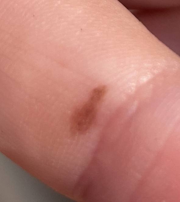
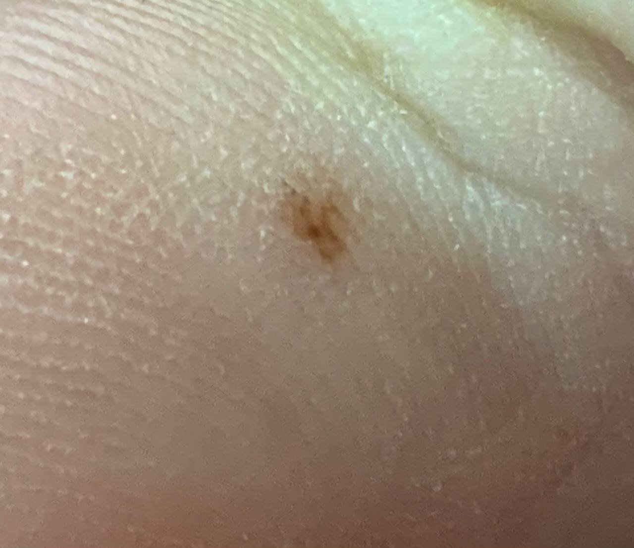
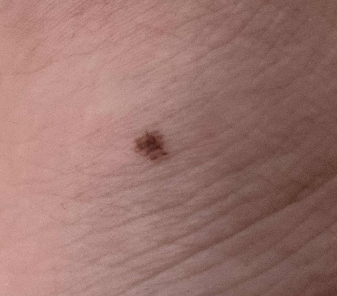
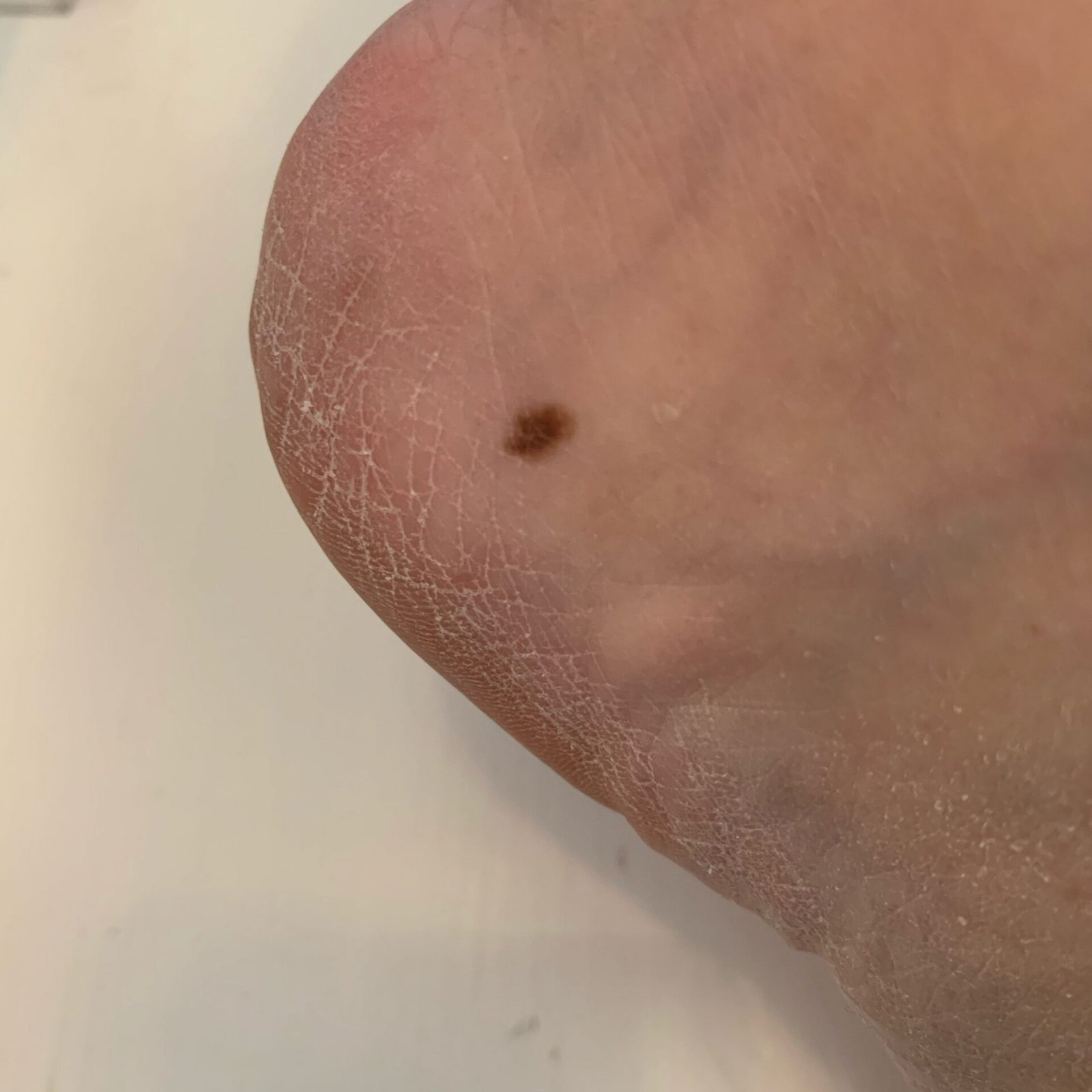
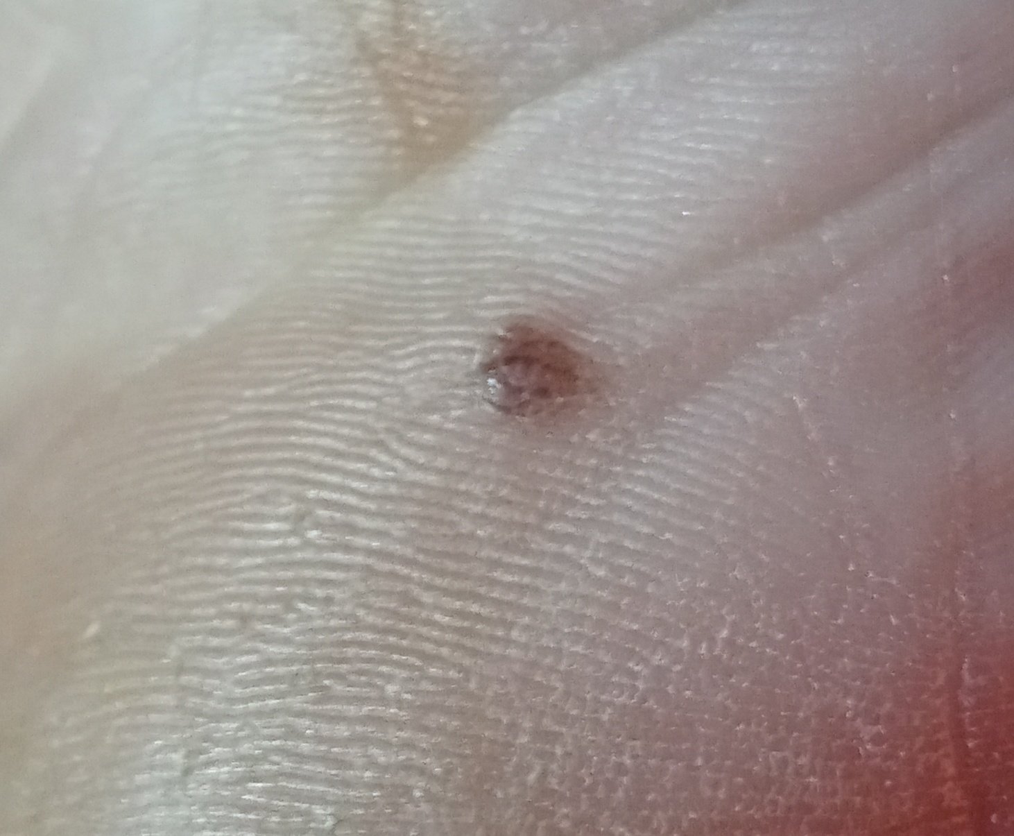
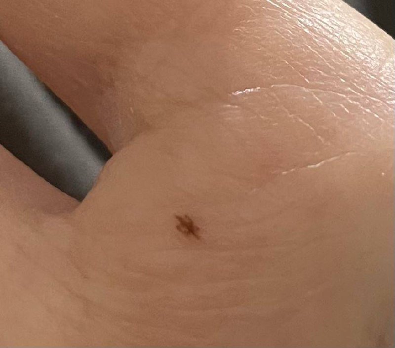
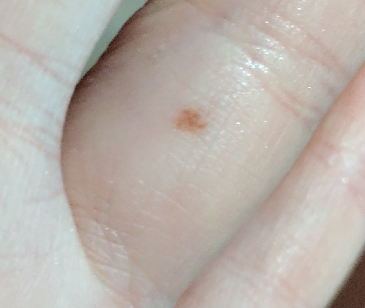
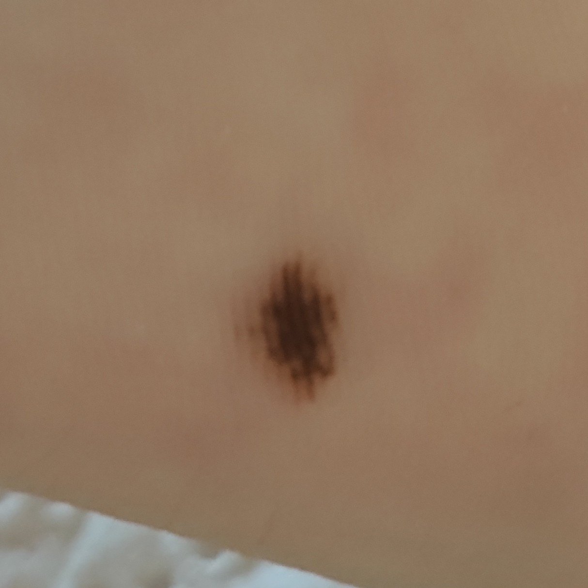
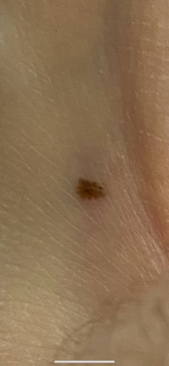
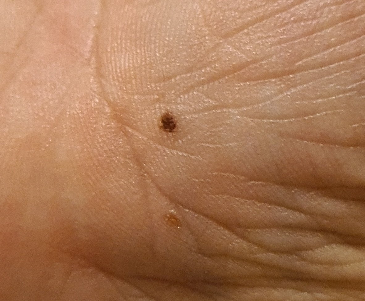
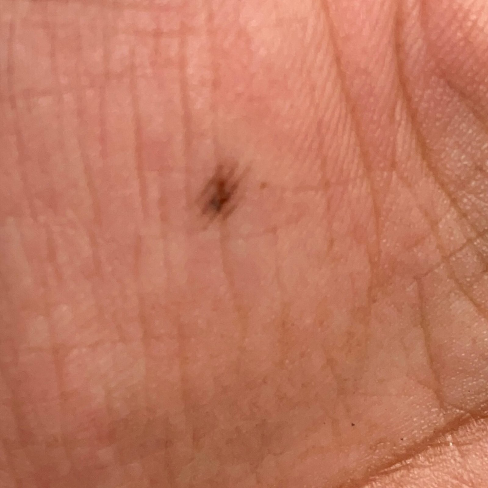
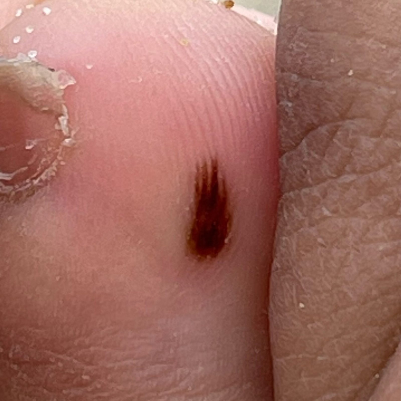
Acral Nevus (also known as benign nevus of the palms and soles, pigmented nevus of the palms and soles) is a benign skin growth that typically appears on the palms of the hands and soles of the feet. This type of nevus can be either acquired or congenital, but having multiple acral nevi on the same person is rare. Acral nevi make up about 5% of all pigmented nevi.
Predisposing Factors
The exact cause of acral nevi is not fully understood, but there are factors that may contribute to their development or increase the risk of neoplasms:
- Genetic Factor: The appearance of acral nevi may depend on genetic predisposition.
- Ultraviolet Radiation: Both artificial and natural sunlight can stimulate the rapid growth of nevus cells and increase melanin production, leading to darkening of the nevus.
- What is not established: Thyroid and adrenal hormones, viral infections, trauma and ionising radiation are sometimes listed as causes of acral nevi. None of them is supported well enough to be presented as a cause.
Diagnosis
The diagnosis of acral nevi is made through clinical examination, which includes a routine physical examination of the nevus and dermatoscopy. If malignancy is suspected, a biopsy may be performed.
Symptoms
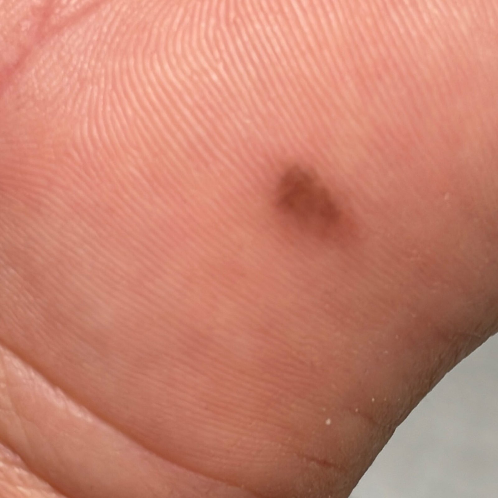
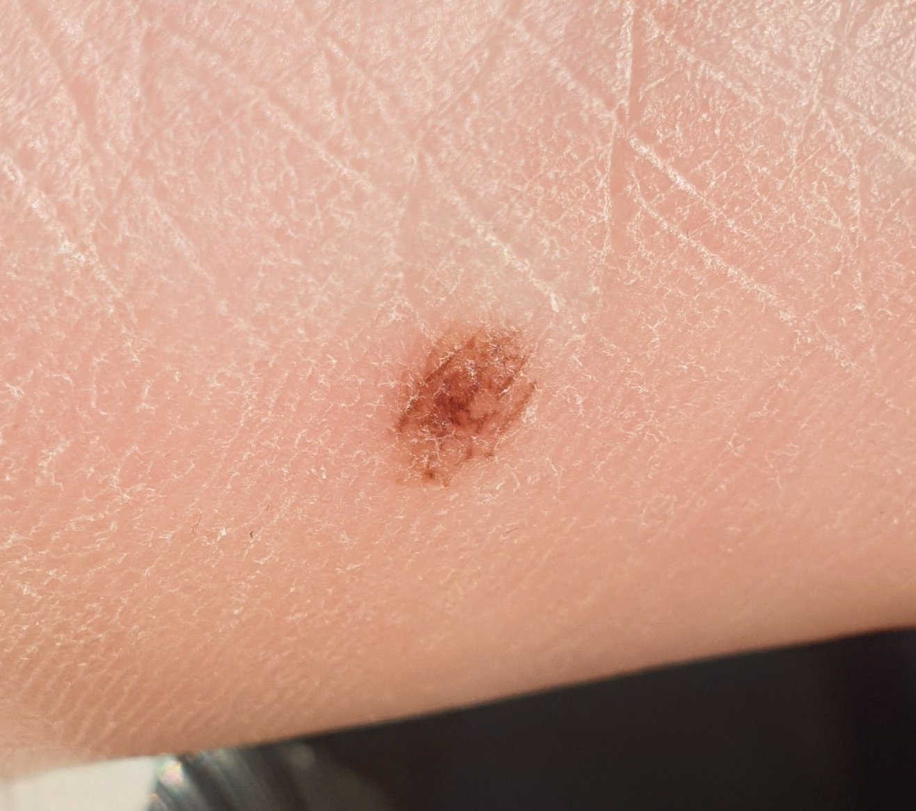
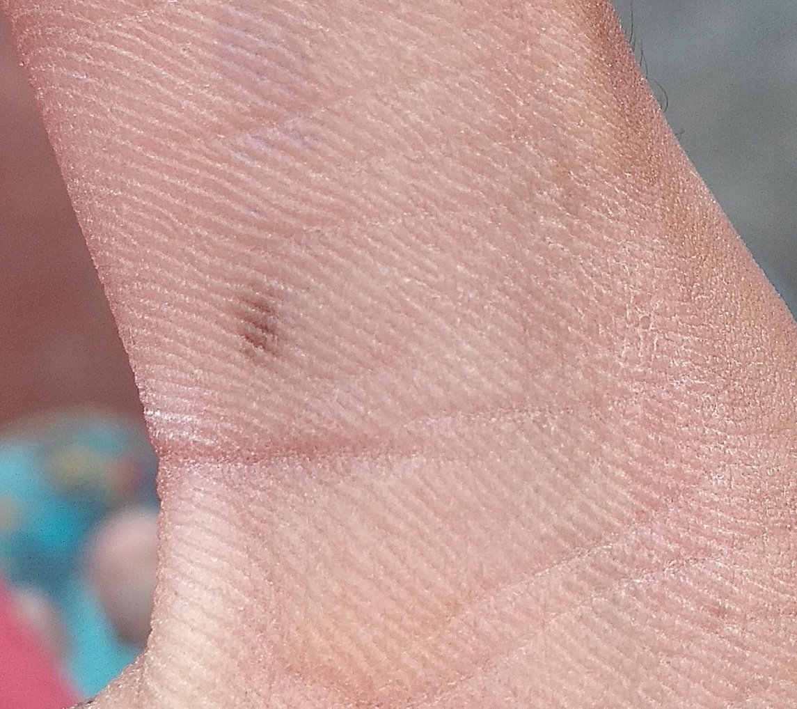
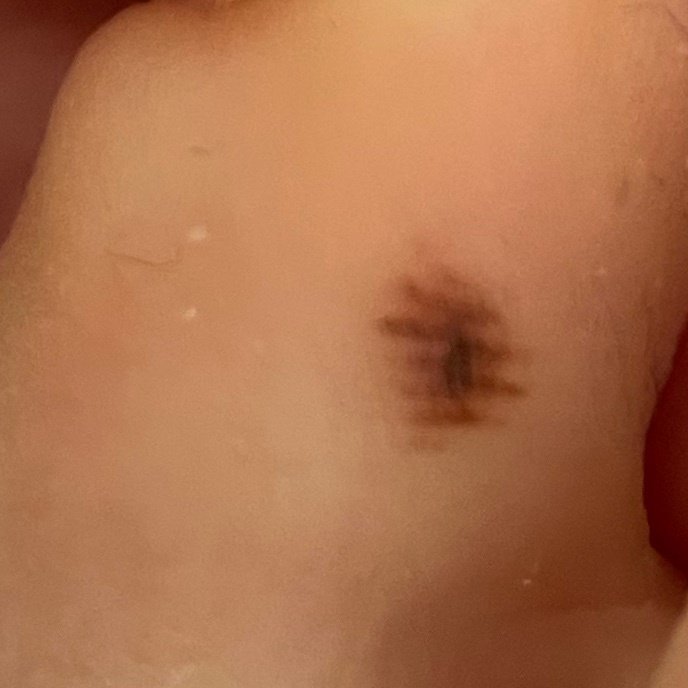
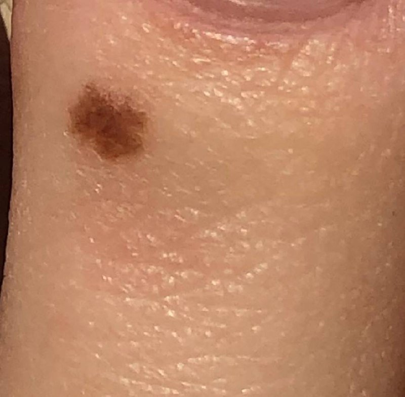
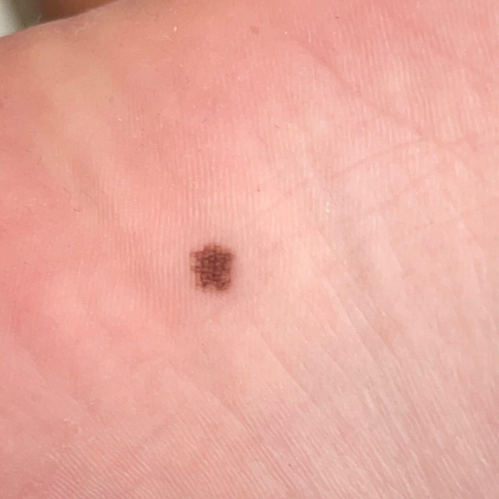
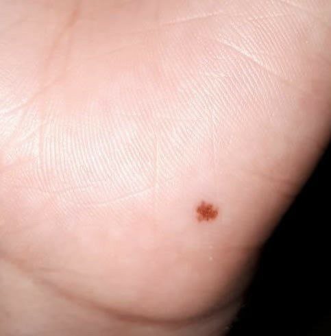
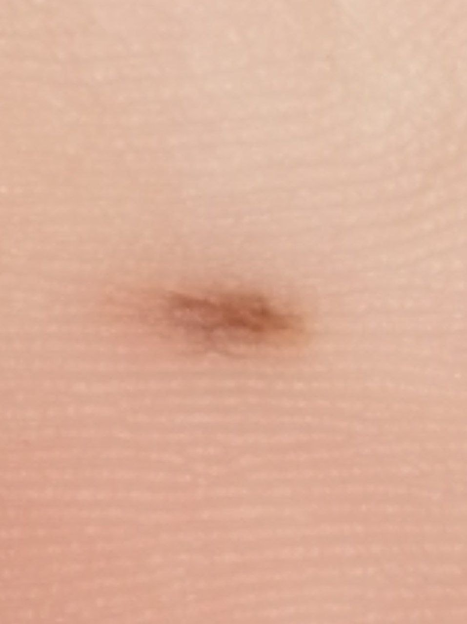
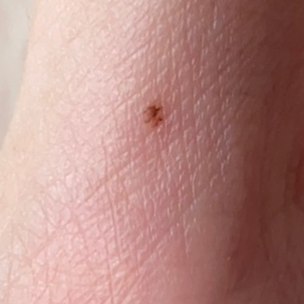
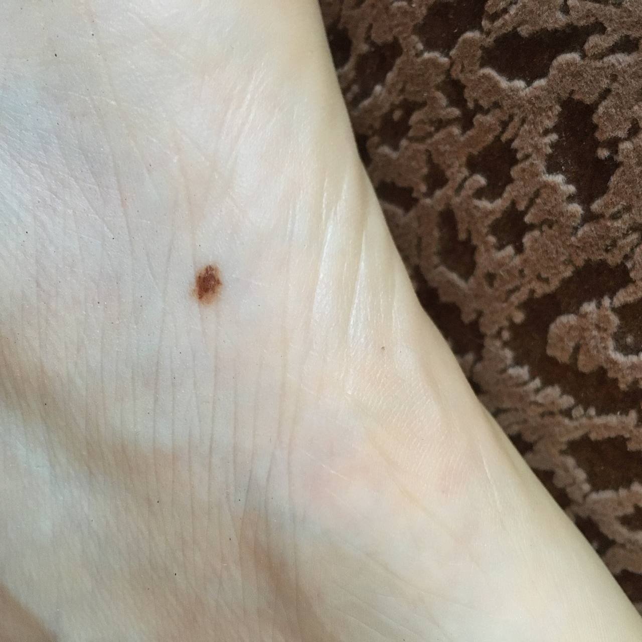
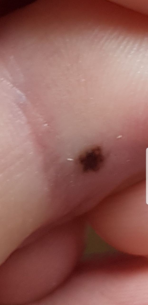
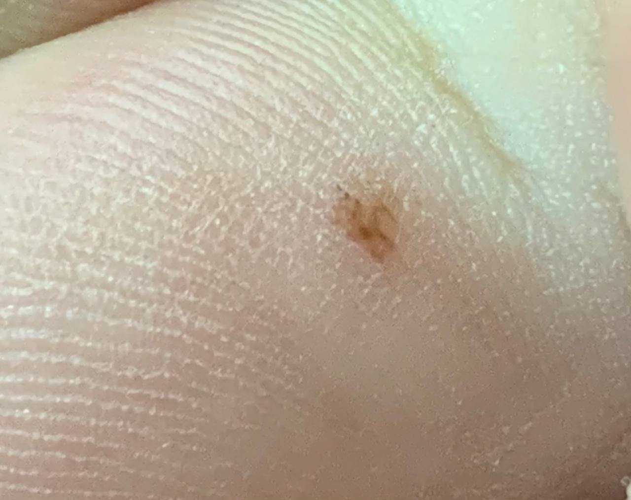
Acral nevi are found on the skin of the palms and soles. On visual examination, a benign acral nevus appears as a pigmented growth that is typically symmetrical, oval or round, with a regular outline. Its edges can look slightly indistinct because the pigment follows the furrows of palmar and plantar skin — but a genuinely irregular border, or marked asymmetry, is not simply a product of that skin pattern. On the palms and soles those features are concerning and need assessment. The texture of the nevus is either the same as the surrounding skin or slightly rough.
The color of acral nevi ranges from yellow-brown to dark brown (almost black), and in a benign lesion the pigment is evenly distributed. Sometimes, the color intensity gradually decreases from the center to the outer edges.
The size of acral nevi usually does not exceed 10 mm, most often ranging from 3 to 5 mm. Upon palpation, the consistency is similar to normal skin, and there are no subjective sensations.
Dermatoscopic Description
On dermatoscopy, an acral nevus appears with the following features:
- Parallel Structure: The linearity of the pattern due to the unique skin structure on the palms and soles.
- Parallel Furrow Pattern: Pigment follows the furrows of the skin. This, together with the lattice-like and fibrillar patterns, makes up the recognised benign arrangement of pigment in an acral nevus; the fibrillar pattern is seen especially on the weight-bearing sole.
- Parallel Ridge Pattern — a warning sign: Pigment running along the ridges rather than the furrows is not a feature of a benign acral nevus. It is associated with acral melanoma and calls for assessment.
- Lighter Scallops: The raised areas (scallops) are lighter than the grooves.
- Wider Scallops: The raised areas are broader than the grooves.
- Visible Duct Openings: Small white dots appear at the tops of the scallops, which are the openings of sweat glands.
- Regular Pattern: There is a consistent arrangement of grooves, scallops, and duct openings.
Differential Diagnosis
Acral nevi need to be differentiated from the following pigmented lesions:
- Subcorneal hematoma
- Skin contamination
- Dysplastic nevus
- Melanoma
Risks
Most acral nevi are benign, but acral melanoma can resemble them clinically, so a newly detected or changing acral lesion should be evaluated by a dermatologist rather than watched.
The signs that matter are changes in the lesion itself: a change in size, shape or colour, growing asymmetry, or a change in the dermatoscopic pattern. Bleeding and ulceration are more concerning still. Non-specific discomfort, by contrast, is not a useful warning sign for melanoma and should not be relied on as one.
Acral lentiginous melanoma is the particular concern on the palms and soles, and it is often recognised late. That is the reason a pigmented lesion in these sites deserves assessment rather than reassurance.
Management
A pigmented lesion on the palm or sole is worth having looked at by a dermatologist rather than monitored at home, because this is the site where acral melanoma occurs and where it is most often recognised late. Once a lesion has been assessed as a typical acral nevus, how it is followed up is decided individually: dermatoscopic monitoring for some lesions, biopsy for those that are atypical, changing or diagnostically uncertain. There is no fixed yearly interval that suits every lesion, and trauma, sun exposure or radiation are not in themselves reasons to seek review.
The specialist will assess whether ongoing monitoring is needed, or if the nevus should be removed. Nevi that are constantly irritated by clothing, jewelry, or occupation may also need to be removed.
Photographic documentation of the nevus can be very helpful for future comparisons, allowing even minor changes to be detected. Mapping of skin lesions is not needed routinely. Mapping remains a reasonable option for someone at high risk — a patient with many atypical or dysplastic nevi, or with a previous melanoma — where it is decided individually.
Treatment
A typical benign acral nevus does not require removal. Where biopsy or excision is indicated — because the diagnosis is uncertain, or because the lesion has changed in a way that raises concern — the tissue should be submitted for histopathology. Classic excision, electrosurgery and radiosurgery are all used.
Destructive methods such as laser removal and cryosurgery should be avoided here. They leave no tissue to examine, and on the palms and soles melanoma has to be excluded before a pigmented lesion is destroyed.
Prevention
The prevention of nevi formation and their malignant transformation involves gentle care of the skin:
- Limit UV exposure (avoid tanning beds and excessive sunbathing).
- Use sunscreen during peak sunlight hours.
- Avoid chronic skin trauma.
- Follow safety protocols when handling skin-damaging agents.
- Maintain personal hygiene and be aware of skin changes.
Regular checks of acral nevi, timely consultation with a specialist when changes occur, and removal of potentially dangerous nevi are essential for maintaining skin health.