
Healthy Nails: No Pathology Detected 💚
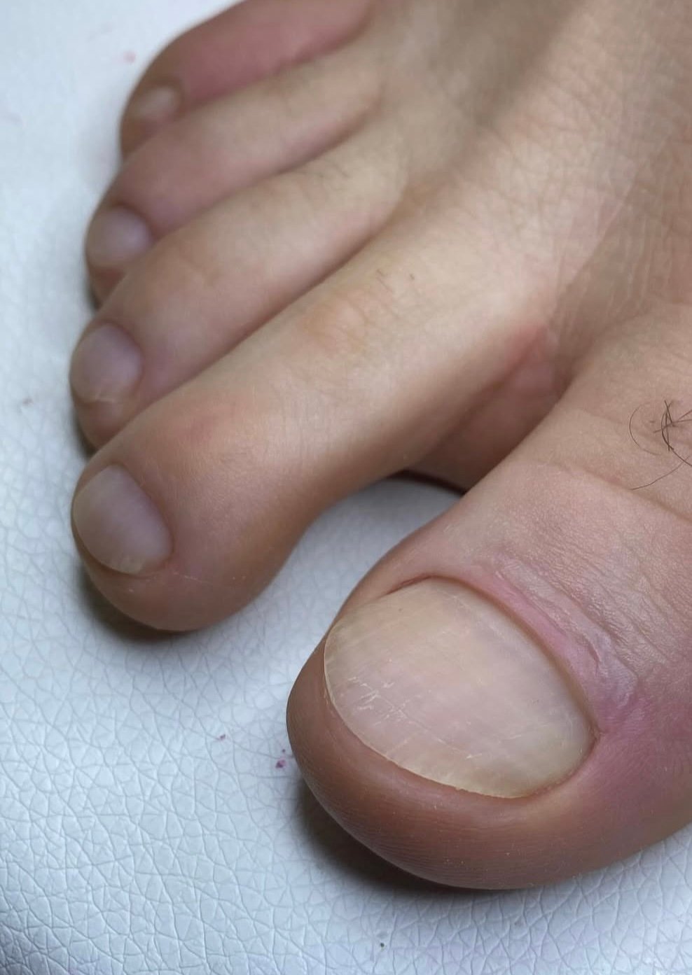
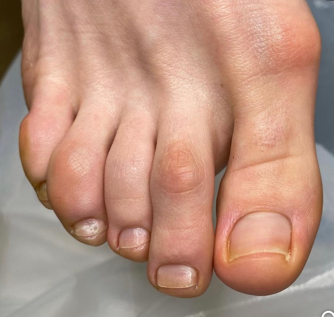
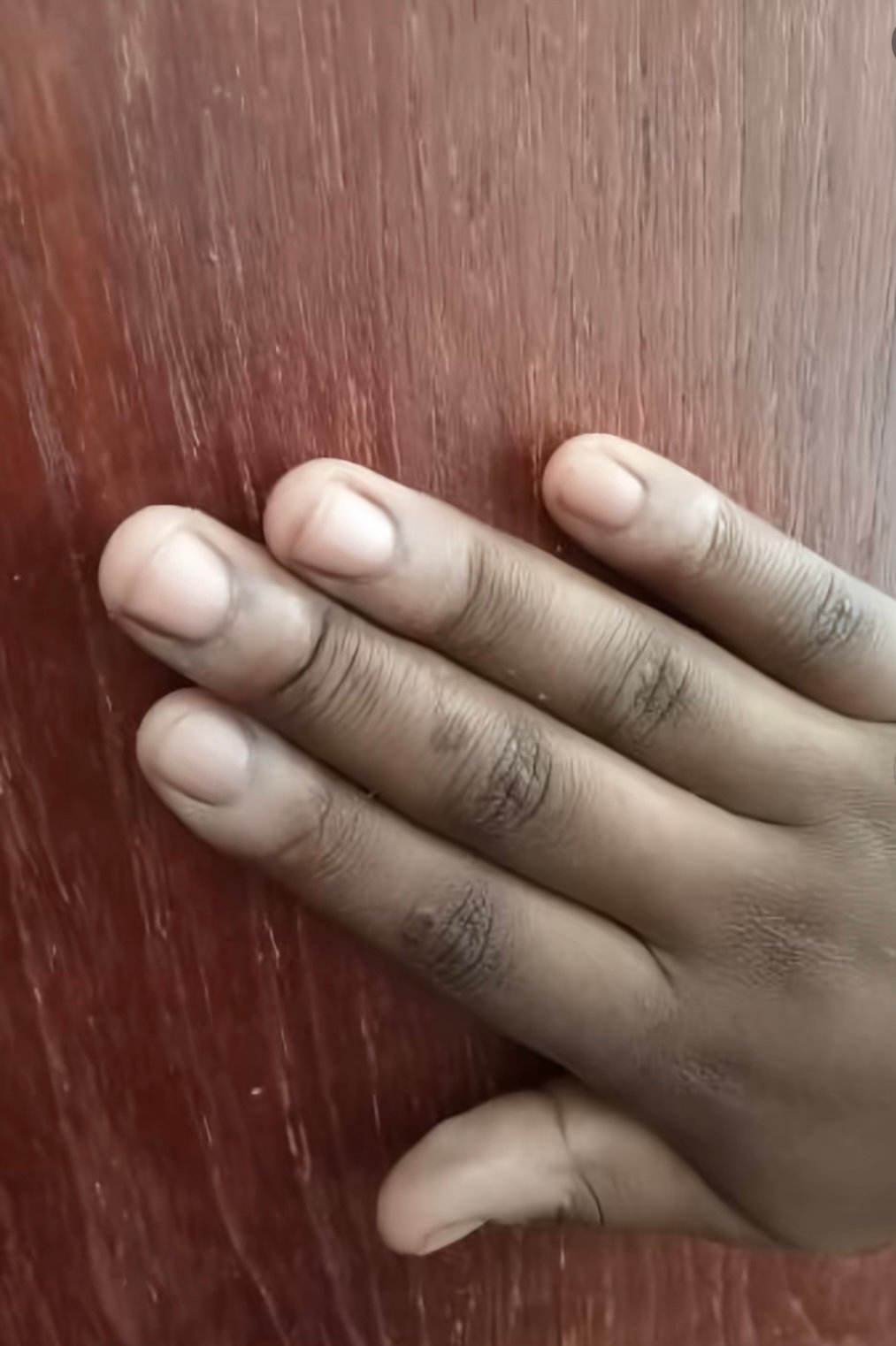
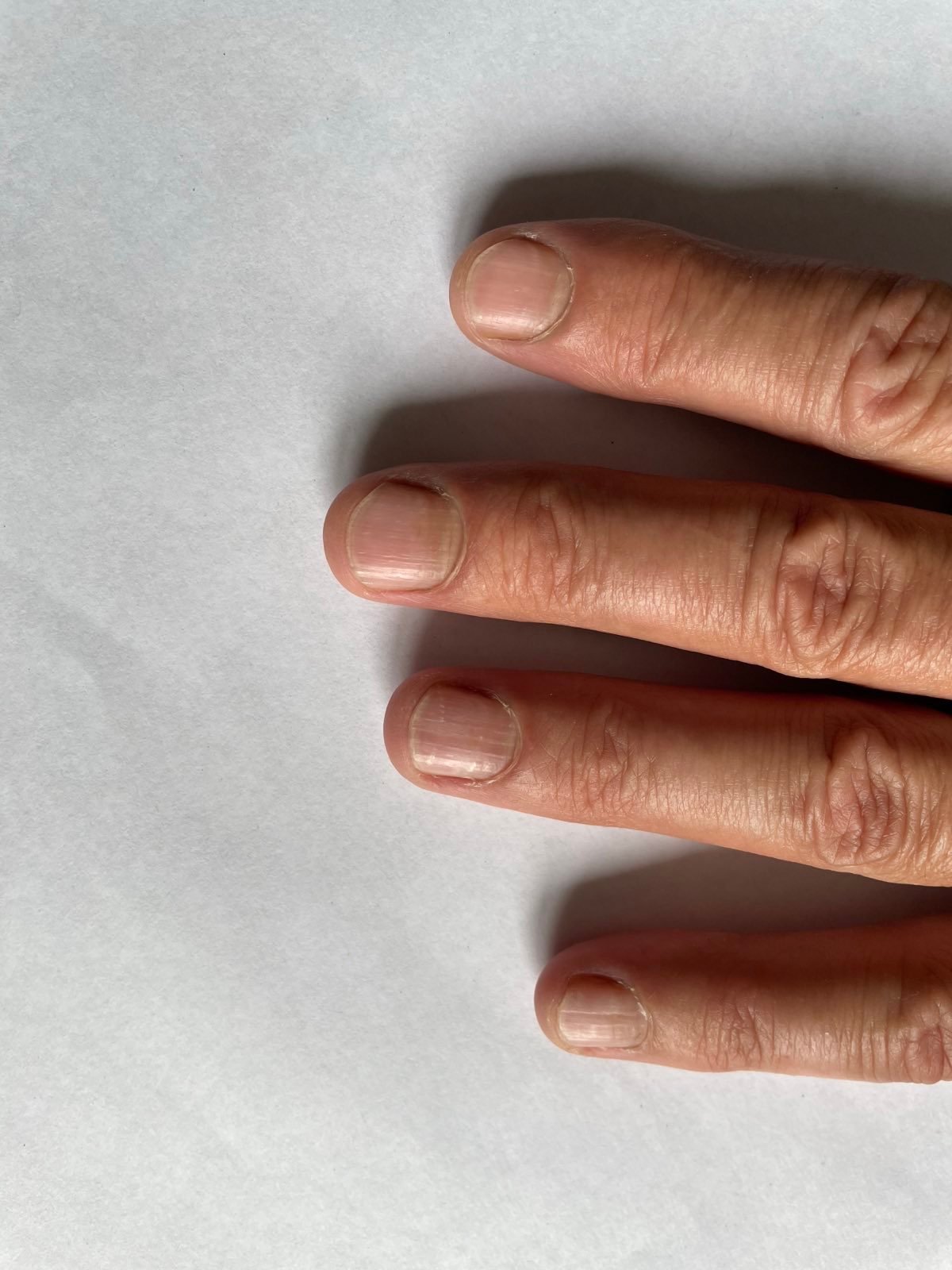
Healthy Nails: What Nails Without Visible Pathology Look Like
This page is the reference point for nail analysis: what a nail looks like when an examination finds nothing that needs treatment. It exists because “no pathology detected” is a frequent result when a nail is photographed, and because nails carry a number of harmless variations that are easily mistaken for disease — alongside a few genuinely important signs that are easily dismissed.
The Anatomy Behind a Normal Nail
The visible nail plate is made of compacted keratin. It grows from the matrix, hidden under the skin at the base, which is where the plate’s thickness and surface are determined — damage there, not to the visible nail, is what produces lasting deformity. The pale half-moon at the base, the lunula, is the visible front edge of the matrix; it is prominent on some people and invisible on others, and both are normal. The nail bed beneath the plate is richly supplied with blood, which is what gives a healthy nail its pink appearance. The cuticle seals the gap between skin and plate and keeps water and microorganisms out.
Fingernails grow roughly 3 mm a month and toenails around 1 mm, which is why a fingernail takes about six months to replace itself and a great toenail can take a year or more. That timescale explains why nail treatment is slow and why improvement is judged by new growth at the base rather than by the appearance of the whole nail.
What a Normal Nail Looks Like
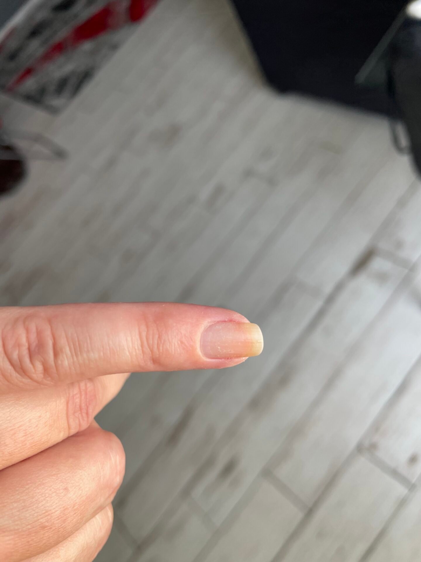
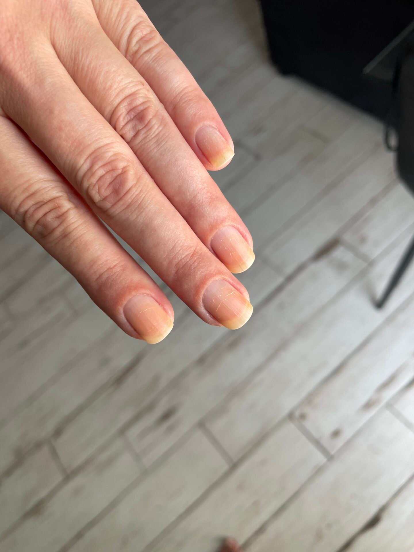
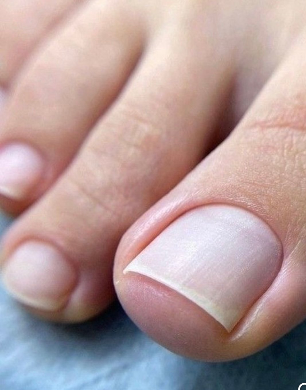
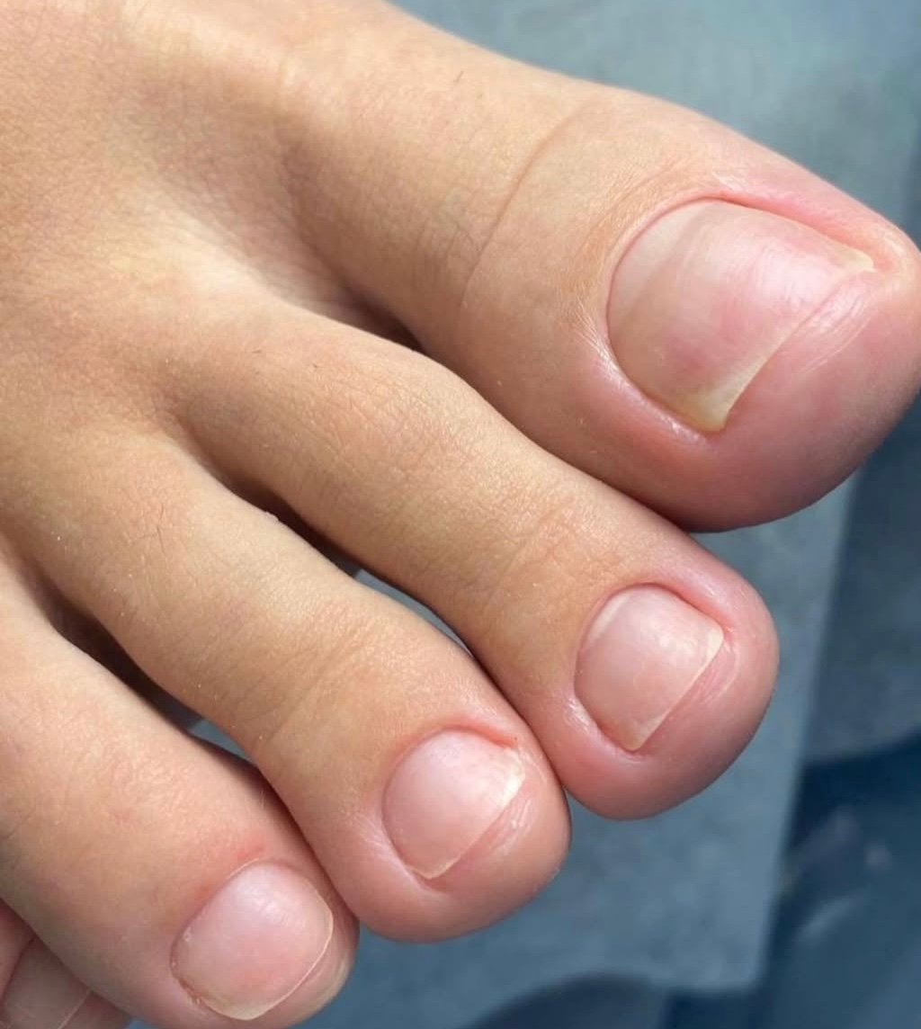
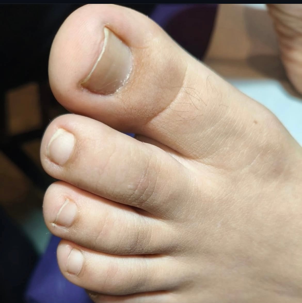
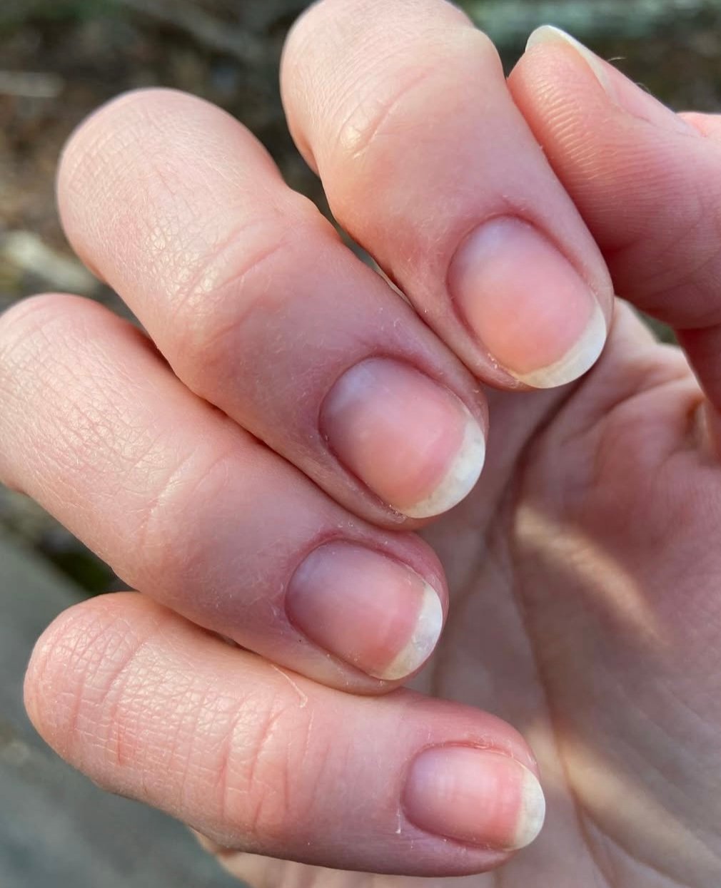
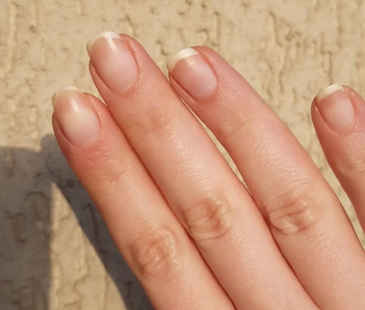
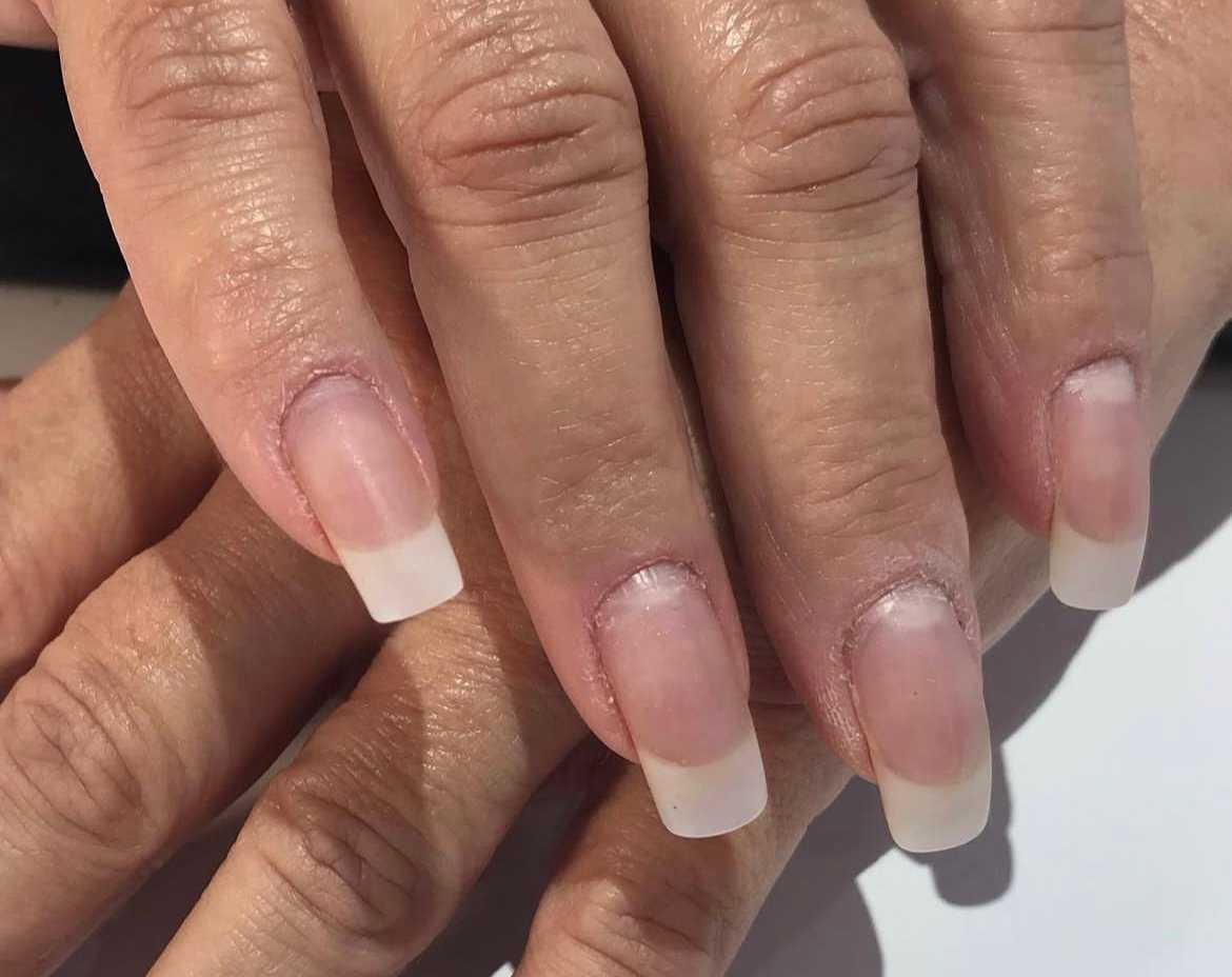
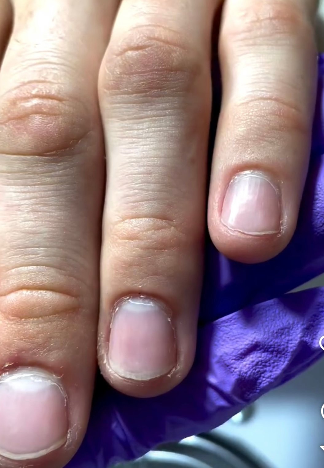
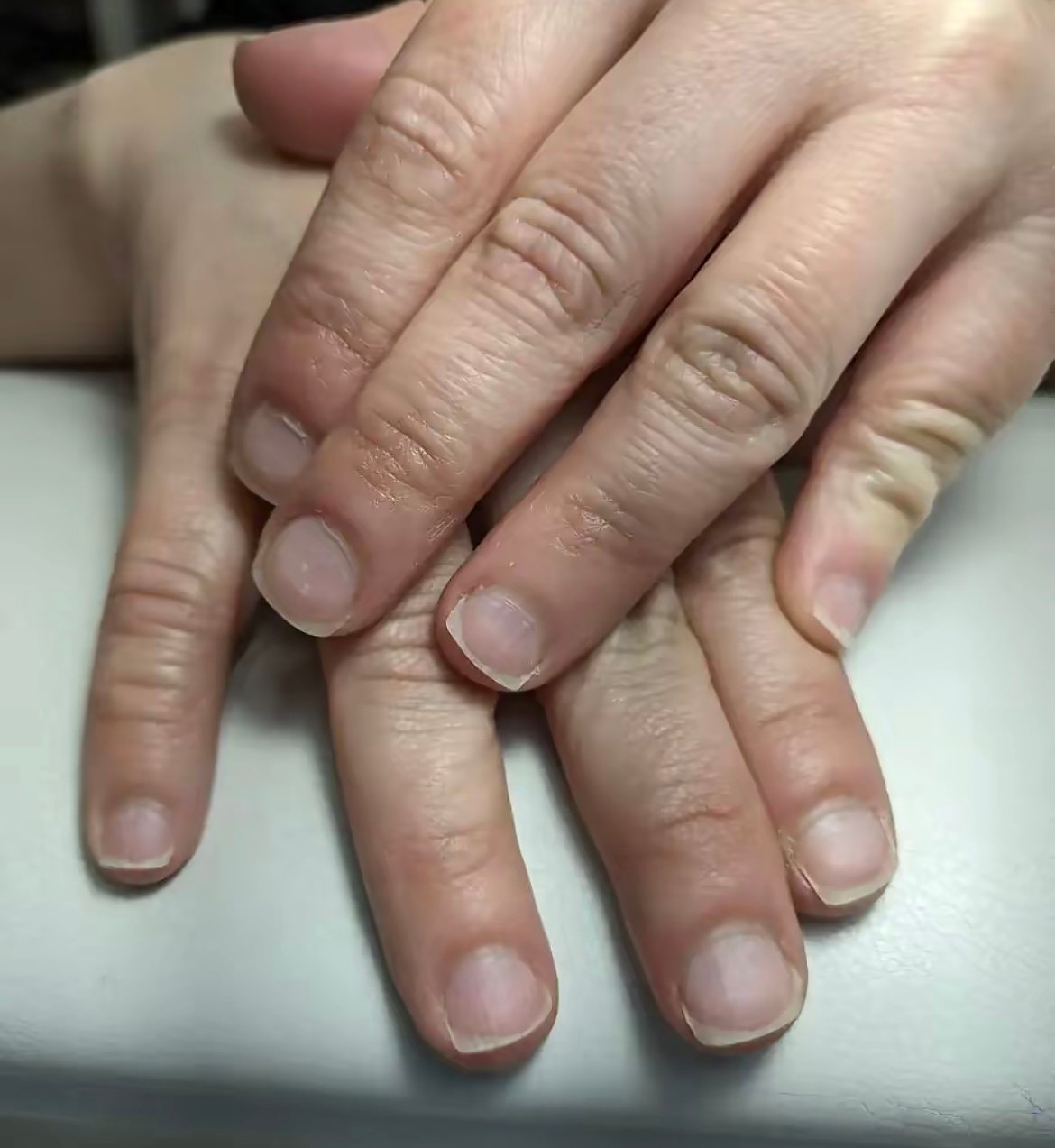
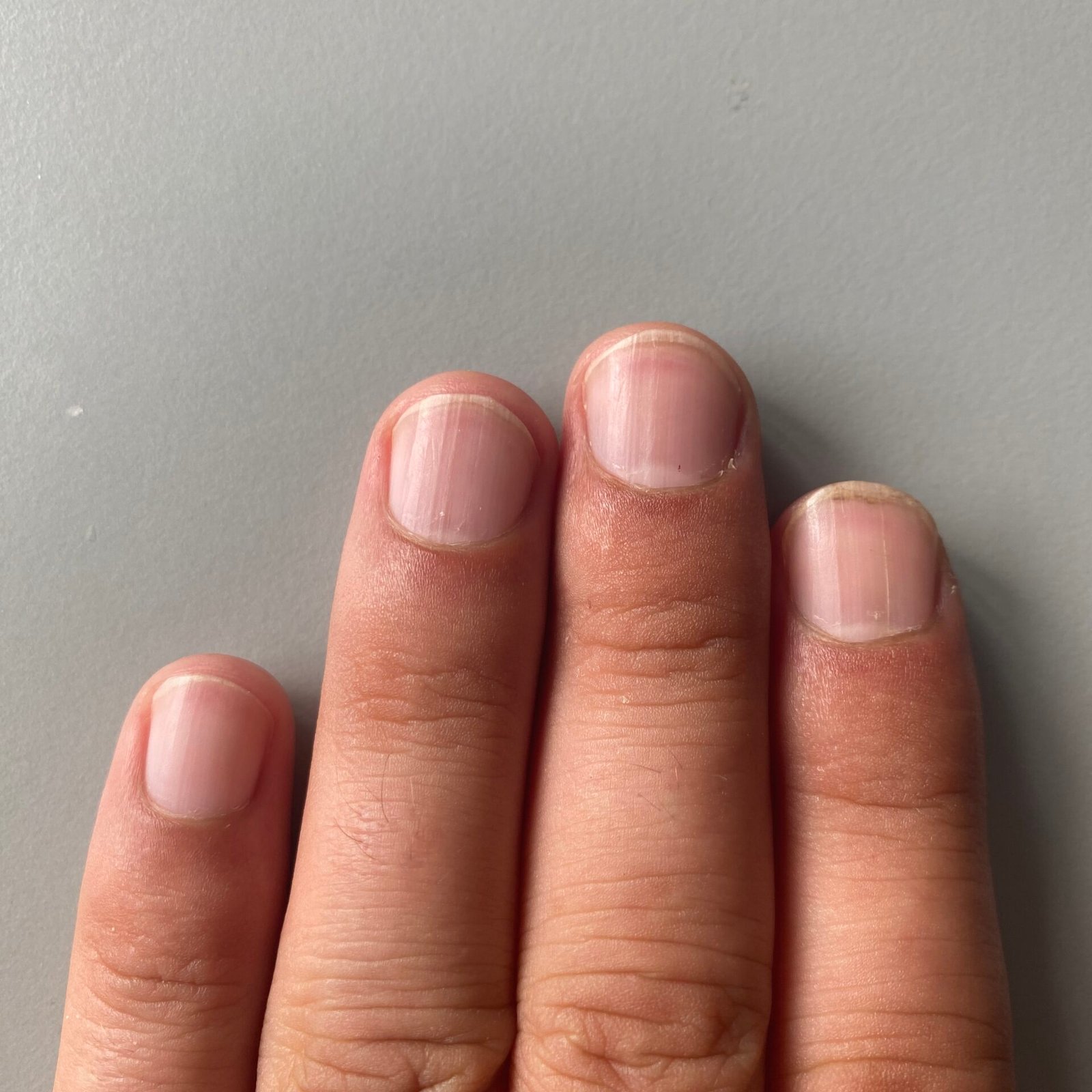
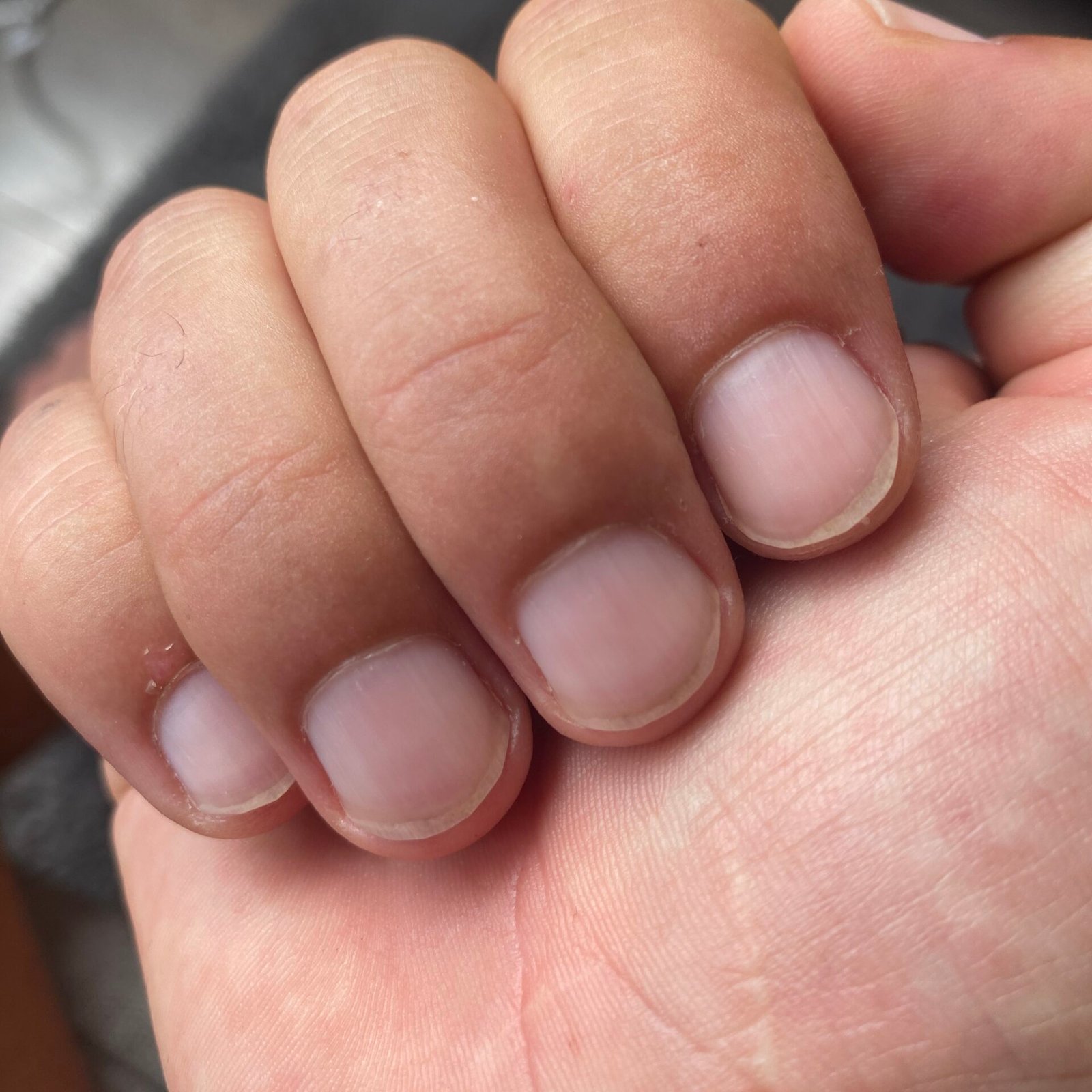
- A smooth, translucent plate through which the pink nail bed shows.
- A white free edge where the plate separates from the bed.
- An even, gentle curve both across and along the nail.
- An intact cuticle with no swelling, redness or tenderness of the surrounding skin.
- Uniform thickness, typically around half a millimetre on fingers and thicker on toes.
- A pink nail bed showing through the plate. Pressing the nail blanches it briefly, and the colour returns as the pressure is released — but how quickly depends on how cold the hands are and how hard you press, so this is a general impression rather than a test with a pass mark.
Normal Variations That Are Not Disease
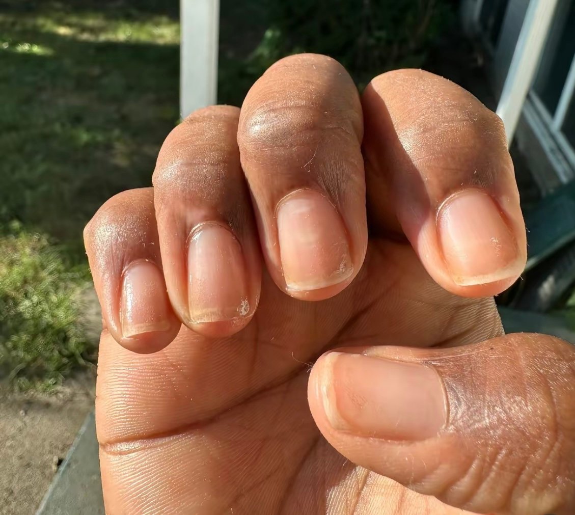
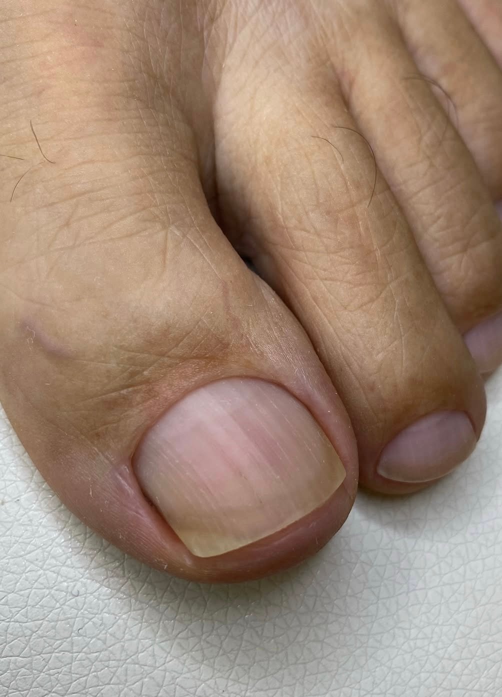
- Fine lengthwise ridges. Shallow vertical lines running from base to tip become more pronounced with age and are a normal finding.
- Small white spots. Scattered white marks (punctate leukonychia) commonly follow minor knocks to the matrix weeks earlier and grow out on their own. They are not a sign of calcium or zinc deficiency. White discolouration that persists, covers much of the nail, or affects several nails at once is a different matter and has other causes, so it is worth showing to a clinician.
- An absent or barely visible lunula, especially on the little finger.
- Mild yellowing after prolonged nail polish use, which resolves during a break from polish.
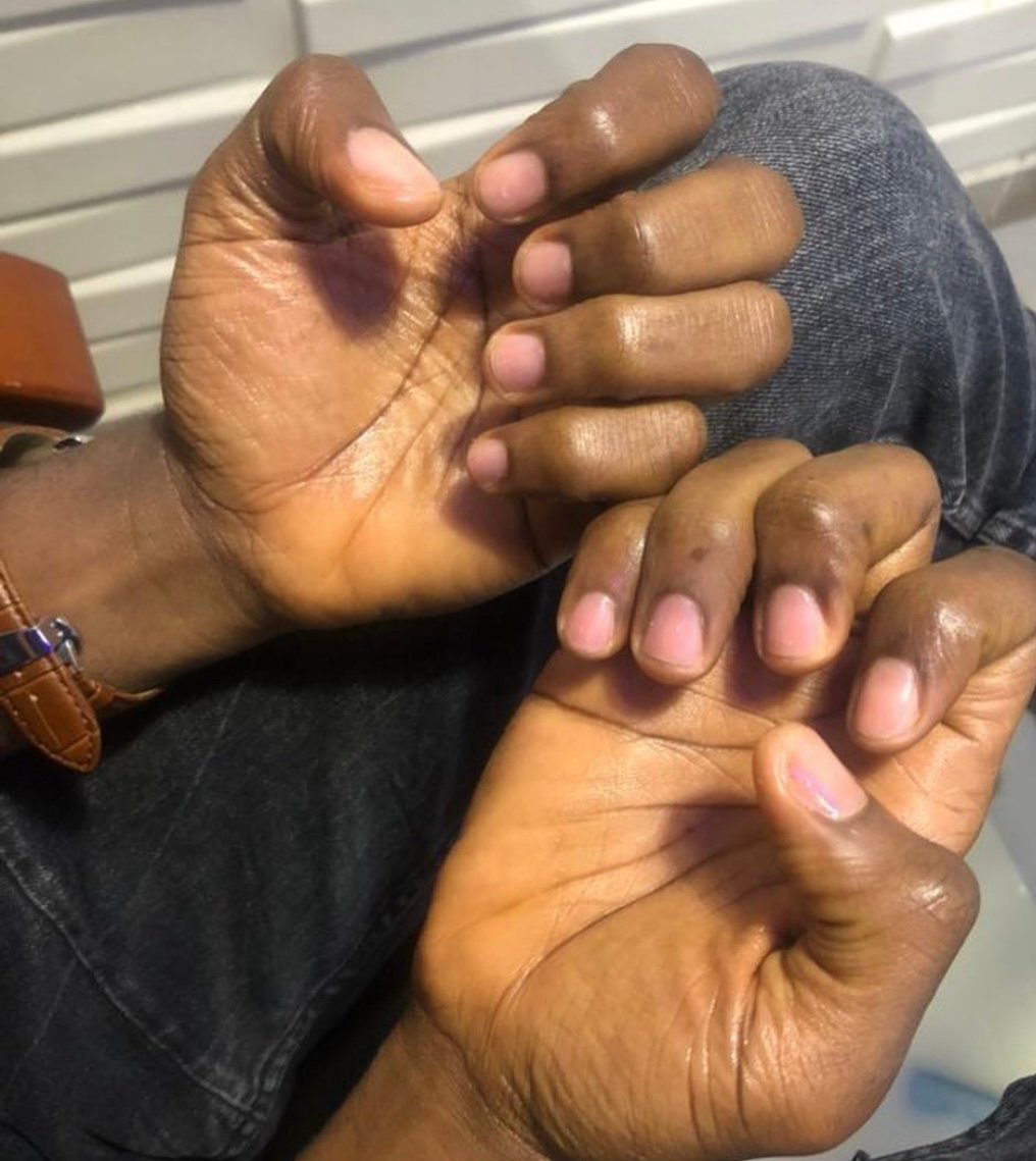
- A darker band or diffuse pigmentation of several nails in people with deeply pigmented skin. Benign racial melanonychia is common and typically involves more than one nail with stable, even bands.
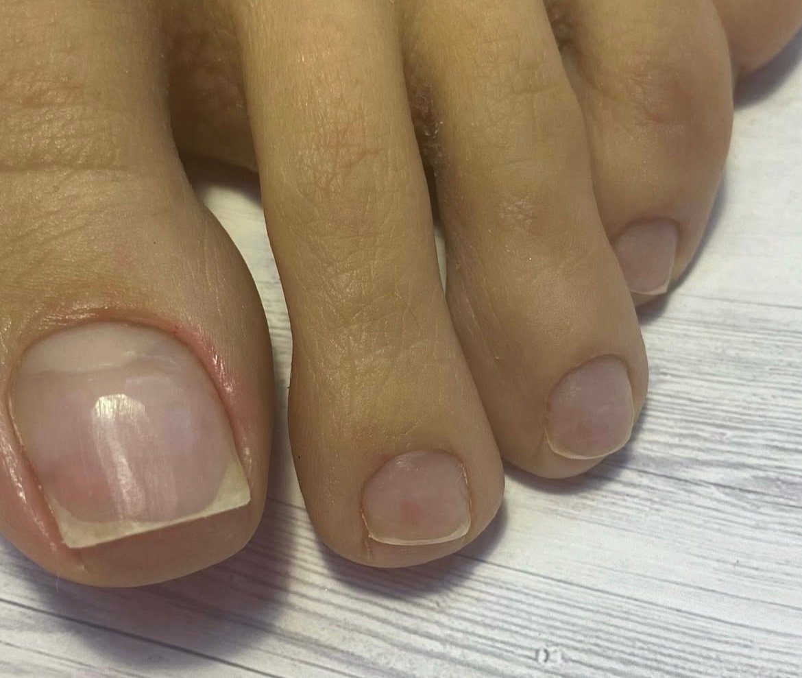
- Slightly different shapes between hands and feet, and thicker, more opaque toenails, particularly the great toe.
What an AI Nail Analysis Can and Cannot Tell You
An image-based analysis assesses the nail in the photograph. Several limits follow from that, and they are worth stating plainly:
- Nail polish, gel and acrylic obscure most of what matters. Colour, surface and the nail bed underneath are hidden, even though the shape of the nail and the skin around it can still be seen. Photograph bare nails.
- One nail is not ten. Fungal infection frequently starts in a single toenail, and pitting from psoriasis may show on only a few nails.
- A normal result does not exclude infection. Distinguishing onychomycosis from other causes of a thickened, discoloured nail generally requires a laboratory sample — microscopy, culture, PAS staining or PCR, depending on local practice, since culture alone misses a substantial share of infections; treatment is long and should not be started on appearance alone.
- It cannot see the matrix, where the origin of a pigmented band or a deformity lies.
Nail Changes That Should Be Assessed in Person
These are not normal variations, and none of them should be ignored because a screening result was reassuring:
- A single dark longitudinal band in one nail, particularly if it is new, widening, irregular in colour, or if the pigment extends onto the surrounding skin or cuticle. This combination can indicate melanoma of the nail unit and warrants prompt dermatological assessment.
- Thickening, crumbling, yellow-brown discolouration or separation of the nail from its bed, most often caused by onychomycosis but also seen in psoriasis and after trauma.
- Pitting, oil-drop patches or crumbling across several nails, which frequently accompanies psoriasis and may appear before skin signs.
- Redness, swelling, warmth or pus around the nail fold, suggesting acute or chronic paronychia.
- Spoon-shaped nails (koilonychia), which can accompany iron deficiency.
- Clubbing — increased curvature with loss of the normal angle at the base — which may reflect a condition of the lungs, heart, liver or bowel and should be investigated.
- Deep transverse grooves (Beau’s lines) across several nails, marking a point at which nail growth was interrupted. Minor trauma or local nail disease can do this; so can serious illness, high fever or chemotherapy, which is why grooves appearing across several nails at once are worth mentioning.
- Sudden separation, bleeding under the nail without an injury, or a growth under or beside the plate.
Caring for Healthy Nails
- Trim toenails straight across and not too short, to reduce ingrowing; fingernails may follow the natural curve.
- Leave the cuticle alone. Cutting or pushing it back aggressively breaks the seal that keeps infection out and is a frequent cause of chronic paronychia.
- Dry thoroughly between the toes after washing, and let footwear dry fully between wears.
- Wear breathable shoes and change socks daily; use sandals in shared showers and pool changing rooms.
- Do not share clippers or files, and make sure a salon sterilises its instruments.
- Give nails breaks from polish and gel, and take care with removal, which does more damage than the polish itself.
- Wear gloves for wet work and cleaning chemicals, and moisturise the nail folds.
- Treat athlete’s foot early, since untreated skin infection is a common route to nail infection.
Healthy nails change slowly, which is their most useful property: a difference you can see from one month to the next is worth showing to a clinician. Stability is reassuring but not decisive — a pigmented band, clubbing, marked deformity or a lump beside the nail deserves assessment even if it has looked the same for years.

Medical review
Board-Certified Dermatologist · Fellow of the American Academy of Dermatology (FAAD)