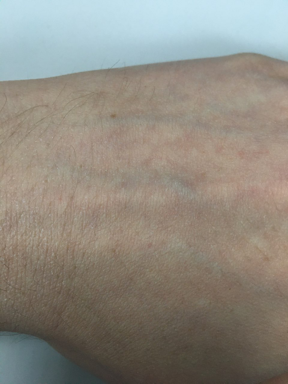
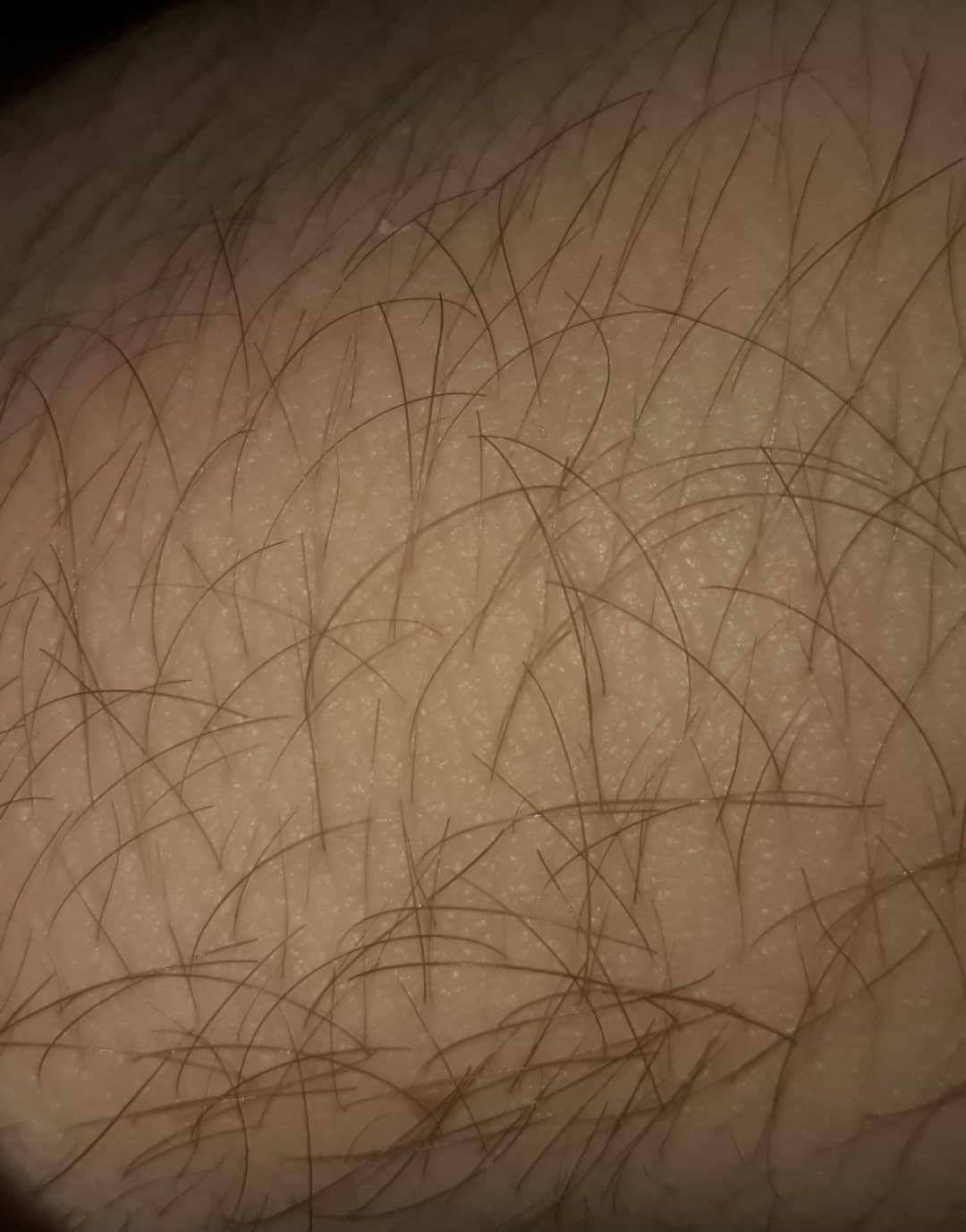
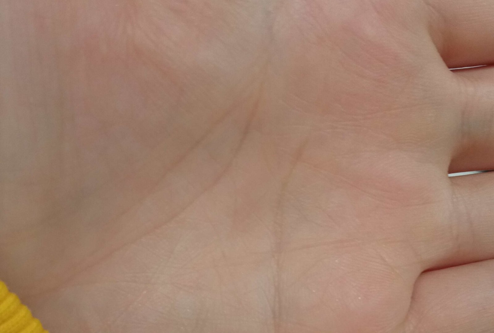
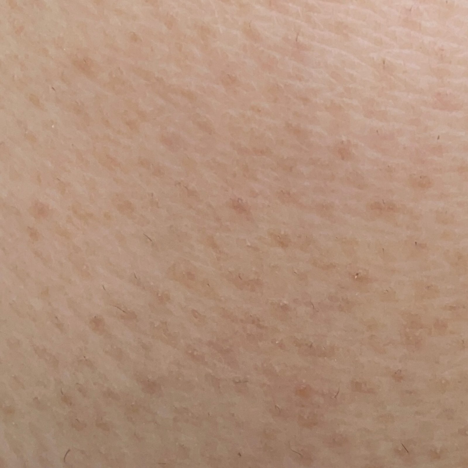
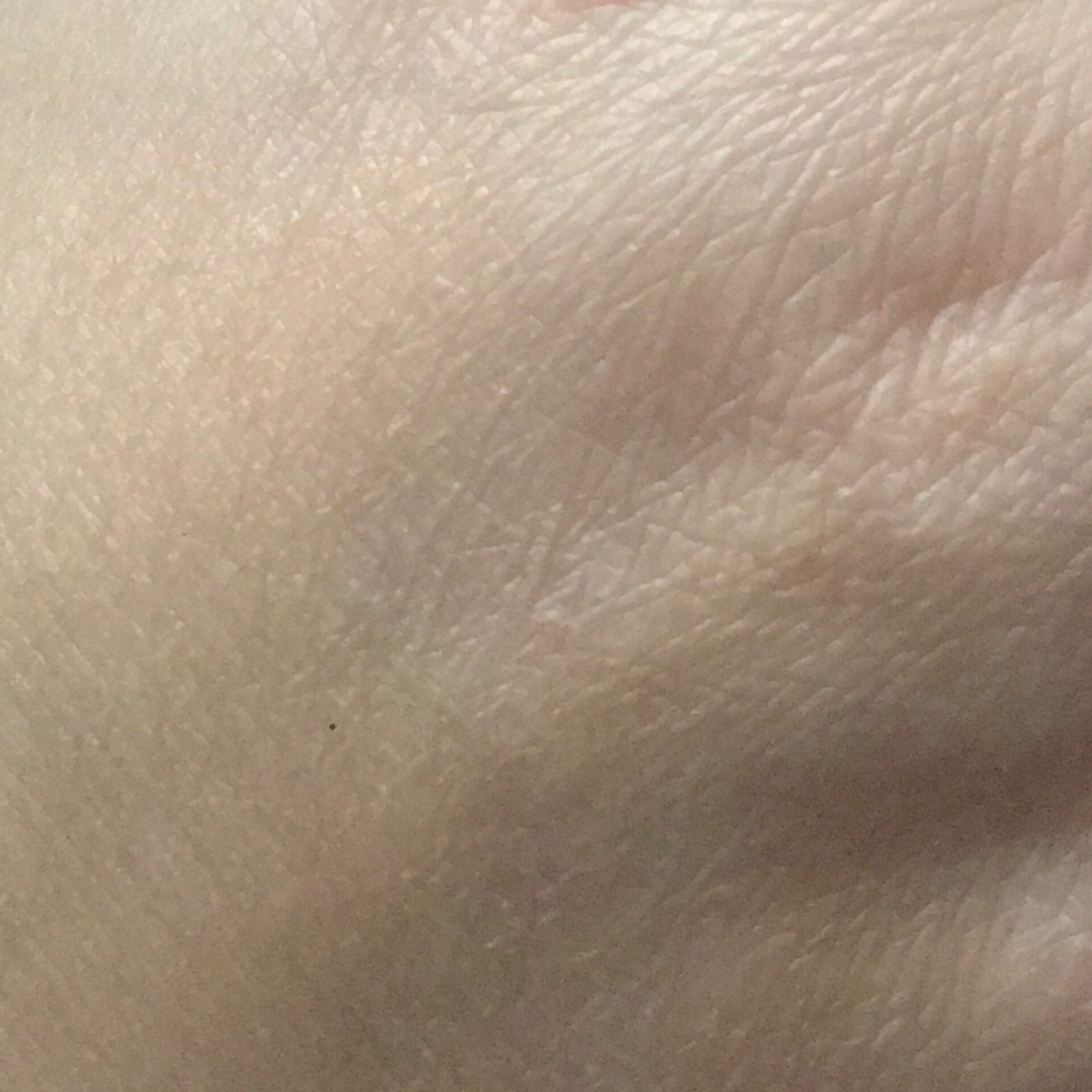
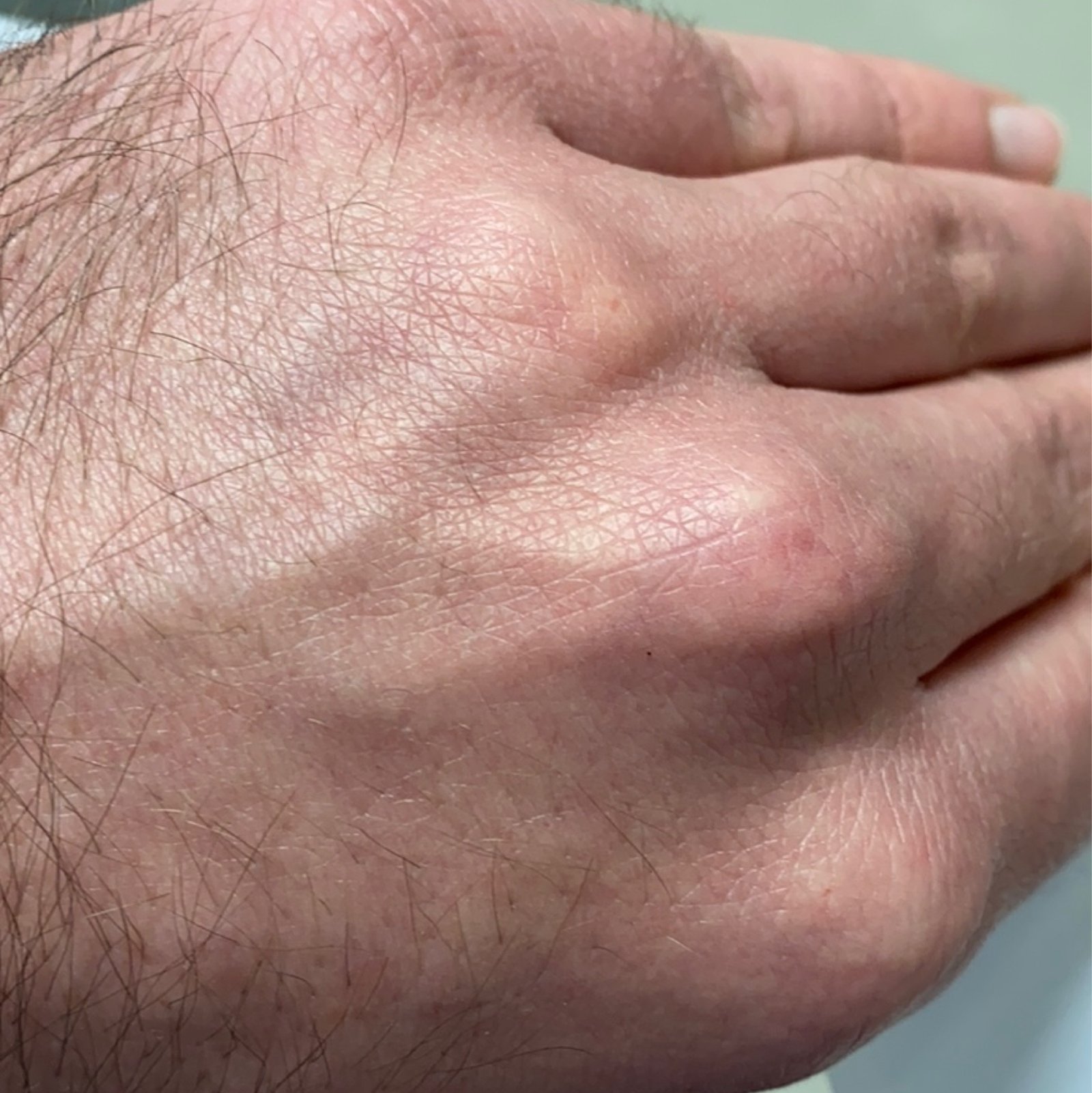
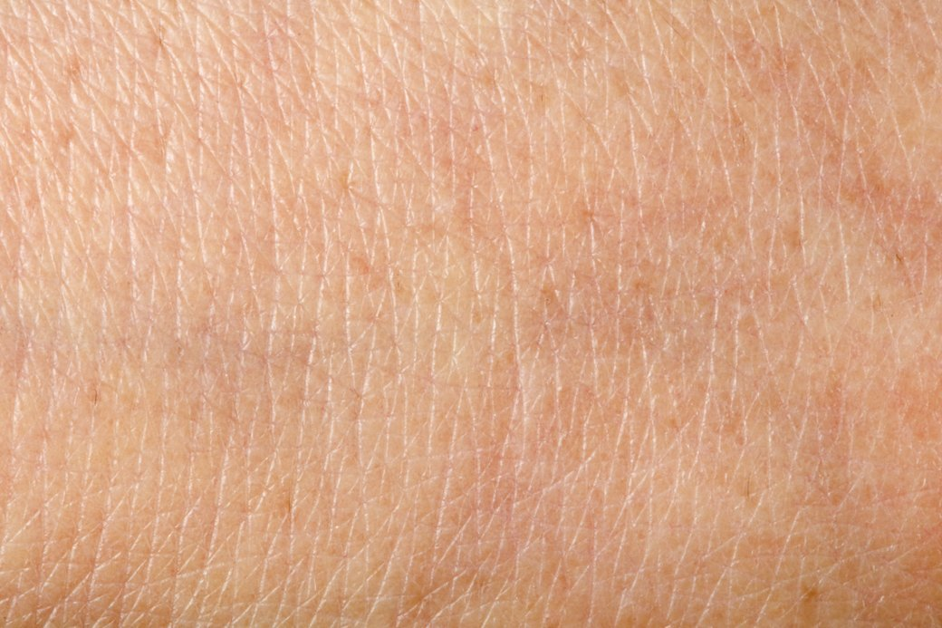
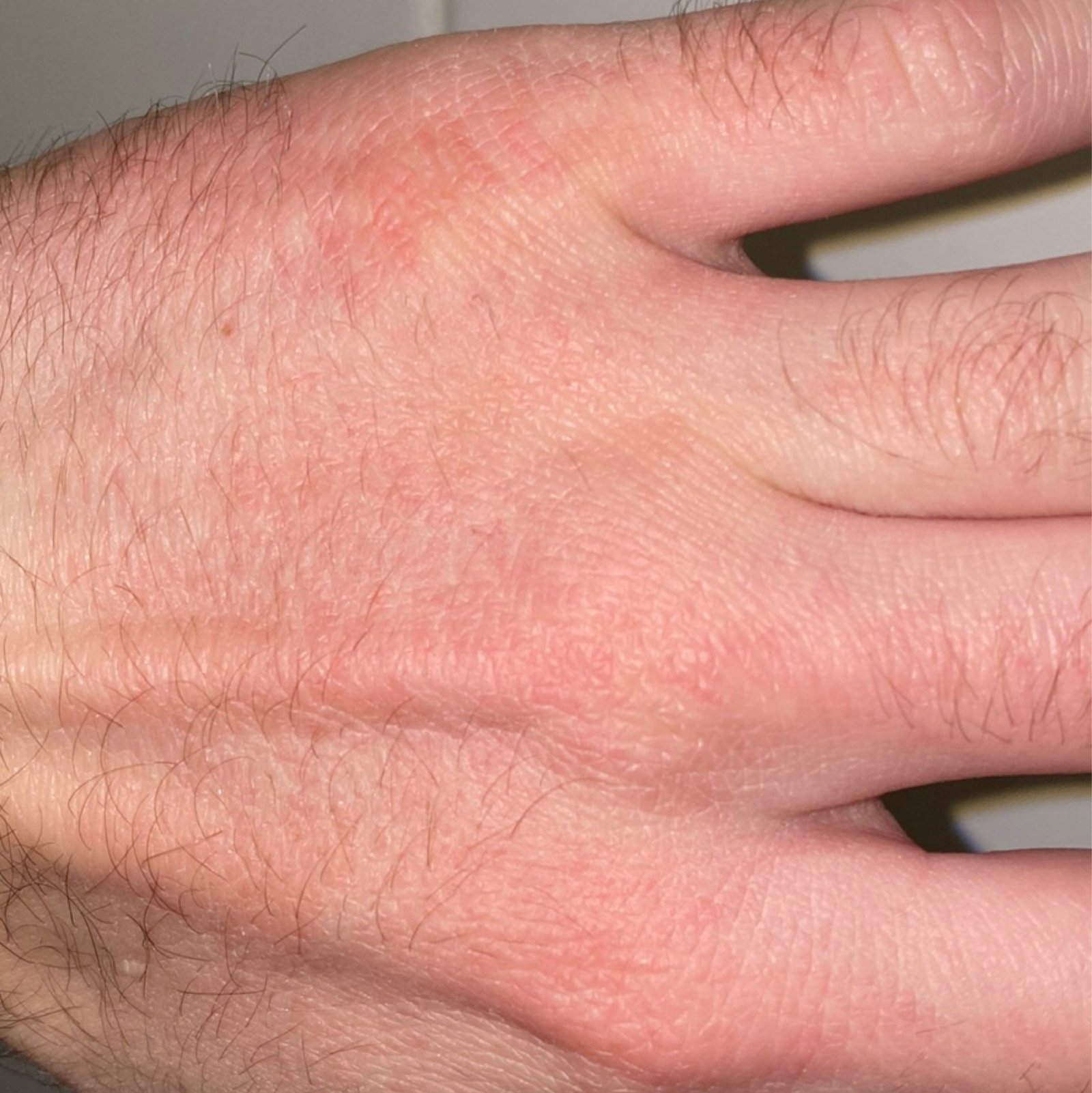
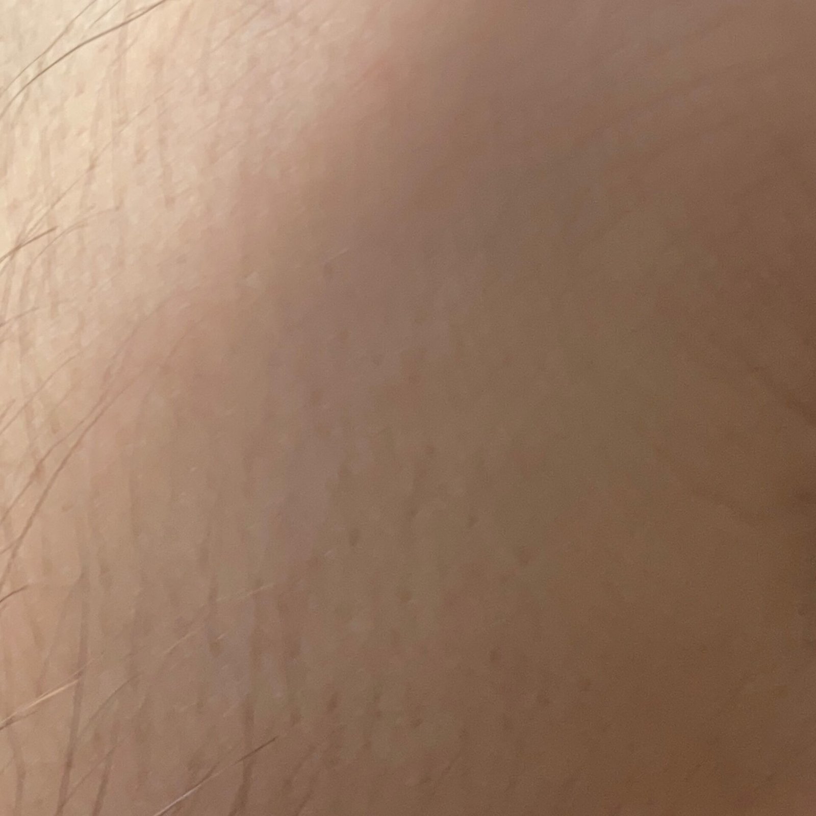
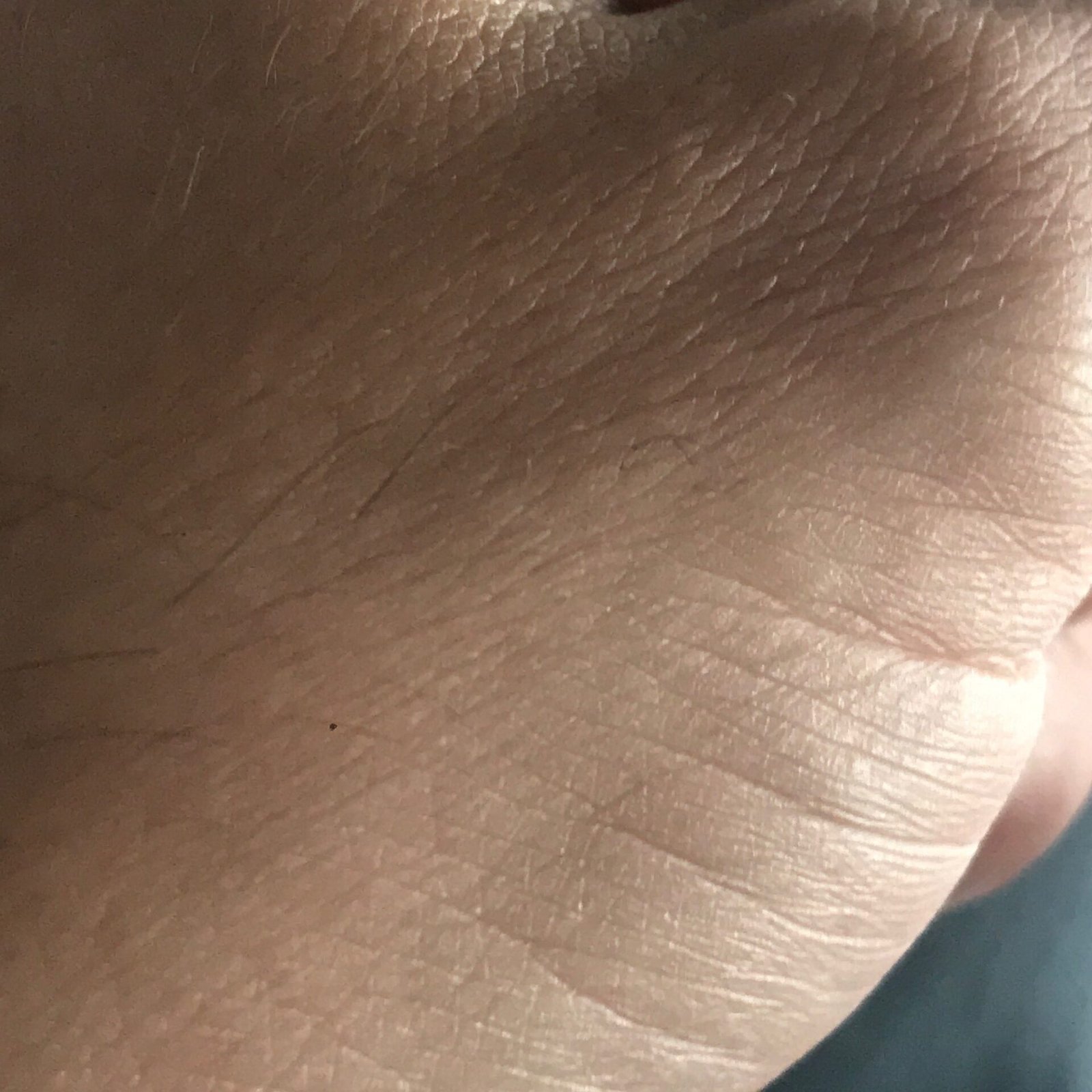
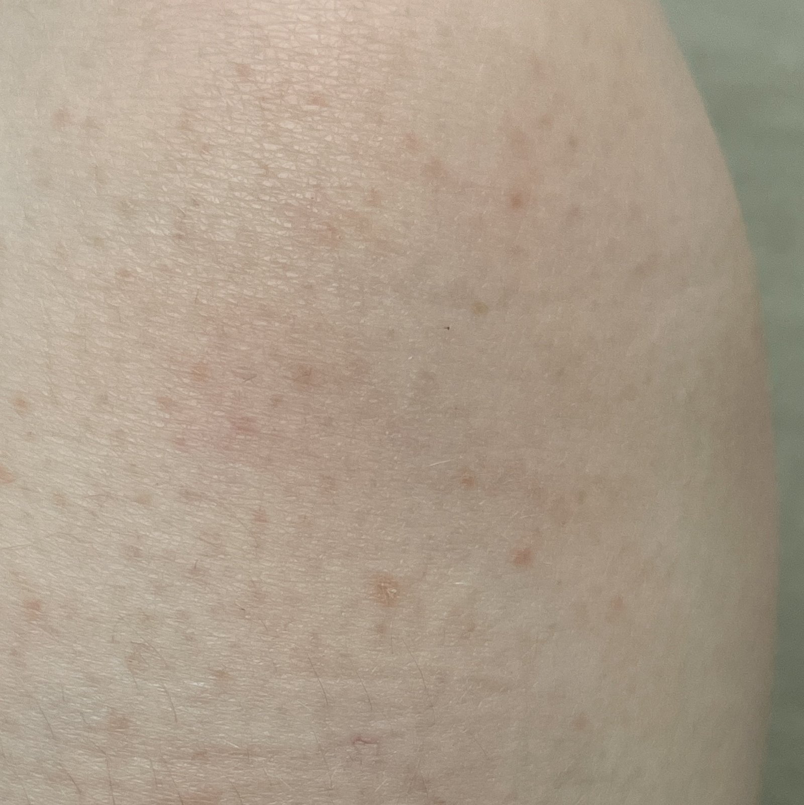
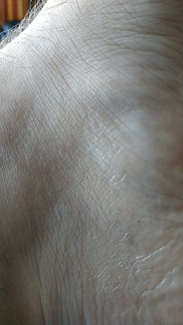
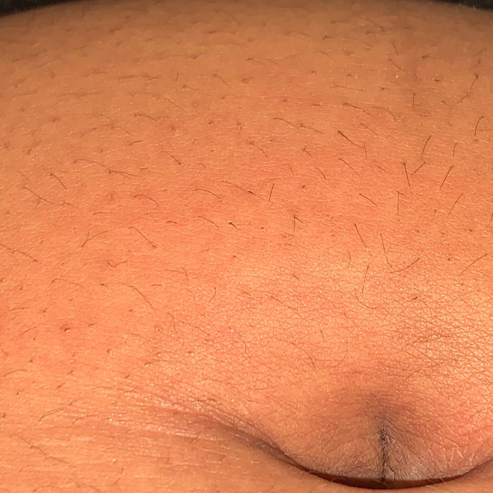
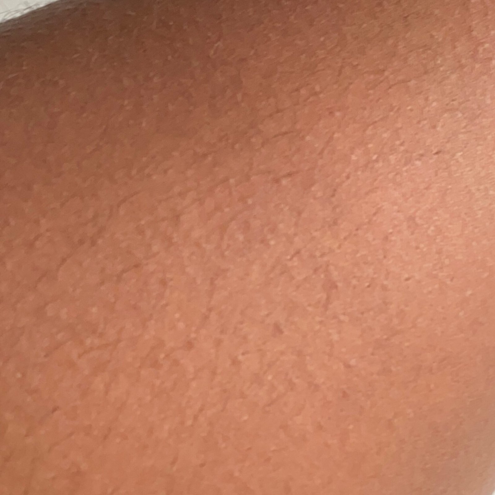
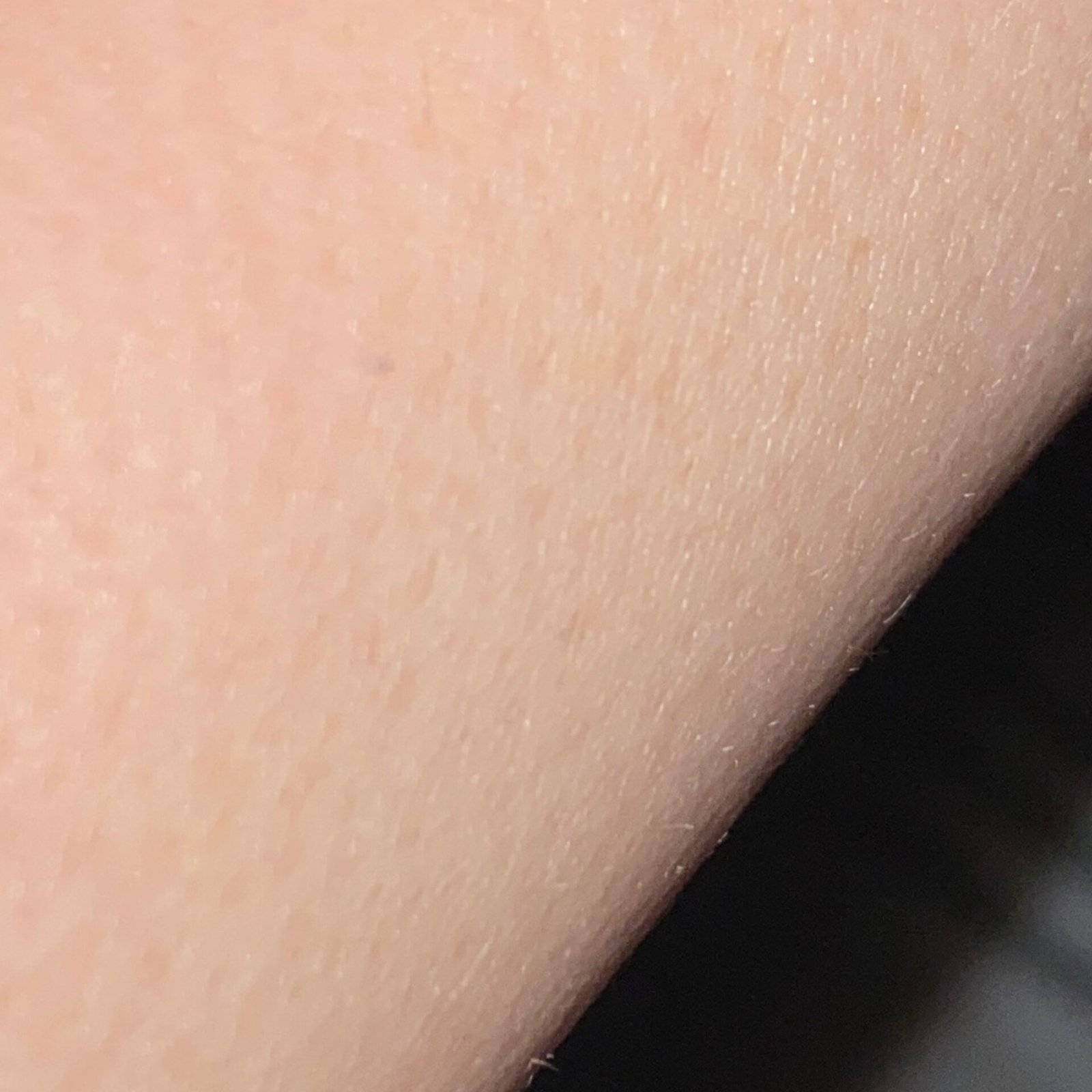
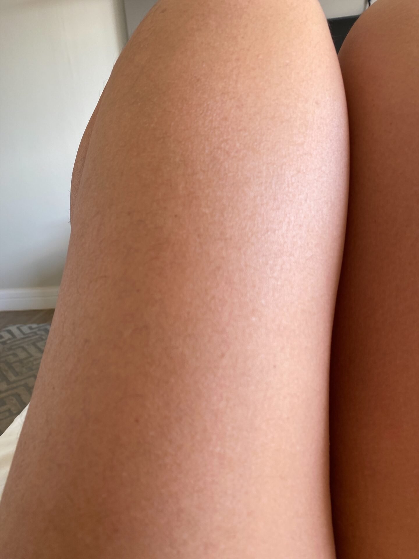
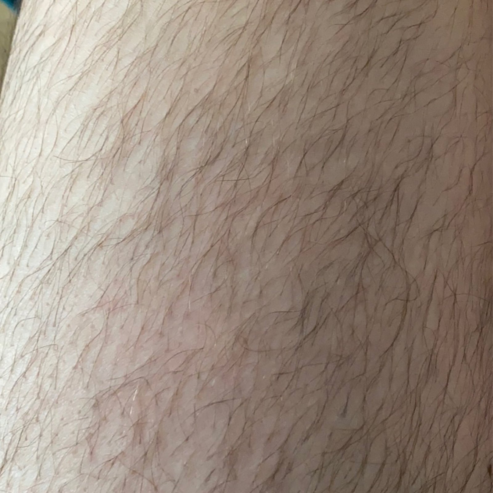
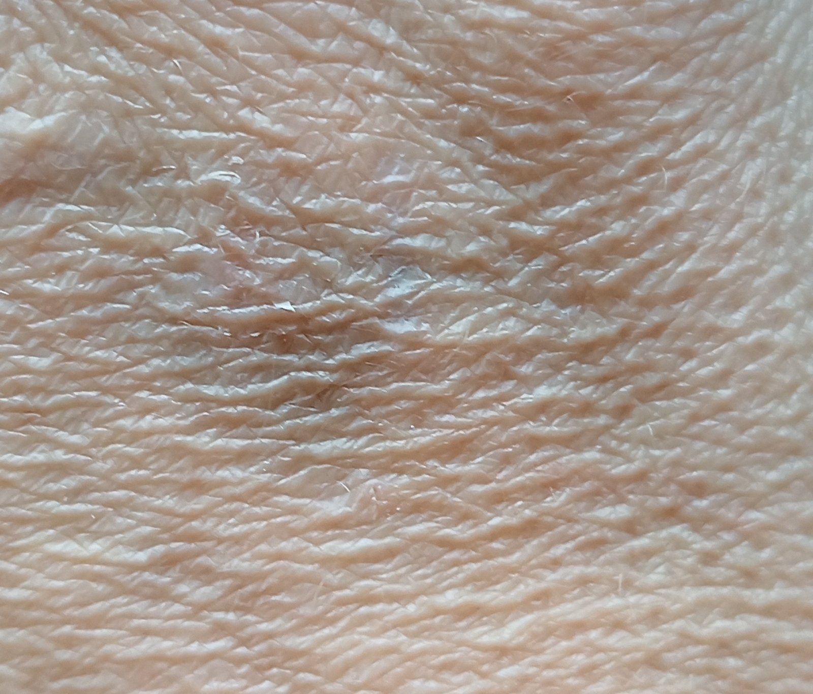
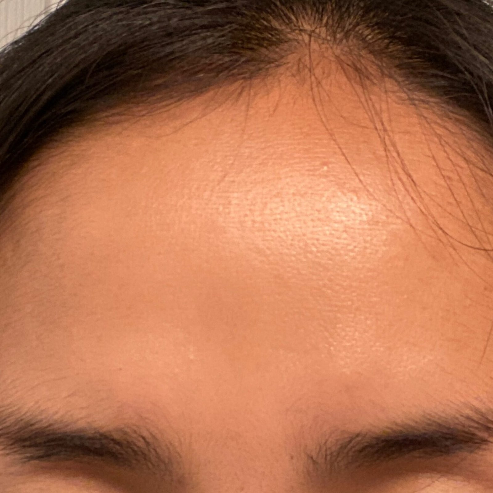
Healthy Skin: No Pathology Detected 💚
Healthy Skin: What Skin Without Visible Pathology Looks Like
Most entries in this atlas describe a disease. This one describes the reference point they are measured against: skin on which an examination finds nothing that requires treatment. It exists because “no pathology detected” is one of the most common results an AI skin analysis can return, and because that result is easy to read as more reassurance than it actually carries.
Healthy skin is not flawless skin. It is skin that is performing its work — keeping water in, keeping irritants and microorganisms out, regulating temperature, and sensing the world — without a lesion, rash or growth that needs attention. Almost everyone’s skin carries marks, and the great majority of them are normal.
What a “No Pathology Detected” Result Means
An AI analysis compares the area you photographed against patterns learned from clinical images. When it reports no pathology, it means that in that photograph, of that area, at that moment, it did not recognise a pattern associated with a skin disease in its training. That is genuinely useful information, and it is also narrower than it sounds.
It does not mean the following, and the distinction matters:
- It is not a full skin examination. One photograph covers one area. A lesion on your back, scalp, between your toes or under a nail was never assessed.
- It is not a diagnosis, and it does not exclude one. No image-based tool detects every lesion. Early changes can be too small, too flat or too subtle in a phone photograph to register, and poor lighting, blur or distance reduce accuracy further.
- It is not a permanent result. It describes today. A mole that is unremarkable now can change over months, and change is the signal that matters most.
- It does not override a symptom. If something itches, bleeds, hurts or will not heal, that symptom carries more weight than a reassuring screening result.
The right way to use this result is as one data point in ongoing self-monitoring, and as a baseline you can compare future photographs against — not as a clearance.
What Normal Skin Looks Like
Healthy skin varies enormously between people and across a single body. The features below are normal findings, not defects:
- Colour variation. Skin tone differs across the body and across the year. Sun-exposed areas are darker; palms, soles and skin usually covered are lighter.
- Freckles and sun-induced pigmentation. Small, flat, evenly coloured spots that darken with sun exposure are common, particularly in lighter phototypes.
- Ordinary moles. Most adults have between ten and forty. A typical benign mole is small, symmetrical, evenly coloured and stable over years, and an individual’s moles tend to resemble one another.
- Visible pores and texture. Pore size is largely inherited and varies by area; the face, chest and back have more sebaceous glands and a different texture from the forearm.
- Fine lines and looser skin with age. Collagen and elastin change over a lifetime; this is not a pathology.
- Small rough patches. Keratosis pilaris — fine bumps on the upper arms or thighs — is extremely common and harmless.
- Skin tags and small benign growths. Soft, skin-coloured tags in the neck, armpits and groin are a normal finding, especially after middle age.
- Mild seasonal dryness. Skin that is tighter or flakier in winter and settles with a moisturiser is functioning normally.
Appearance also differs by skin phototype. Darker skin is less prone to sunburn but is not exempt from skin cancer, and melanoma in darker skin more often arises on the palms, soles and under the nails — areas people are least likely to check. Redness, which is a familiar sign of inflammation in lighter skin, may present as a violet, grey or darker brown discolouration in deeply pigmented skin, and is easy to miss on both a photograph and a casual glance.
What Is Not a Normal Finding
Regardless of any screening result, the following warrant an in-person assessment:
- A mole that is changing in size, shape, colour, height or sensation. Change over weeks or months is more meaningful than any single feature.
- A lesion that stands out from your others — the “ugly duckling”. Because a person’s moles usually look alike, one that does not belong deserves attention.
- Asymmetry, an irregular or blurred border, more than one colour, or a diameter beyond about 6 mm — the familiar ABCDE features, with E for evolving carrying the most weight.
- A sore that has not healed within about four weeks, or that heals and reopens.
- Any lesion that bleeds, crusts repeatedly, itches persistently or is painful.
- A new dark streak under a fingernail or toenail, particularly a single one, or pigment spreading onto the surrounding skin.
- A rash that is spreading, blistering, or accompanied by fever or feeling unwell.
Who Should Be Checked Even With Normal-Looking Skin
Some people carry enough risk that professional skin examination is worth arranging whatever the skin looks like. How often, and whether it is needed at all, is decided individually and varies between national guidelines — it is a conversation to have with a clinician rather than a fixed schedule. Discuss a schedule with a dermatologist if you have a personal or family history of melanoma or other skin cancer, a very large number of moles or atypical moles, a history of severe sunburns or sunbed use, fair skin that burns easily, long-term immunosuppression including after an organ transplant, or previous radiotherapy to an area of skin.
Keeping Skin Healthy
- Sun protection. The most effective single measure. Use broad-spectrum SPF 30 or higher on exposed skin, seek shade in the middle of the day, cover up, and do not use sunbeds. This applies to every skin tone.
- Gentle cleansing. A mild cleanser and lukewarm water. Over-washing, hot water and harsh scrubs damage the skin barrier.
- Moisturise after washing, while the skin is still slightly damp, particularly in dry or cold conditions.
- Do not smoke, and keep alcohol within recommended limits; both affect skin healing and ageing.
- Sleep, nutrition and stress influence barrier function and inflammatory skin conditions.
- Check your own skin regularly. Once a month, in good light, with a mirror for the back and a partner’s help where needed. Include the scalp, behind the ears, between the fingers and toes, the soles and the nails. Photographs taken from the same distance make change easier to spot than memory does.
A result of “no pathology detected” is a good outcome and a reasonable reason to relax — but the value of skin screening comes from repetition. The purpose of knowing what your skin looks like when it is healthy is to recognise, quickly, the day something changes.