Milium (ICD-10: L72) 💚
Milium Cyst: A Superficial Epidermal Keratin-Filled Lesion
Milium cyst, commonly referred to as “milia” or “milk spots,” is a small, white to yellowish, dome-shaped skin cyst that forms in the upper layers of the epidermis. These lesions are small keratin-filled cysts—keratin being a protein produced by skin cells—enclosed beneath the surface of the skin. Primary milia are thought to arise from the pilosebaceous unit or other epidermal structures; unlike closed comedones they are true cysts rather than plugged follicles and do not result from sebum obstruction.
While milia are generally benign and painless, their appearance—particularly when clustered around the eyes, nose, or cheeks—can raise cosmetic concerns. They are most frequently seen on the face but can occur on any area of the body. Milium cysts can appear as single or multiple elements and are often mistaken for other types of cysts or acne lesions.
Milia are commonly observed in newborns as a physiological response to immature skin and disappear within a few months without intervention. However, a second peak of incidence is seen during adolescence and early adulthood, particularly in females. In adults, milia can appear spontaneously or be triggered by skin trauma, chronic irritation, or inappropriate cosmetic use. They may also be secondary to dermatoses or certain dermatological procedures such as laser resurfacing or dermabrasion.
Predisposing Factors: What Causes Milium Cysts to Form?
The exact mechanism of milium cyst formation is not fully established. Milia are small cysts lined by a thin epidermis-like wall and filled with laminated keratin; primary milia are thought to originate from the pilosebaceous unit or other epidermal structures, while secondary milia arise where the skin has been injured. Either way the result is keratin enclosed within a closed epithelial sac that sits just beneath the surface and cannot drain, forming a distinct, superficial cyst.
Primary milia most often appear without any identifiable cause, while secondary milia follow injury to or disease of the skin. The circumstances recognized in association with milia include:
- Chronic photodamage: Milia are sometimes seen within severely sun-damaged skin, where the epidermis and its appendages are structurally altered.
- Prolonged topical corticosteroid use: Long-term application of potent topical steroids to facial skin has been associated with milia.
- Blistering skin disorders: Conditions such as bullous pemphigoid, epidermolysis bullosa, or porphyria cutanea tarda commonly leave milia behind as blisters heal.
- Burns, scars, and grafts: Milia frequently develop within healing burns, surgical scars, or skin grafts as the damaged epidermis repairs itself.
- Rare inherited syndromes: Multiple or eruptive milia are an occasional feature of genetic conditions such as Bazex–Dupré–Christol or Rombo syndrome.
- Mechanical trauma and skin injury: Procedures like chemical peels, laser therapy, or prolonged friction may result in secondary milia (also referred to as Balser’s pseudomilia).
- Occlusion and chronic irritation: Heavy occlusive products or long-standing friction have been reported alongside milia, although the evidence for a causal role is limited.
The practical distinction is between primary milia, which arise spontaneously and usually have no identifiable trigger, and secondary milia, which follow blistering, trauma, or procedures affecting the epidermis. This distinction matters for counseling and for what to look for clinically, rather than for the removal technique, which is the same in both cases.
Diagnostics: How Milium Cysts Are Identified
The diagnosis of milium cysts is typically made during a clinical examination. A trained dermatologist can usually identify milia based on their distinct visual characteristics and anatomical location. Medical history is also important to determine whether the milia are primary (spontaneous, often congenital or idiopathic) or secondary (associated with skin trauma, underlying disease, or medical procedures).
In uncertain cases or when the lesions appear atypical, additional diagnostic steps may be warranted:
- Dermatoscopy: A non-invasive technique used to closely examine the surface and subsurface features of skin lesions, helping to differentiate milia from other cystic or pigmented growths.
- Histological examination: A biopsy may be performed in rare cases to confirm diagnosis, especially when the lesion is solitary, persistent, or mimics neoplastic skin pathology.
Proper diagnosis is essential mainly to rule out similar-appearing lesions and to determine the best course of treatment; typical milia call for no further investigation, which is reserved for atypical lesions.
Symptoms: What Milium Cysts Look and Feel Like
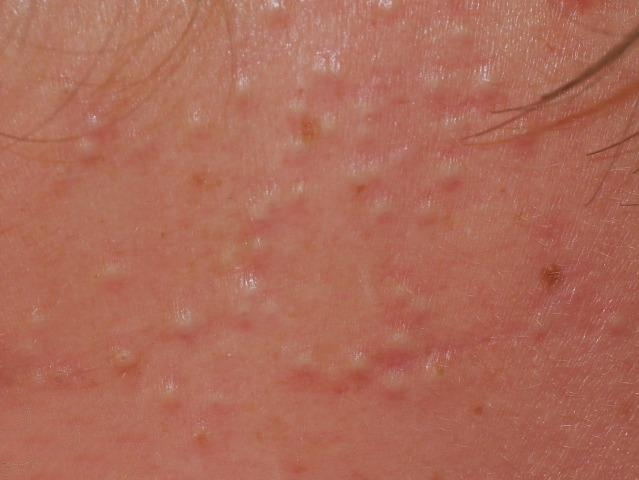
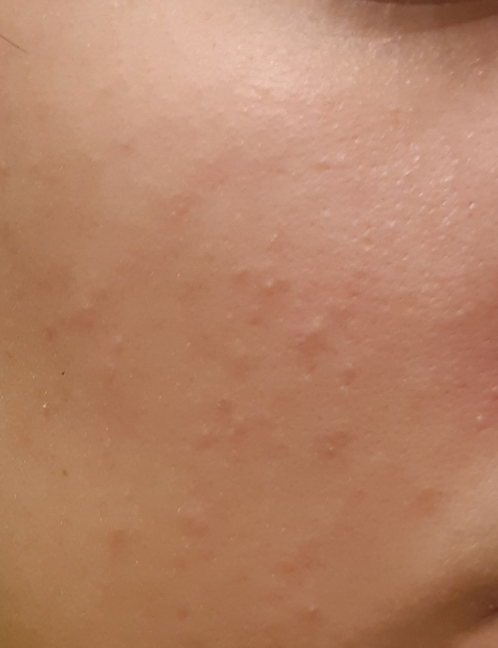
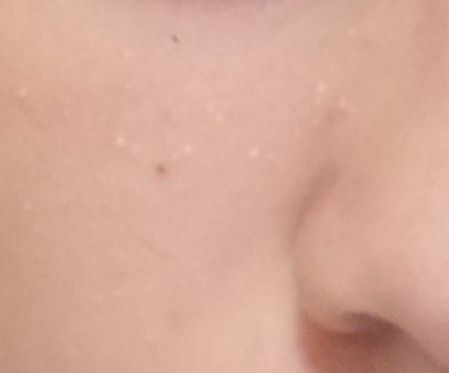
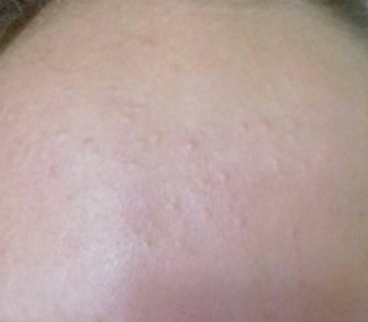
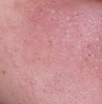
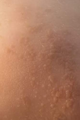
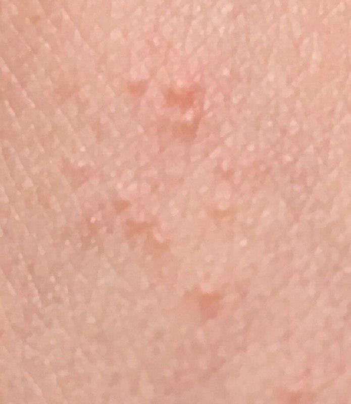
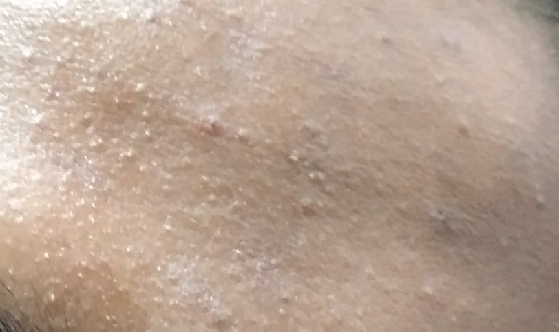
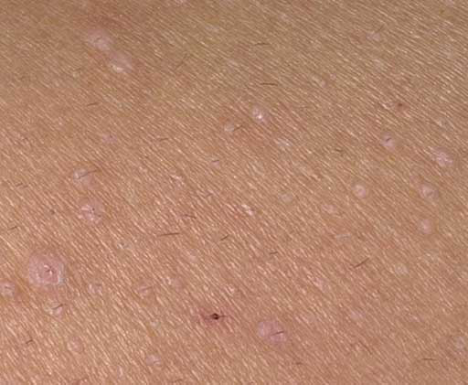
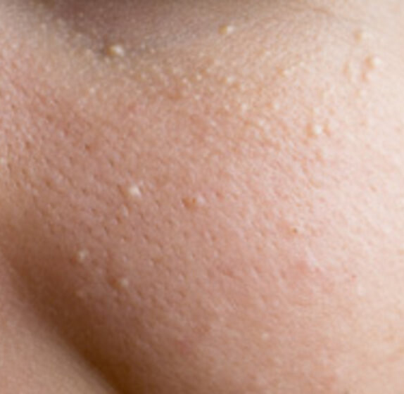
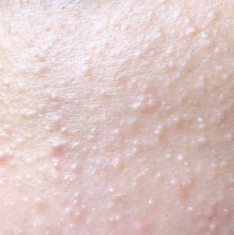
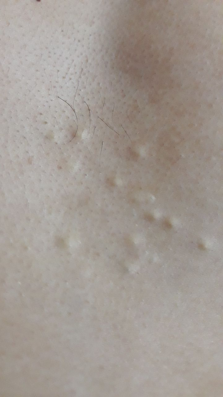
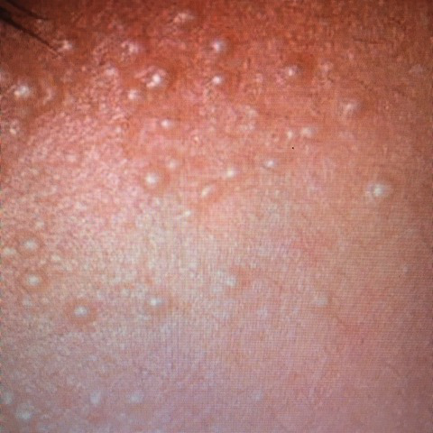
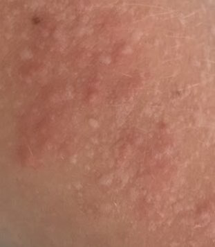
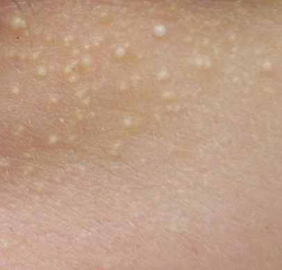
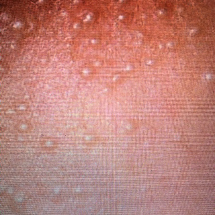
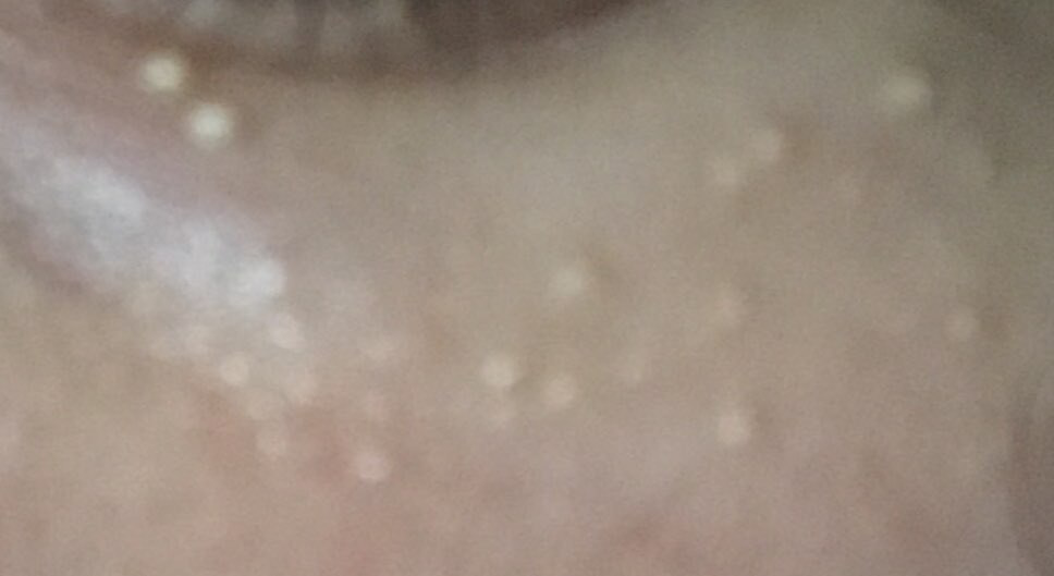
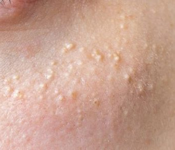
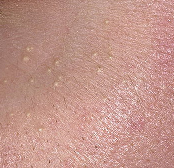
Milium cysts are typically small, white or yellowish papules that measure about 1–2 mm in diameter. They are well-circumscribed, firm, and dome-shaped, and they protrude slightly above the skin’s surface. Milia are most commonly located on the face—particularly around the eyes (periorbital region), nose, chin, forehead, and cheeks—but may also appear on the upper torso or genital region.
Clinical characteristics include:
- Shape and surface: Symmetrical, round or oval lesions with a smooth, stretched epidermal layer. The normal skin texture is lost in the area of the cyst due to tension on the surface.
- Color: White to pale yellow, due to keratin contents visible through the thin overlying epidermis.
- Skin reaction: No surrounding inflammation or erythema unless the lesion is irritated or secondarily infected.
- Hair growth: Unaffected. Milia do not interfere with hair growth in the surrounding skin.
- Sensation: Milia are typically asymptomatic—there is no pain, itching, or discomfort. They are found incidentally or due to cosmetic concern.
In some cases, particularly with multiple lesions, milia may cover several square centimeters of skin. However, even in such presentations, the lesions remain isolated and do not coalesce.
Dermatoscopic Description
Dermatoscopy enhances visualization and supports differential diagnosis. Typical dermatoscopic features of milium cysts include:
- Central white to yellow globular structure: Representing the keratin-filled cyst cavity.
- Clear demarcation: A well-defined boundary between the central lesion and the surrounding healthy skin.
- Compression sign: Gentle pressure flattens the lesion slightly due to its soft, pliable contents and superficial placement.
These dermatoscopic characteristics help distinguish milia from comedones, molluscum contagiosum, or other cystic and nodular lesions.
Differential Diagnosis: Conditions to Rule Out
Although milia have a characteristic appearance, several other dermatologic conditions may resemble them. Differential diagnosis includes:
- Closed comedones: While also white and dome-shaped, comedones are associated with sebaceous glands and are not true cysts.
- Molluscum contagiosum: Viral papules with central umbilication, typically clustered and contagious.
- Papillomas: Skin-colored growths that have a rougher surface and often appear on friction-prone areas.
- Sebaceous gland nevi: Congenital hamartomas that may present as yellow plaques, especially on the scalp.
- Non-inflammatory acne: Characterized by comedones, usually with an oily skin background and wider pore involvement.
Risks: Why Milium Cysts Should Be Monitored
Milium cysts are benign and non-life-threatening, and in most people they are isolated, incidental findings that call for no investigation of systemic disease. Only an unusual presentation—lesions appearing suddenly in large numbers, in an unexpected distribution, or alongside blistering—means the wider clinical context deserves a closer look.
Risks associated with untreated or improperly treated milia include:
- Cosmetic impact: Facial milia can affect self-esteem, particularly when they are numerous or located in prominent areas like the eyelids or nose.
- Misdiagnosis: Mistaking milia for acne or trying to extract them forcefully may cause skin trauma or secondary infection.
- Progression to inflammatory conditions: Rarely, milia may become secondarily inflamed, especially when scratched or exposed to harsh chemicals.
- Atypical presentations: Sudden, widespread, or unusually distributed milia in adults may warrant evaluation for an underlying blistering or inherited disorder, depending on the clinical context.
Tactics: When to Consult a Specialist
Although milia in newborns typically resolve on their own, persistent, multiple, or cosmetically concerning lesions in adolescents or adults should be evaluated by a dermatologist or cosmetologist. A professional consultation is essential when:
- Milia do not resolve spontaneously after several months.
- New lesions appear rapidly or recur after removal.
- The skin around milia becomes inflamed or painful.
- There is a history of skin injury, burns, or resurfacing procedures.
Once the diagnosis is clear, typical milia need no ongoing surveillance; treatment addresses the cosmetic concern, and review is reserved for lesions that are atypical, persistent, or recurrent.
Treatment: Professional Removal and Prevention of Recurrence
Because milium cysts are closed keratin-filled sacs, topical creams and over-the-counter products cannot clear them; sterile extraction is the standard treatment for bothersome lesions. Self-removal is not recommended due to the risk of skin trauma, infection, and scarring.
Professional treatment options include:
- Mechanical extraction: Performed under sterile conditions using a fine lancet or extractor; generally preferred for typical superficial milia.
- Laser ablation: Carbon dioxide (CO2) lasers vaporize the keratinous material with precision and serve as an alternative in selected cases, such as lesions at delicate sites.
- Radiofrequency or electrocautery: Thermal destruction of the cyst wall and contents; a further option, chosen by lesion number and location.
- Topical retinoids or keratolytics: Sometimes useful where milia are multiple or recurrent; they are not routinely required after an uncomplicated removal.
After removal, ordinary gentle skincare is usually all that is needed; further preventive measures matter mainly where milia have been recurrent.
Prevention: How to Minimize the Risk of Milium Cysts
Although not all cases of milia can be prevented, the risk of recurrence can be reduced through consistent skin care and healthy lifestyle habits:
- Use non-comedogenic skincare: Avoid thick creams and occlusive products, especially around the eyes.
- Practice gentle exfoliation: Periodic use of mild chemical exfoliants helps maintain skin turnover.
- Sun protection: Daily use of broad-spectrum SPF can prevent UV-induced epidermal thickening.
- Avoid skin trauma: Refrain from picking, squeezing, or irritating the skin.
- Treat the underlying skin problem: Where milia follow blistering, a burn, or a procedure, managing that process is what limits further lesions.
- Review when something changes: Seek assessment for lesions that are atypical, widespread, persistent, or recurrent, rather than on a schedule.
With an accurate diagnosis and, where the lesions are bothersome, straightforward professional removal, milium cysts are readily managed—and for most people they remain a purely cosmetic matter.