
Erysipelas (ICD-10: A46) 🚨
Erysipelas: A Sharply Bordered Bacterial Infection of the Skin
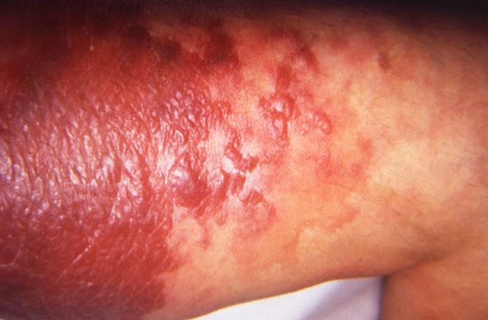
Erysipelas is an acute bacterial infection of the upper layers of the skin and its superficial lymphatic vessels, caused in most cases by Streptococcus pyogenes. It produces a bright red, hot, tender area, classically raised with a border you can trace with a finger — the feature that has given it its nickname, St Anthony’s fire. That appearance is typical rather than invariable: the edge is sometimes indistinct or flat, and the absence of a sharp raised border does not exclude the diagnosis.
Cellulitis involves deeper tissue and classically fades into surrounding skin. In practice the two sit on a continuum — the border is sometimes indistinct or flat — and no treatment decision should rest on this feature alone. Both are bacterial infections requiring antibiotics.
The illness usually announces itself before the skin does. Fever, shivering and feeling unwell commonly precede the rash by several hours, and a patient may describe going to bed well and waking up ill. Onset is abrupt, and it progresses over hours rather than days.
The lower leg is now the most common site. The face, where a symmetrical butterfly pattern across the cheeks and nose is the classic description, is affected less often than older accounts suggest.
Predisposing Factors: Why Erysipelas Develops
The bacteria need a way in. A portal of entry is often found, though it may be small enough to overlook, and sometimes none is identified even after a careful look — which does not mean the examination was inadequate.
- A break in the skin: Cracks between the toes from athlete’s foot are a common and readily treatable portal in leg erysipelas, though not the cause of every episode. Cuts, insect bites, leg ulcers, eczema and injection sites all serve the same purpose.
- Lymphoedema: Both a cause and a consequence. Impaired lymphatic drainage predisposes to infection, and repeated infection can damage the lymphatics further, though how much any single episode contributes varies.
- Venous insufficiency and the fragile skin that accompanies it. See venous eczema.
- Excess body weight, which is consistently associated with leg involvement.
- Diabetes and impaired immunity.
- A previous episode, which is among the strongest predictors of another.
Diagnostics: How Erysipelas Is Diagnosed
The diagnosis is clinical: the appearance, the sharply defined raised border, the abrupt onset and the systemic symptoms together. No test confirms it reliably, and treatment is not delayed waiting for one.
Blood tests show inflammation and help judge severity. Blood cultures are positive in only a minority of cases and are taken for severe illness, systemic upset, impaired immunity or an unusual exposure, according to local protocols. A swab is useful when there is an obvious wound or entry point. Ultrasound is not part of the routine assessment. It is used where there is a specific clinical suspicion of an abscess or of a deep vein thrombosis, both of which can mimic or accompany erysipelas.
Marking the edge of the redness with a pen and dating it is a simple and genuinely useful practice: it makes it possible to tell the next day whether the infection is advancing or retreating.
Symptoms: What Erysipelas Looks Like
- A sharply demarcated, raised red plaque whose border can be felt as a step. In deeper skin tones the colour change may read as violet, dusky brown or simply darker, and the warmth, swelling and tenderness carry more weight than the colour.
- Heat and tenderness over the affected area.
- Swelling, sometimes with an orange-peel texture to the skin.
- Fever, chills, shivering and malaise, frequently preceding the skin signs.
- Swollen, tender lymph nodes draining the area, and sometimes red streaking towards them.
- Blisters over the surface in more severe episodes.
- One side only, in most cases. When both legs are red, venous eczema, dependent redness and lipoedema are the usual explanations — though bilateral infection does occur, so this points a direction rather than excluding one.
Differential Diagnosis: Ruling Out Other Conditions
- Cellulitis: Deeper, with a less defined edge and less elevation. The two are frequently indistinguishable and are managed similarly.
- Venous eczema: The most consequential mimic. It is usually bilateral, itchy rather than painful, without fever, and it does not respond to antibiotics. Recurrent “cellulitis” of both legs deserves a second look.
- Deep vein thrombosis: Swelling and pain with less pronounced redness and no sharp border; the two can coexist.
- Necrotising fasciitis: Rare, but the diagnosis that must not be missed. Pain out of proportion to the appearance, rapid progression over hours, dusky or grey skin, blisters with dark fluid, numbness over the area, or a patient who looks far more unwell than the skin suggests — any of these needs immediate emergency assessment and a surgical opinion. Their absence does not exclude an early necrotising infection.
- Allergic contact dermatitis: Itchy and without fever.
- Acute gout or septic arthritis, when redness overlies a joint.
Risks: Why Erysipelas Needs Prompt Treatment
Most episodes respond well to antibiotics. The reasons to start them quickly are what happens otherwise.
Infection can extend into deeper tissue, form an abscess, or enter the bloodstream and cause sepsis, which is more likely in older people and those with other illness. Rarely, what begins as an apparent erysipelas turns out to be necrotising fasciitis, where hours matter.
The longer-term problem is the cycle it sets up. Each episode damages the lymphatic vessels, that damage causes swelling, and swollen skin is more prone to the next infection. After several episodes this can establish permanent lymphoedema, which is difficult to reverse — which is why treating the entry point, and preventing recurrence, is as much a part of care as the antibiotic course.
Tactics: When and How to Seek Treatment
Erysipelas needs medical assessment, not self-treatment. Contact a doctor the same day if an area of skin becomes red, hot, swollen and tender, especially with fever or shivering.
Seek emergency care if pain is severe and out of proportion to what is visible, if the area is spreading rapidly over hours, if the skin turns dusky, grey or blistered with dark fluid, if there is numbness over the area, or if you feel very unwell, confused or faint.
Two practical points: photograph or mark the edge so that change can be judged, and elevate the affected limb, which helps with the swelling. Elevation is supportive care, not treatment — it should accompany medical assessment and antibiotics rather than become a reason to wait.
Treatment: Modern Strategies for Erysipelas
- Antibiotics: Penicillin or a related agent is a usual first choice given the organisms involved, with alternatives for allergy. Which drug, whether by mouth or by drip, and for how long all follow local guidance, resistance patterns, allergy and severity rather than a single fixed regimen; most people are treated at home, while severe illness or significant other conditions may warrant intravenous treatment.
- Elevation of the limb, which reduces swelling and discomfort and is easy to under-do.
- Pain relief and fluids.
- Treating the entry point: Clearing athlete’s foot, dressing an ulcer, managing eczema. Skipping this is a common reason the infection returns.
- Reassessment if it is not improving, generally within two or three days, or sooner if it is getting worse. Redness may extend slightly in the first day before it retreats, which is not by itself a sign of failure.
- Managing lymphoedema afterwards. Compression is generally avoided while the limb is acutely painful and inflamed, and introduced once the infection has settled, provided the arterial circulation allows it.
- Preventive antibiotics for people with repeated episodes, which reduce recurrence while they are taken. They are considered for recurrent disease once the modifiable risk factors have been addressed, since dealing with the entry point and the swelling comes first. There is no universal episode count that triggers them: the threshold, the choice of drug and the duration follow local guidelines and an individual assessment.
Prevention
- Treat athlete’s foot and keep the skin between the toes dry, which is the most actionable single measure for leg erysipelas.
- Moisturise dry or cracked skin, particularly on the lower legs.
- Care for wounds and ulcers properly rather than improvising.
- Manage lymphoedema and venous disease, including wearing compression where it has been prescribed.
- Address weight and diabetes control where relevant.
- Act early on a recurrence, since people who have had erysipelas often recognise the onset before it is visible.
Erysipelas usually resolves without trouble when it is treated early. The work that prevents the next episode — the entry point, the swelling, the skin barrier — is the part most often left undone.
Image: CDC / Dr. Thomas F. Sellers, Emory University, Public Health Image Library #17528.

Medical review
Board-Certified Dermatologist · Fellow of the American Academy of Dermatology (FAAD)