Lentigo Melanoma (ICD-10: C43) 🚨
Lentigo Melanoma
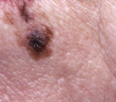
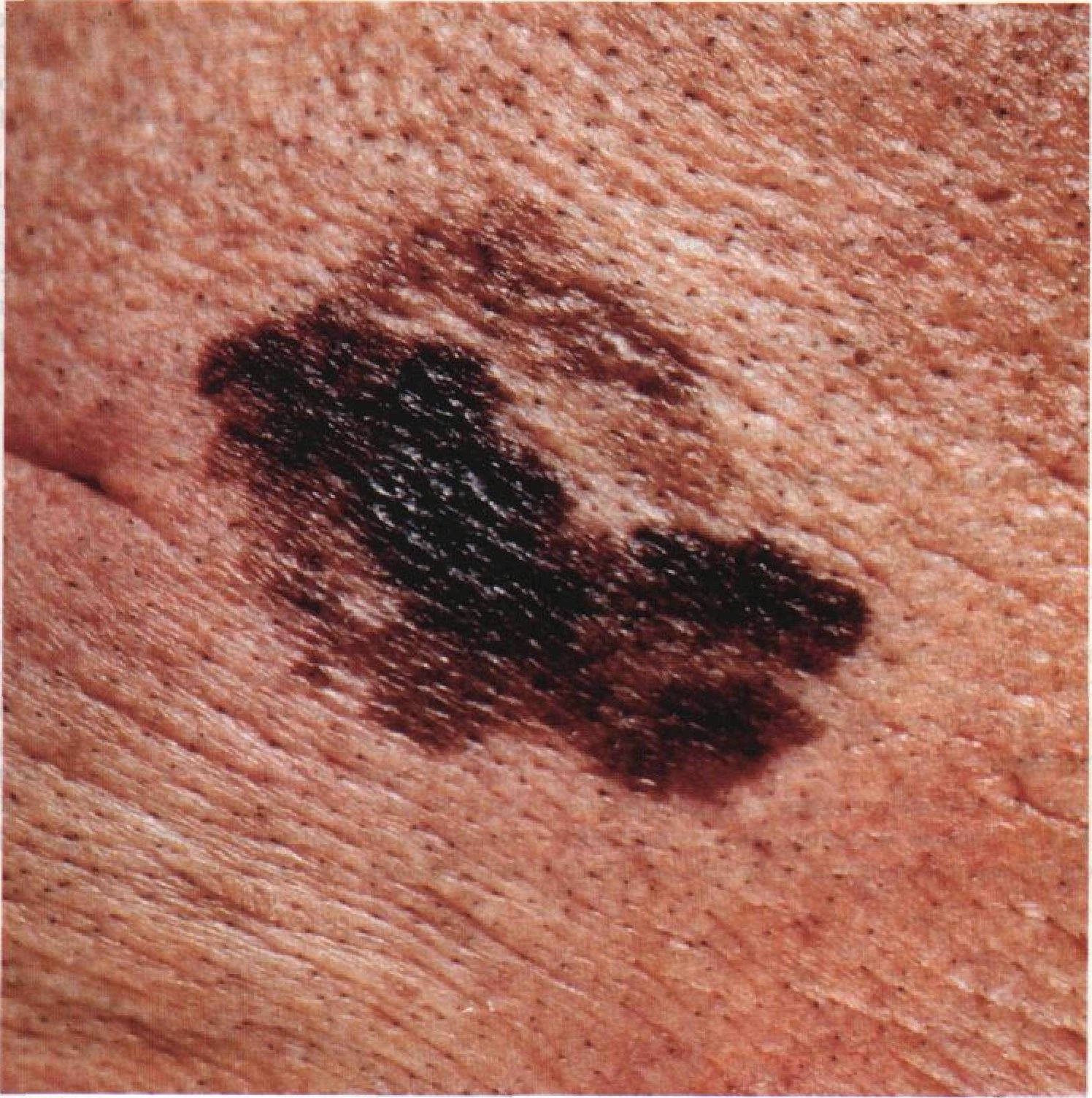
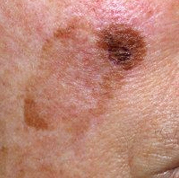
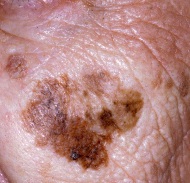
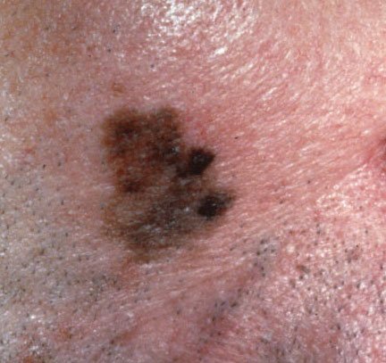
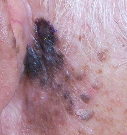
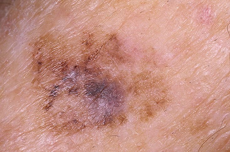
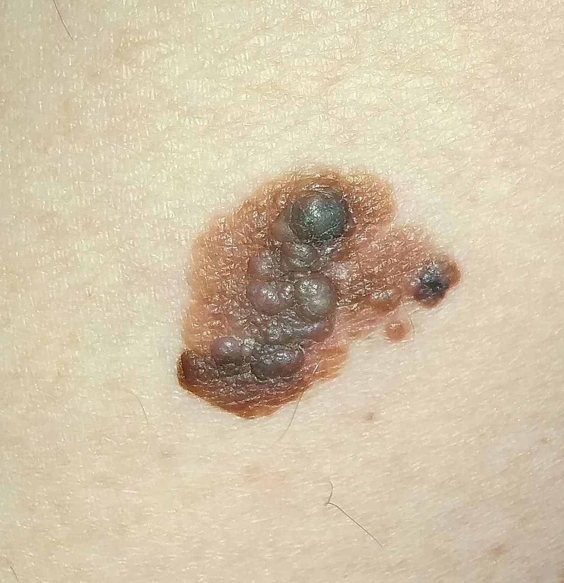
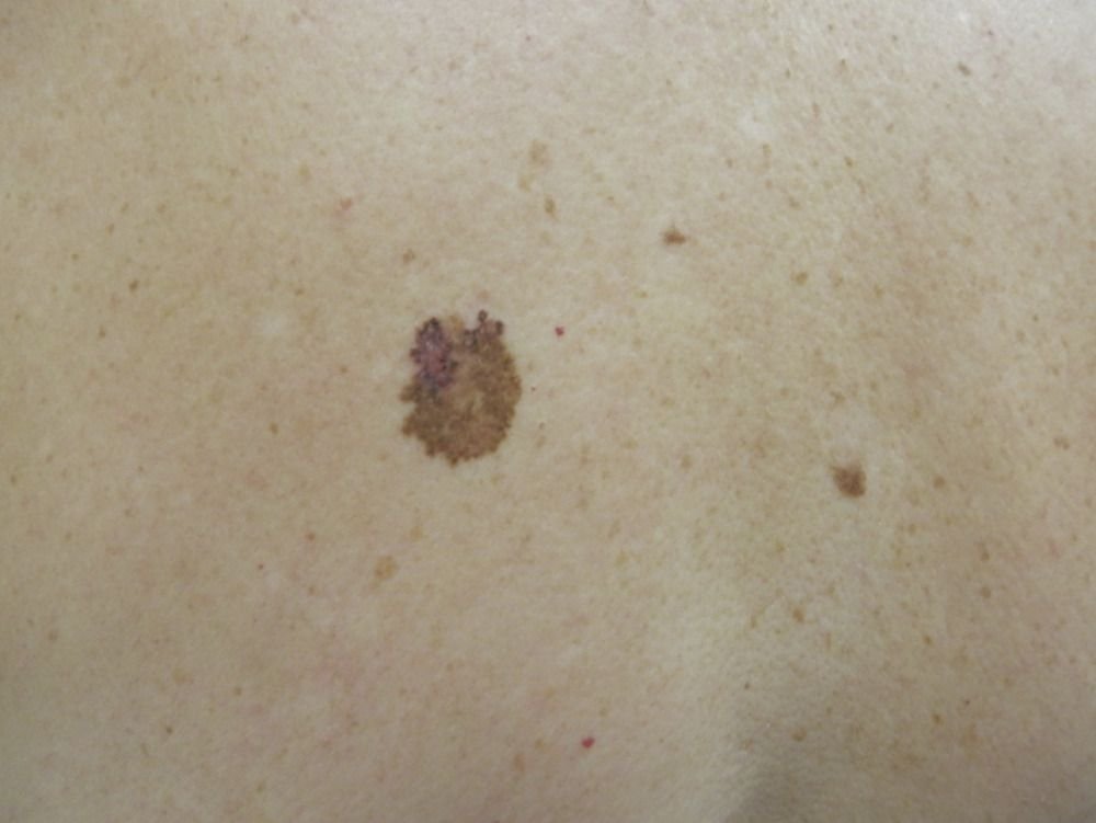
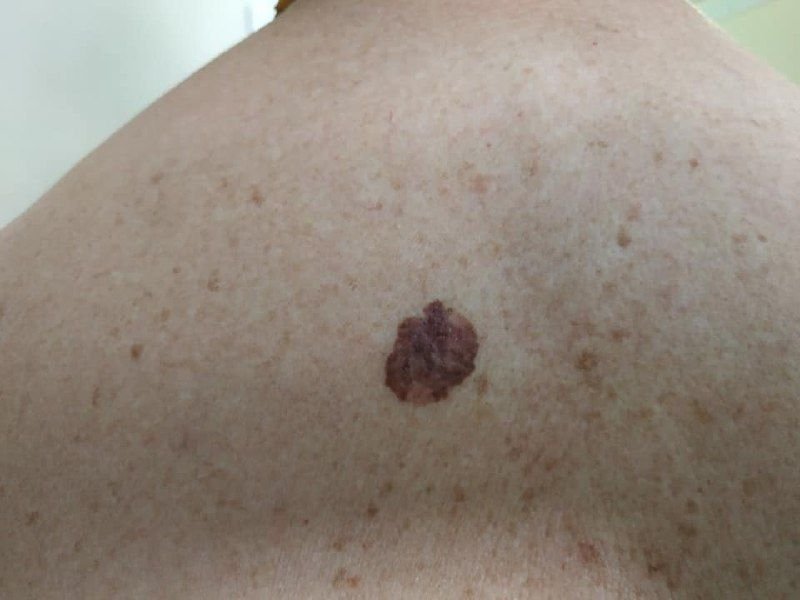
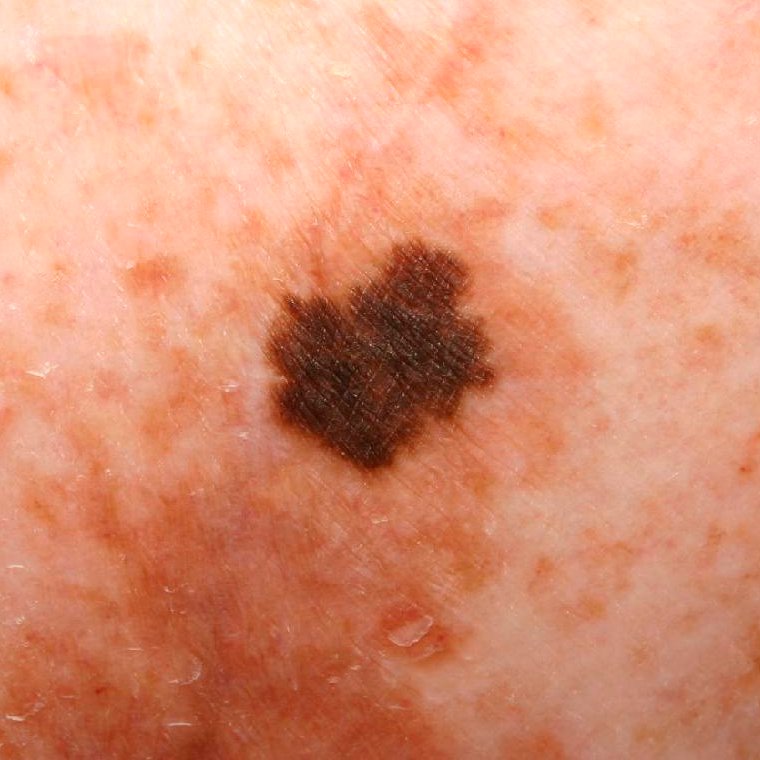
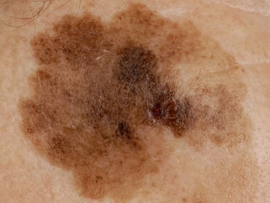
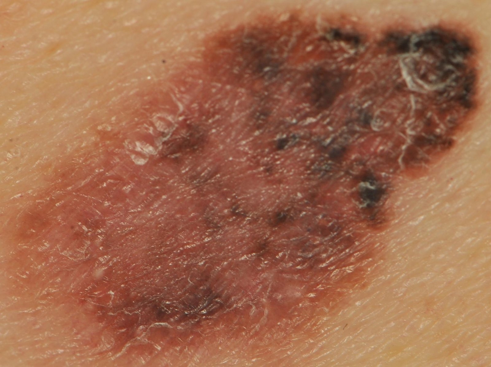
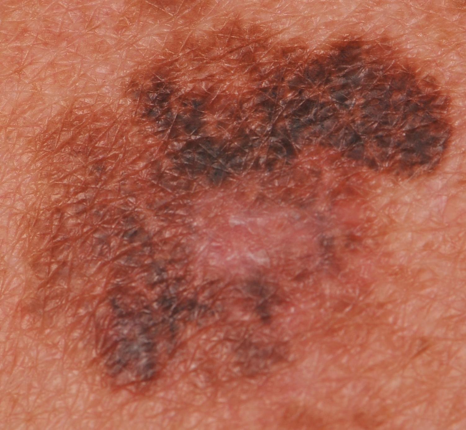
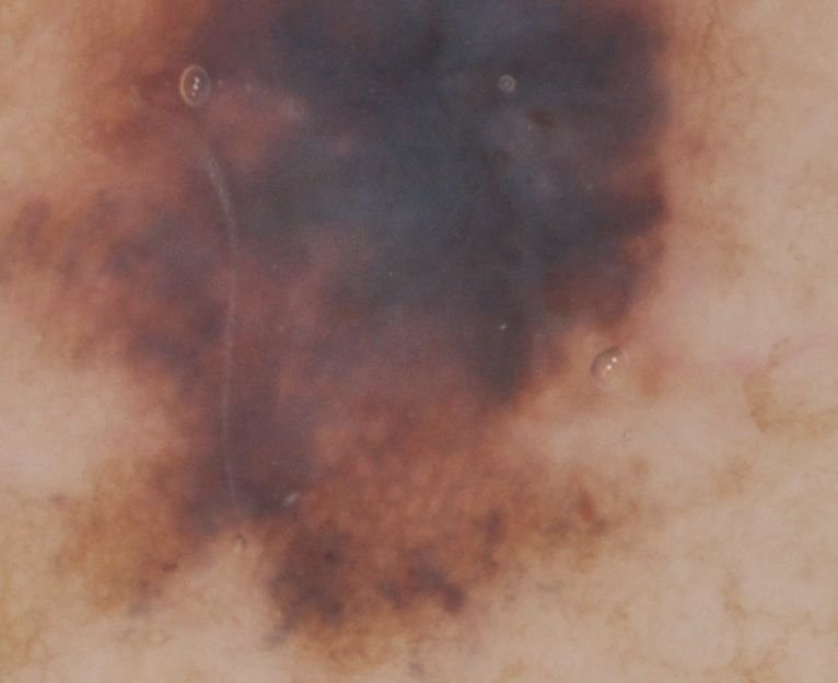
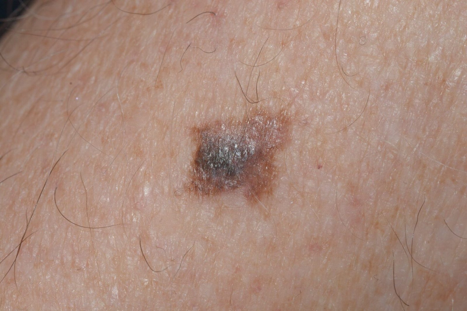
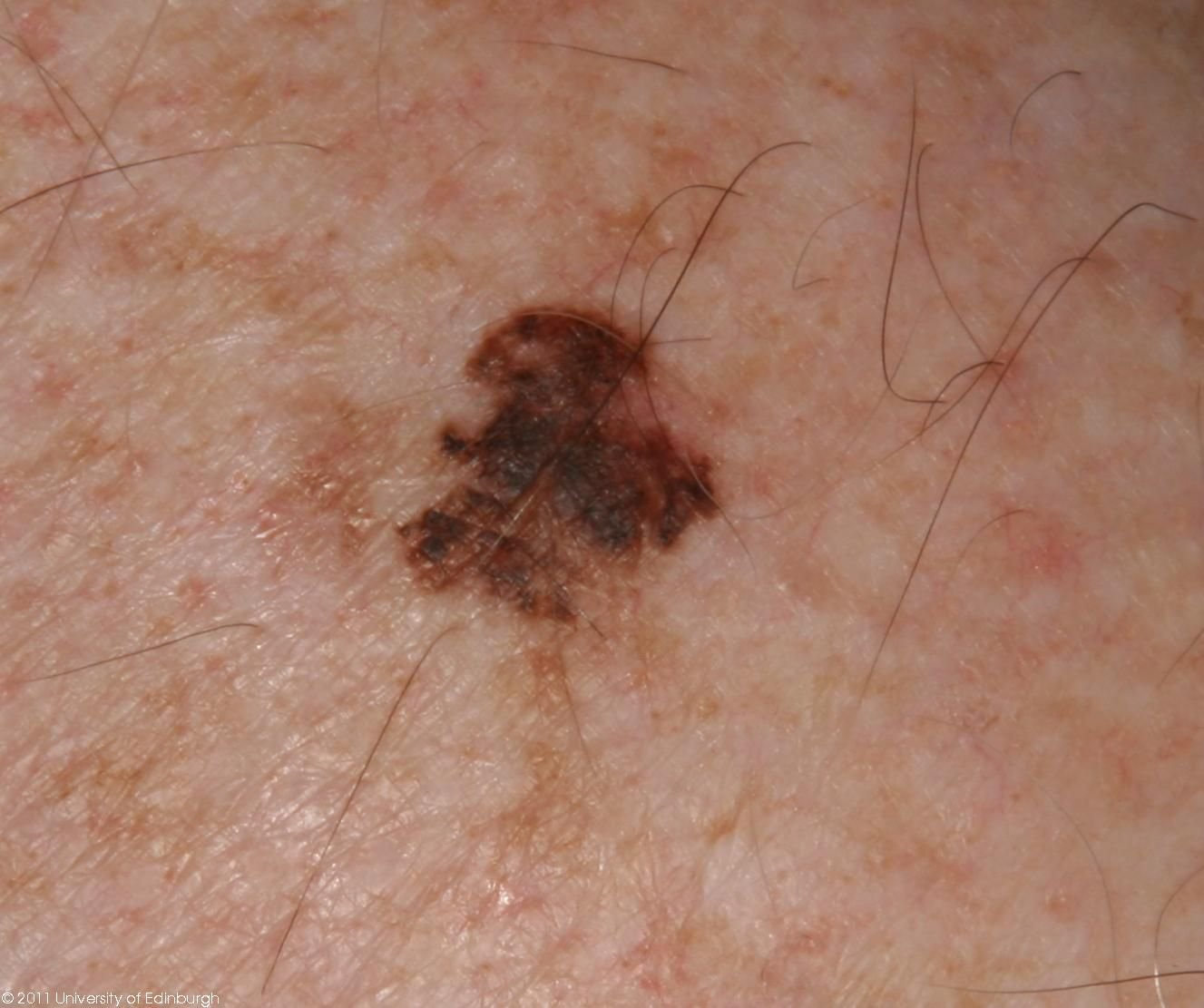
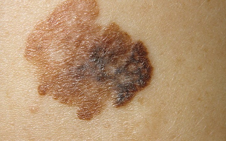
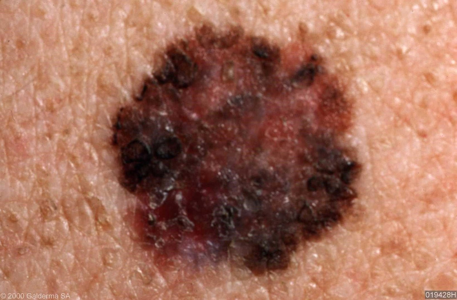
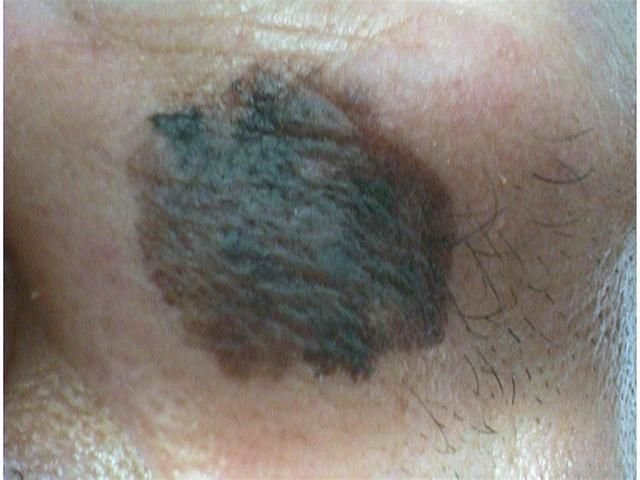
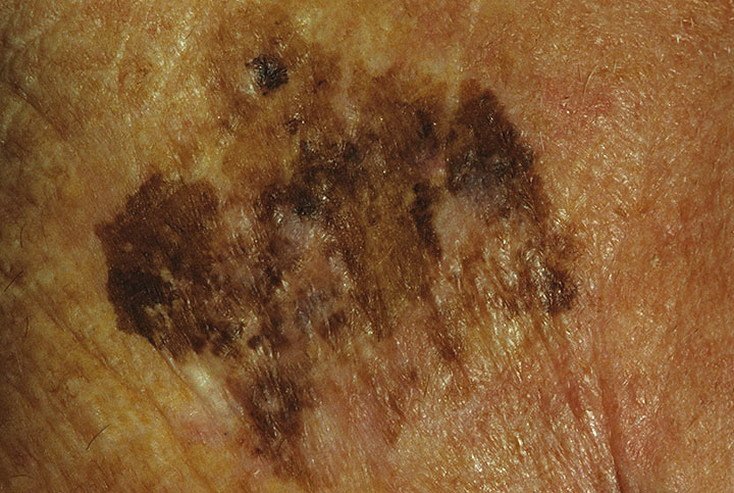
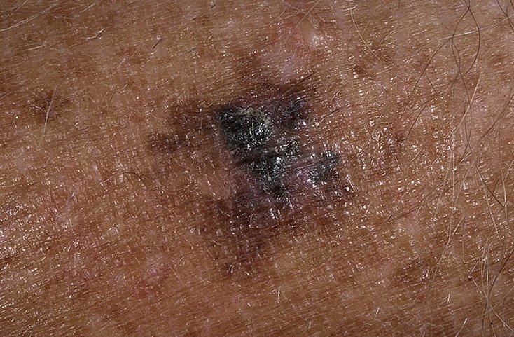
Lentigo maligna melanoma is a distinct form of malignant melanoma that originates from melanocytes — the pigment-producing cells responsible for melanin synthesis in the skin. This tumor most often arises in individuals over the age of 50 and typically affects areas chronically exposed to ultraviolet radiation, such as the face, neck, ears, and scalp. Lentigo maligna melanoma develops from lentigo maligna (also referred to as Dubreuilh’s melanosis), a long-standing pigmented lesion on sun-damaged skin. Lentigo maligna is itself a melanoma — melanoma in situ, confined to the epidermis — rather than a precancerous change that may or may not become one. Lentigo maligna melanoma is what it becomes once the tumour invades beyond the epidermis. Although it represents a relatively small percentage of all melanoma cases — about 5% — this subtype is considered clinically significant due to its potential for local invasion, recurrence, and eventual metastasis.
Aggressiveness and Metastatic Potential
Unlike nodular melanoma, which is known for rapid vertical growth, lentigo maligna melanoma expands slowly in a radial pattern along the skin’s surface, and it may do so for years before invading. That slower growth pattern is characteristic of the subtype. Once invasion has occurred, however, the outlook is governed by the same factors as in any melanoma — above all the Breslow thickness, together with ulceration, mitotic activity and nodal status. The tumour can then reach nearby lymph nodes or distant organs such as the lungs, liver, bones or brain, which is why early diagnosis and appropriate treatment matter.
Predisposing Factors
Lentigo maligna melanoma is rarely diagnosed in younger individuals and is closely associated with chronic UV damage. Risk factors that contribute to its development include:
- Prolonged exposure to sunlight or artificial UV sources (such as tanning beds).
- Fair complexion — particularly individuals with Fitzpatrick skin types I or II, pink freckles, or a tendency to burn easily.
- Light eye color (blue, gray, or green) and hair color (blonde or red), both of which are indicators of UV sensitivity.
- A history of repeated sunburns, especially in childhood and adolescence (under the age of 14).
- A personal or family history of melanoma or atypical nevi.
- The presence of pre-existing lentigo maligna, which is melanoma in situ and from which invasive disease develops.
- Age-related reduction in immune surveillance, particularly after 50 years of age.
Diagnosis
The diagnostic process begins with a detailed skin examination by a dermatologist, including dermatoscopic analysis. Lesions suspected of being lentigo maligna melanoma are assessed using the ABCDE rule, which helps identify signs of malignancy:
- A – Asymmetry: One half of the lesion does not mirror the other.
- B – Border: Edges appear irregular, poorly defined, or blurred.
- C – Color: Uneven pigmentation with multiple shades of brown, black, gray, blue, pink, or white.
- D – Diameter: Typically larger than 6 mm, often exceeding 10 mm due to delayed detection.
- E – Evolution: Noticeable changes in size, shape, color, surface texture, or symptom onset over time.
Confirmation of the diagnosis is only possible through histopathological evaluation of a biopsy sample. Advanced imaging (e.g., ultrasound, CT, MRI, or PET) may be required to assess for metastasis.
Symptoms and Clinical Presentation
In the early stages, lentigo maligna melanoma may appear as a slowly expanding, irregularly pigmented macule. Initially asymptomatic, it may go unnoticed for years. Over time, however, the lesion can darken, develop uneven borders, and change in texture. Symptoms and visible changes that often prompt medical attention include:
- Gradual enlargement of a previously stable pigmented patch.
- Presence of multiple pigment colors within the lesion.
- Loss of normal skin texture and skin markings over the area.
- Hair loss on the lesion site.
- Subjective sensations such as itching, burning, or tingling.
- Appearance of satellite lesions nearby (suggesting dermal spread).
- Swollen or firm regional lymph nodes (potential metastasis).
- Ulceration, oozing, or bleeding in advanced cases.
Dermatoscopic Features
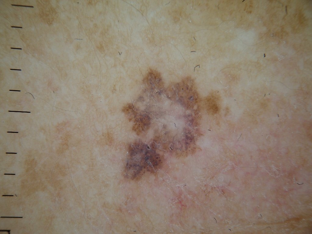
Dermatoscopy significantly enhances diagnostic accuracy by revealing characteristic subsurface features of lentigo maligna melanoma. Common findings include:
- Atypical pigment network: Irregular, broken, or thickened pigment lines.
- Irregular streaks and peripheral globules: Common along lesion margins.
- Blue-white veil: Associated with dermal invasion and regression.
- Hypopigmented zones: Areas that appear lighter or scar-like, indicating regression.
- Peripheral radial projections: Suggest outward tumor spread.
- Multicolor appearance: Three or more distinct pigment shades in one lesion.
- Atypical vascular patterns: Especially in later stages, with linear or dotted blood vessels.
Differential Diagnosis
Several skin lesions can mimic lentigo maligna melanoma, making differential diagnosis critical. Conditions to consider include:
- Congenital dermal melanocytosis
- Dysplastic or atypical nevi
- Blue nevus
- Spitz nevus
- Simple lentigo or solar lentigo
- Pigmented basal cell carcinoma
- Thrombosed hemangioma
Risks and Prognosis
Although melanoma comprises a smaller fraction of skin cancer cases overall, it is responsible for a disproportionately high number of skin cancer-related deaths. Lentigo maligna melanoma, in particular, may go undetected for years due to its slow onset and resemblance to benign pigmented lesions. The longer the diagnosis is delayed, the greater the risk of dermal invasion and metastatic spread.
When detected and treated early — before the vertical growth phase — lentigo maligna melanoma has a comparatively favorable outcome. However, once metastasis occurs, the prognosis becomes significantly poorer. Regular monitoring and early intervention are essential to reduce mortality.
Management Strategy
Suspected lentigo maligna melanoma should be urgently referred for specialist evaluation. A complete excisional biopsy gives the most reliable tissue sample, but it is not always practical: these lesions are often large and sit on the face, where removing the whole thing before a diagnosis exists is hard to justify. In that situation a partial, incisional biopsy of the most suspicious area is an appropriate way to establish the diagnosis, with definitive margin-controlled or surgical treatment following once it is known what the lesion is.
Staging investigations are not performed on every case. Which ones are appropriate — clinical palpation of the nodes, ultrasound, or cross-sectional imaging such as CT, MRI or PET — depends on the stage of the tumour and on the clinical findings.
Ongoing surveillance with digital dermatoscopy and periodic full-body skin exams is strongly recommended for patients with high-risk lesions or a history of melanoma.
Treatment
The cornerstone of treatment is wide surgical excision with histologically confirmed clear margins. In cases where regional lymph nodes are involved, a lymphadenectomy may be performed. For metastatic disease, treatment options are tailored to the individual and may include:
- Immunotherapy: Checkpoint inhibitors like pembrolizumab or nivolumab.
- Targeted therapy: For tumors with mutations such as BRAF.
- Chemotherapy: Used less frequently today but still considered in selected cases.
- Radiation therapy: Primarily for palliation or as part of multimodal treatment.
Important: Superficial destructive methods such as cryotherapy or laser therapy are avoided because they do not guarantee complete tumor clearance and prevent proper histological assessment; full-thickness excisional biopsy is generally preferred for accurate staging.
Prevention
Preventing lentigo maligna melanoma involves long-term vigilance and minimizing UV damage. Recommended strategies include:
- Applying broad-spectrum SPF 30+ sunscreen daily and reapplying every two hours when outdoors.
- Wearing protective clothing, wide-brimmed hats, and UV-blocking sunglasses.
- Avoiding peak sun exposure (10 a.m. to 4 p.m.) and tanning beds.
- Regularly examining the skin for changes in pigmented lesions.
- Scheduling annual dermatological checkups, especially for those with a history of skin cancer.
- Avoiding trauma or irritation to existing moles or pigmented areas.
Early recognition and prompt removal of suspicious lesions are crucial steps in preventing disease progression and improving long-term outcomes.