
White spots and patches on skin: pictures and possible causes
This tool is for informational purposes only and does not constitute medical advice. Always consult a qualified dermatologist.
The pictures and answers on this page are free. SkinAI Lab is a paid AI analysis; pricing is shown before you scan. The check does not cover every cause on this page, so a result cannot rule one out.
What causes white spots on the skin?
White spots or patches are often areas of skin that have lost pigment; others are small raised bumps. Causes range from harmless sun-related spots to a common skin infection that usually needs treatment, vitiligo and, more rarely, conditions that can be serious. Pictures can help you describe what you see, but they cannot tell you which condition you have. First, should you see a doctor?
- See a doctor about a pale or reddish patch that feels numb or where you have lost feeling, an itchy white patch on your genitals or skin, a new or changing spot, or a new or changing mole. All the signs
- Often harmless: white sun spots and milia are harmless, and pityriasis alba usually clears over time. Check the signs first, and see a doctor if you are not sure what the spots are.
See a doctor — don’t rely on a photo check — if you have:
- A pale or reddish skin patch that feels numb or where you have lost feeling
- An itchy white patch on your genitals or skin
- A lump, thickening or an ulcer that does not go away in an area of lichen sclerosus
- A new area of skin that is hard, thick and shiny
- A new or changing spot, or a white spot with painful, bleeding or itchy skin
- A new or changing mole, or a mole that is painful, itchy, inflamed, bleeding or crusty — don’t wait 4 weeks for these
- A newly noticed pale ring around a mole, especially in an adult, or a ringed mole with an asymmetric, multicoloured or changing centre
- If you have psoriasis: joint pain, swelling, or morning stiffness lasting more than about 30 minutes
- If you have lichen sclerosus: treatment that is not helping, or pain when going to the toilet or having sex
- A growth that is getting bigger or has changed colour or texture, or a growth or area of skin that hurts, itches, bleeds, crusts or scabs for more than 4 weeks
White spots aren’t typically dangerous, says Cleveland Clinic, but some rare causes can be, so new or changing spots are worth having checked.
Protect pale patches from the sun. Skin without pigment is more vulnerable to sunburn, so use a sunscreen with a high SPF.
Sources: WHO — Leprosy · NHS — Lichen sclerosus · DermNet — Morphoea · Cleveland Clinic — White spots on your skin · NHS — melanoma symptoms · NHS — skin cancer symptoms · NHS — Vitiligo · Atlas — Psoriasis vulgaris · Atlas — Halo nevus
White spots on brown, black and tanned skin
Pale patches often show more on darker or tanned skin. Our atlas describes vitiligo as most obvious against darker or tanned skin, and white sun spots and pityriasis alba can also be more noticeable on skin of colour. Tinea versicolor patches tend to be paler than the skin around them on brown or black skin; on white skin they are usually pink, red or pale brown. Lichen nitidus bumps can look paler or darker on dark skin. People with white, brown or black skin can get lichen sclerosus. Several of the photos below show darker skin for this reason.
Sources: the atlas articles linked in the cards below · DermNet — Idiopathic guttate hypomelanosis · DermNet — Pityriasis alba · NHS — Pityriasis versicolor · DermNet — Lichen nitidus · NHS — Lichen sclerosus
Possible causes: examples with pictures
Grouped by features described in the atlas articles, not by every way a condition can look. Groups overlap and cannot identify or rule out a condition, so look through both. Each description is quoted word for word from the atlas article; open a card for the full article and all its photos.
Pale or white patches
The tinea versicolor picture is a black-and-white photo from the CDC Public Health Image Library (ID 1688, public domain), added to the atlas gallery after the dermatologist’s review. It cannot show the colour differences described in the quote.
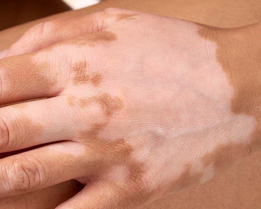
Vitiligo
“Milky-white patches with a sharp border, most obvious against darker or tanned skin.”
Read the article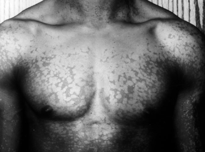 25 photos
Tinea versicolor (pityriasis versicolor)
“The hallmark of pityriasis versicolor is the development of well-demarcated, scaly, discolored patches that vary in hue—from pink, tan, and light brown to hypopigmented or chalky white, especially in darker-skinned individuals.”
Read the article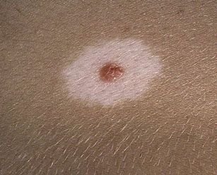 28 photos
Halo nevus (Sutton’s nevus)
“a benign skin neoplasm that typically appears as a raised spot surrounded by a rim of hypopigmented skin”
“A halo can rarely form around a melanoma, and it is the central lesion that needs looking at.”
Read the articleRaised bumps or white scale
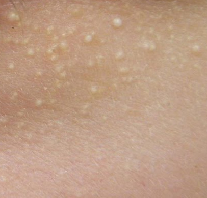
On dark skin, lichen nitidus bumps can look paler than the skin around them (DermNet).
 20 photos
Milia (milium cysts)
“Milium cysts are typically small, white or yellowish papules that measure about 1–2 mm in diameter.”
Read the article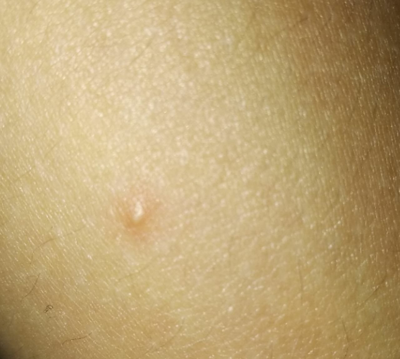 24 photos
Closed comedones (whiteheads)
“Closed comedones typically present as small, round, or oval skin-colored or slightly pale bumps that protrude just slightly above the skin’s surface.”
Read the article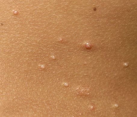 24 photos
Molluscum contagiosum
“Each lesion has a central depression or dimple and may contain a waxy, white core.”
Read the article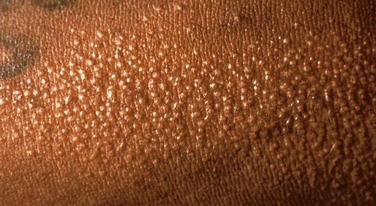 20 photos
Lichen nitidus
“Lichen nitidus is a rare, non-infectious dermatological condition that manifests as numerous tiny, shiny papules on the skin.”
Read the article 29 photos
Psoriasis (psoriasis vulgaris)
“Psoriasis vulgaris typically presents with erythematous plaques covered by thick, white or silvery scales.”
Read the articleOther possible causes
Not in our atlas as separate articles, so described from the sources linked.
- White sun spots (idiopathic guttate hypomelanosis). Small, smooth, flat, pale or white spots, usually 2 to 5 mm across, most often on sun-exposed forearms and shins and the V of the chest. They become more common with age. They are harmless and usually need no treatment, but they do not regain their colour on their own, and sun protection is recommended. DermNet — Idiopathic guttate hypomelanosis · Schweiger Dermatology — White spots on skin
- Pityriasis alba. A mild type of eczema mainly seen in children and teenagers aged 3 to 16. It causes pale patches with fine scale, most often on the cheeks and chin, which show more in summer when the skin around them tans. It clears after about a year on average, from a few months up to two or three years, and the colour gradually returns to normal. DermNet — Pityriasis alba
- Pale marks after inflammation (post-inflammatory hypopigmentation). Our atlas article on vitiligo describes these as “Pale areas left behind by eczema, psoriasis or injury; the border is indistinct and the colour usually returns over months, though recovery can be slow or incomplete.” Skin treatments such as freezing (cryotherapy), chemical peels and laser can also leave pale or white marks. Rao et al., J Clin Med 2023 — Post-inflammatory hypopigmentation · DermNet — Leukoderma
- Lichen sclerosus. Itchy white patches that are smooth or crinkled and easily damaged. They can appear anywhere, but most often around the vulva, the anus, the foreskin and the end of the penis. It can affect people of all ages, including children, and is much more common in women over 50. It increases the risk of cancer on the vulva, penis or anus. The risk is low, but see a doctor about a lump, a thickening or an ulcer that does not go away. NHS — Lichen sclerosus
- Chemical (contact) leukoderma. White patches of skin caused by contact with a chemical. They start where the chemical touched the skin, and small white spots can then appear beyond that area. Colour may come back slowly if the chemical can be avoided. DermNet — Leukoderma
- Morphea. A rare condition in which an area of skin becomes hard, thick, shiny and ivory white, sometimes with a purple border. It often begins in childhood. DermNet — Morphoea · Cleveland Clinic — White spots on your skin
- Discoid lupus. Long-lasting scaly patches, most often on the scalp, face and ears, that can scar and leave paler or darker skin and permanent hair loss. It is more common in skin of colour. DermNet — Discoid lupus
- Leprosy (Hansen disease). A pale or reddish patch with a definite loss of feeling is one of its main signs. It does not spread through casual contact such as shaking hands or sharing meals. It is curable, and early treatment can help prevent disability. WHO provides the medicines free of cost. It still occurs in more than 120 countries, and in 2024 Brazil, India and Indonesia each reported more than 10,000 new cases. WHO — Leprosy
How selected causes differ
A summary of selected features from our atlas articles and the sources on this page. Many conditions overlap, so this table cannot tell you which one you have. Tap a condition to see its picture or description.
| Condition | What it usually looks like | How it feels | Where it often appears |
|---|---|---|---|
| Vitiligo | Milky-white patches with a sharp border | Usually not itchy, scaly or painful; itch can occur | Around the eyes, mouth and nostrils, fingers, wrists, elbows, knees, ankles |
| Tinea versicolor | Flat, round patches that can join up: paler than the skin around them on brown or black skin; pink, red or pale brown on white skin | May feel scaly, sometimes itchy | Chest, upper back, upper arms, neck, tummy |
| White sun spots | Smooth, flat, pale or white spots, usually 2 to 5 mm across | Usually no symptoms | Sun-exposed forearms and shins, V of the chest |
| Pityriasis alba | Pale patches with fine scale, usually with poorly defined edges; mainly children and teenagers | Fine scale; little or no itch | Cheeks and chin; also neck, shoulders, upper arms |
| After inflammation | Pale areas with an indistinct border, after eczema, psoriasis or an injury | Not stated in our sources | Where the skin was inflamed or injured |
| Lichen sclerosus | Itchy white patches, smooth or crinkled, easily damaged | Itchy; may bleed or hurt if rubbed or scratched | Most often around the vulva, anus, foreskin and end of the penis; can appear anywhere |
| Halo nevus | A mole with a pale ring around it | Usually no symptoms; mild itching can occasionally occur | Most often the trunk |
| Milia | White or yellowish dome-shaped bumps, about 1 to 2 mm | Firm, painless | Most often the face: around the eyes, nose, cheeks |
| Closed comedones | Small, round or oval, skin-coloured or slightly pale bumps | Usually painless; firm to the touch | Forehead, temples, cheeks, chin, upper back, neck, chest |
| Molluscum contagiosum | Small, firm, pink or skin-coloured bumps with a central dimple, which may have a waxy white core | Smooth and shiny; may itch | Children: face, trunk, arms, legs. Adults: lower abdomen, groin, genitals, thighs |
| Lichen nitidus | Tiny shiny bumps, 1 to 2 mm, usually skin-coloured; can look paler or darker on dark skin | Usually no symptoms; can itch | Forearms, backs of the hands, penis, chest, abdomen, buttocks |
| Psoriasis | Raised patches with clear edges, covered by thick white or silvery scales | Thick scale; itch in about 60% of people | Elbows, knees, scalp, lower back |
Sources: the atlas articles linked in the cards above · NHS — Pityriasis versicolor · DermNet — Idiopathic guttate hypomelanosis · DermNet — Pityriasis alba · NHS — Lichen sclerosus · DermNet — Lichen nitidus
Can a white spot be skin cancer?
The growths and patches of skin cancer can vary in colour, size and texture, so colour alone cannot tell you. See a doctor about a new or changing mole, or a mole that is painful, itchy, inflamed, bleeding or crusty — don’t wait 4 weeks for these. Also see a doctor about a growth that is getting bigger or has changed colour or texture, or a growth or area of skin that hurts, itches, bleeds, crusts or scabs for more than 4 weeks.
Source: NHS — Non-melanoma skin cancer
 32 photos
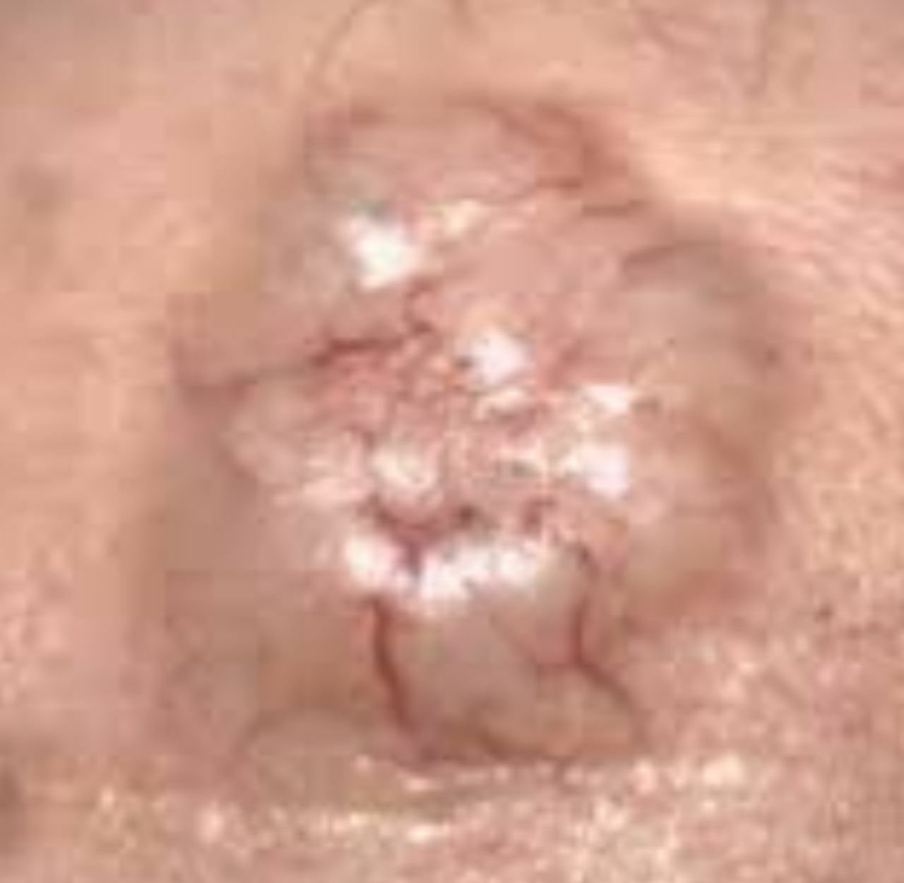
Basal cell carcinoma
“The color can vary depending on the lesion’s stage: early forms are pink to red, while older or necrotic areas may appear white, yellowish, or gray.”
Read the article 32 photos
Melanoma
“C – Color: Uneven pigmentation with multiple shades of brown, black, or other colors like blue, pink, or white.”
Read the article
Medical review of the atlas articles above
Board-Certified Dermatologist · Fellow of the American Academy of Dermatology (FAAD)
Source for the advice on moles above: NHS — melanoma symptoms
Terms used in the quotes
- Hypopigmented
- Lighter in colour than normal skin.
- Papule
- A small, solid, raised bump on the skin that has a border with edges that are easy to see.
- Cyst
- A closed, sac-like pocket of tissue that can form anywhere in the body.
- Lesion
- An area of abnormal or damaged tissue caused by injury, infection, or disease.
- Erythematous
- Red; erythema is redness of the skin.
- Plaque
- In medicine, a small, abnormal patch of tissue on a body part or an organ.
- Neoplasm
- An abnormal mass of tissue. Neoplasms may be benign (not cancer) or malignant (cancer).
- Necrotic
- Relating to necrosis, the death of living tissues.
Source: NCI Dictionary of Cancer Terms.
White spots on skin: FAQ
Why do I have white spots on my skin?
White spots or patches are often areas of skin that are missing pigment. Causes that Cleveland Clinic lists among the most common are white sun spots (idiopathic guttate hypomelanosis), tinea versicolor, vitiligo, pityriasis alba and milia. Pictures and descriptions cannot tell you which one you have, so have new or changing spots checked by a doctor. Source: Cleveland Clinic — White spots on your skin.
What are small white spots on the skin?
That depends on the cause. Flat spots 2 to 5 mm across on sun-exposed arms and legs can be white sun spots. Raised white or yellowish bumps about 1 to 2 mm across can be milia. Other small pale or skin-coloured bumps on this page are closed comedones, molluscum contagiosum and lichen nitidus. See a doctor if you are not sure what the spots are. Sources: DermNet — Idiopathic guttate hypomelanosis, the atlas articles in the cards above.
Are white spots on the skin a sign of cancer?
Cleveland Clinic says white spots aren’t typically dangerous, but some rare causes can be. The atlas describes older areas of basal cell carcinoma that may appear white, and melanomas can include white among several colours. Lichen sclerosus raises the risk of some genital and anal cancers, though the risk is low. Rarely, a blood cancer called cutaneous T-cell lymphoma can cause itchy, discoloured skin, and one uncommon form causes pale patches. Our atlas says vitiligo does not turn into skin cancer, but Cleveland Clinic advises people with pigment problems to watch closely for any changes to their skin and to protect it from the sun. See a doctor about a new or changing mole, or a mole that is painful, itchy, inflamed, bleeding or crusty — don’t wait 4 weeks for these. Also see a doctor about a growth that is getting bigger or has changed colour or texture, or a growth or area of skin that hurts, itches, bleeds, crusts or scabs for more than 4 weeks. Sources: Cleveland Clinic — White spots on your skin, NHS — Lichen sclerosus, Rao et al., J Clin Med 2023 — Post-inflammatory hypopigmentation, NHS — melanoma symptoms, NHS — skin cancer symptoms, Atlas — Basal cell carcinoma, Atlas — Melanoma, Atlas — Vitiligo.
What are white sun spots?
White sun spots is a common name for idiopathic guttate hypomelanosis: small, flat, pale or white spots on sun-exposed skin such as the forearms and shins. Sun exposure over time and ageing are thought to play a part. They are harmless and usually need no treatment, but they do not regain their colour on their own. Sun protection is recommended. If they bother you, a dermatologist can explain the options. Sources: DermNet — Idiopathic guttate hypomelanosis, Schweiger Dermatology — White spots on skin, Cleveland Clinic — White spots on your skin.
How can I tell vitiligo from other white patches?
You cannot be sure without a medical check. Our atlas describes vitiligo as milky-white patches with a sharp border, typically not itchy, scaly or thickened, though itch can occur. It describes tinea versicolor as pale or fawn patches with fine scale, pityriasis alba as faint pale patches with fine scale, usually on the cheeks of children, and pale marks after inflammation as having an indistinct border. A doctor can examine the skin, and may use a special lamp (Wood’s lamp) to see how much pigment has been lost. Sources: atlas article on vitiligo, NHS — Vitiligo.
Can a fungal infection cause white patches on the skin?
Yes. Tinea versicolor (pityriasis versicolor) is a common skin infection caused by a fungus that normally lives on the skin. On brown or black skin the patches tend to be paler than the skin around them; on white skin they are usually pink, red or pale brown and may not tan. It does not spread from person to person. The NHS advises not to diagnose it yourself and to see a GP or pharmacist if you are worried. It can take a few months for the colour to return after treatment, and the patches can come back. Source: NHS — Pityriasis versicolor.
What causes dry or scaly white patches?
Among the causes on this page, pityriasis alba has fine scale and looks drier in winter, tinea versicolor patches may feel scaly, and psoriasis has thick white or silvery scales on raised patches. Other conditions can look similar, so see a doctor if you are not sure what the patches are. Sources: DermNet — Pityriasis alba, NHS — Pityriasis versicolor, the atlas article on psoriasis.
What causes raised white spots on the skin?
Among the causes on this page are milia, closed comedones (whiteheads), molluscum contagiosum and lichen nitidus. Forcing milia out can damage the skin, according to our atlas, and molluscum contagiosum can spread through contact. See a doctor if a growth is getting bigger or has changed colour or texture. Sources: the atlas articles in the cards above, NHS — skin cancer symptoms.
Why does my child have white spots on the skin?
Possible causes on this page include pityriasis alba, which mainly affects children and teenagers, molluscum contagiosum, and milia, which are common in newborns and disappear within a few months. Vitiligo and lichen sclerosus can also affect children. Ask a doctor about white patches on a child if you are not sure what they are, and always about itchy white patches on the genitals. Sources: DermNet — Pityriasis alba, NHS — Vitiligo, NHS — Lichen sclerosus, the atlas articles in the cards above.
How do I get rid of white spots on my skin?
Treatment depends on the cause, so find out what the spots are first. White sun spots usually need no treatment. Pityriasis alba usually clears over time. Tinea versicolor is treated with antifungal medicines, and the colour can take months to return. The white patches of vitiligo are usually permanent, though treatments can reduce how they look. Our atlas advises seeing a clinician when pale patches appear, particularly if they are spreading, because recently lost colour tends to return more readily, though this is not guaranteed. Do not squeeze, pick, cut or try to extract spots or bumps yourself. Sun protection matters, because skin without pigment burns more easily. Sources: DermNet — Idiopathic guttate hypomelanosis, DermNet — Pityriasis alba, NHS — Pityriasis versicolor, NHS — Vitiligo, Atlas — Vitiligo, Atlas — Milia (milium cysts), Atlas — Closed comedones, Atlas — Molluscum contagiosum.
When should I worry about white spots on my skin?
See a doctor about a pale or reddish patch that feels numb or where you have lost feeling, an itchy white patch on your genitals or skin, a new area of skin that is hard, thick and shiny, or a new or changing spot. See a doctor about a new or changing mole, or a mole that is painful, itchy, inflamed, bleeding or crusty — don’t wait 4 weeks for these. Also see a doctor about a growth that is getting bigger or has changed colour or texture, or a growth or area of skin that hurts, itches, bleeds, crusts or scabs for more than 4 weeks. Sources: WHO — Leprosy, NHS — Lichen sclerosus, DermNet — Morphoea, Cleveland Clinic — White spots on your skin, NHS — melanoma symptoms, NHS — skin cancer symptoms.
For a clear photo: daylight, close and in focus, no filters. Upload a photo for an AI skin check. The check does not cover every cause on this page, so a result cannot rule one out. AI output is not a medical diagnosis. If you have any of the signs above, see a doctor instead.
Check my skin ↑SkinAI Lab is a paid AI analysis; pricing is shown before you scan.


Prepared by the SkinAI Lab editorial team · Updated . Apart from the quoted atlas text, this page has not been reviewed by a doctor. It is general information, not medical advice.