
Infective Eczematoid Dermatitis (ICD-10: L30) 🚨
Infective Eczematoid Dermatitis: Inflammation Around a Focus of Infection
Infective eczematoid dermatitis — the condition this page was previously titled microbial eczema — is not a primary form of eczema. It is an eczematous reaction in the skin surrounding a localised, chronically discharging focus of infection, driven by the persistent infection and by irritation from the discharge itself. It is worth saying at the outset that the term is used variably: many English-speaking clinicians would describe the same cases as secondarily infected eczema, infected dermatitis, or simply dermatitis around a wound or ulcer, and it is not a uniformly defined diagnosis. The usual underlying sources are an infected wound, a chronic ulcer, a draining sinus, or chronic discharge from the ear; the dermatitis then develops in the skin around them. What the label points at is a useful clinical observation — that the eczema sits around a microbial focus, and that treating the skin without addressing that focus tends to disappoint.
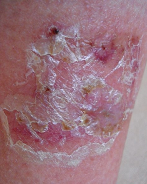
The appearance is suggestive rather than specific. Patches are well demarcated, often asymmetrical, studded with pustules at the edge, and covered with yellow or honey-coloured crust over a moist, weeping base — findings that also occur in impetigo, in infected wounds and in allergic contact dermatitis. They arise where the barrier is already broken and bacteria are established — around a wound or surgical scar, at the edge of a leg ulcer, around a stoma or fistula, behind the ear in chronic otitis externa, at the corner of the mouth, or on a leg affected by venous disease.
Staphylococcus aureus is the organism most often recovered and streptococci the next most common, but skin of this kind is colonised whether or not it is infected, and what grows on a swab varies with the site and the underlying wound. A positive culture does not establish that the organism is causing the eczema. One proposed mechanism is that bacterial products act as superantigens, provoking a disproportionate immune response in the surrounding skin, which would explain why the inflammation extends well beyond the focus — but this remains a model rather than a settled explanation.
The terminology varies by tradition: some clinicians reserve “infective eczematoid dermatitis” for this reaction pattern, others use it for any secondarily infected eczema, and it appears more in some European literature than in English-language dermatology. The practical point survives the disagreement: where a bacterial focus sits at the centre of the eczema, treating the skin alone is unlikely to resolve it.
Predisposing Factors: Why Infective Eczematoid Dermatitis Develops
- A pre-existing break in the skin: A wound, ulcer, burn, fissure, scratch or surgical site provides both the entry point and the reservoir.
- Chronic discharge: Wound exudate, ear discharge, fistula output or saliva at the corners of the mouth keeps the surrounding skin macerated and colonised.
- Venous insufficiency: Lower-leg skin that is already congested and inflamed is one of the commonest sites.
- Existing eczema: Atopic skin carries higher rates of S. aureus colonisation, which makes this reaction more likely.
- Occlusion and moisture: Dressings, plaster casts, adhesive tapes and skin folds all create conditions that favour bacterial growth.
- Impaired immunity or diabetes, which raise the likelihood that colonisation becomes established infection.
- Poorly fitting appliances, such as stoma bags or hearing aids, which combine friction, occlusion and moisture.
Diagnostics: How Infective Eczematoid Dermatitis Is Diagnosed
The diagnosis is clinical, and it rests on recognising the relationship between the eczema and a nearby focus. The most useful question is not what the patch looks like but what it is next to.
A bacterial swab guides antibiotic choice where treatment is needed, but it does not by itself make the diagnosis: skin of this kind is colonised whether or not it is infected, and a positive culture is not automatically a reason to prescribe. Direct microscopy excludes a fungal infection when the picture is atypical. Patch testing deserves consideration in recurrent cases, particularly around ulcers and ears, where contact allergy to dressings, antiseptics or topical antibiotics is common and easily mistaken for persistent infection.
Biopsy is reserved for a persistent solitary plaque that does not respond, where another diagnosis has to be excluded.
Symptoms: What Infective Eczematoid Dermatitis Looks Like
- Sharply bordered plaques that stop abruptly rather than fading into normal skin.
- Yellow or honey-coloured crust over a moist surface.
- Pustules, which may be concentrated at the advancing edge of the patch. This is suggestive rather than diagnostic.
- Weeping and oozing, with dressings or clothing sticking to the surface.
- Itch combined with soreness, rather than itch alone.
- Proximity to a focus: A wound, ulcer, ear canal, stoma, fistula or fissure sitting at or near the centre of the affected area.
- Satellite patches: Smaller eczematous areas sometimes appearing at a distance, including on the opposite limb, understood as a reaction to the primary focus.
Differential Diagnosis: Ruling Out Other Conditions
- Allergic contact dermatitis: Extremely common in exactly these settings, because these sites receive repeated topical products. It may replace the diagnosis or, more often, sit on top of it.
- Impetigo: Golden crusting that spreads rapidly, typically in children, without the underlying eczematous plaque.
- Cellulitis: Spreading warmth, pain, tenderness and systemic upset. It needs prompt medical treatment, and rapidly spreading redness, pain out of proportion, feeling unwell or any suspicion of deeper infection should be assessed urgently.
- Nummular eczema: Coin-shaped and usually symmetrical, without a focus of infection at its centre.
- Tinea: An active scaly edge with central clearing; microscopy distinguishes them.
- Cutaneous lymphoma: Considered for a solitary plaque that persists without explanation.
Risks: Why Infective Eczematoid Dermatitis Should Be Treated
Left untreated, the process tends to extend rather than settle, and the broken skin can progress to cellulitis, which is a more serious problem requiring systemic treatment. Where the focus is a leg ulcer, the surrounding eczema slows healing and enlarges the wound.
There is also a risk created by treatment itself. Skin of this kind receives antiseptics, antibiotics and dressings repeatedly, and it sensitises readily: contact allergy to those products is a frequent complication and one of the main reasons a case appears resistant. Each new topical product applied without review adds to that risk.
Finally, repeated courses of antibiotics prescribed for colonisation rather than genuine infection contribute to resistance without helping the skin.
Tactics: When and How to Seek Treatment
See a clinician if eczema develops around a wound, ulcer, discharging ear, stoma or fistula, if a patch is crusted, weeping or studded with pustules, or if a treated area is not improving. Seek care urgently if redness spreads, the area becomes hot and painful, or there is fever or feeling unwell — that suggests cellulitis rather than eczema.
The judgement that matters most here belongs to a clinician: whether the bacteria present are driving the inflammation or merely living there. That distinction determines whether antibiotics help, and it is not something appearance alone reliably settles.
Treatment: Modern Strategies for Infective Eczematoid Dermatitis
- Treat the underlying focus: The step that most often changes the outcome. Manage the ulcer, the ear discharge, the fissure or the appliance problem. The surrounding dermatitis frequently settles with it, though it may persist where venous disease, contact allergy or another cause is also at work.
- Antimicrobial treatment where the skin is clinically infected: The decision rests on clinical assessment rather than on how large the patch is or on a swab result alone. Routine topical antibiotics are avoided because of resistance and the risk of contact allergy; systemic antibiotics are reserved for clinically significant infection, following local guidance.
- Topical corticosteroids: A short course alongside antimicrobial treatment settles the surrounding eczematous inflammation. They are an adjunct, not a substitute for treating the infection. Suppressing the inflammation without addressing the focus gives short-lived improvement.
- Gentle cleansing to remove crust so that topical treatment reaches the skin. Antiseptics are used selectively rather than routinely; repeated use irritates the skin and is itself a cause of contact allergy.
- Emollients, using simple preparations with few potential allergens.
- Appropriate dressings that manage exudate without occluding an infected surface.
- Reviewing every product in use when a case is not improving, and patch testing where contact allergy is suspected.
Prevention: Reducing Recurrence
- Care for wounds and ulcers properly, with appropriate dressings and review rather than improvised treatment.
- Manage discharge at source, and follow the care plan for that specific site — some wounds, stomas and ear conditions need a balance of moisture rather than simply keeping the area dry.
- Check the fit of appliances, hearing aids and dressings, since friction and leakage are recurring causes.
- Maintain the skin barrier around known problem sites with regular emollient.
- Limit the number of topical products applied to chronically inflamed skin, and avoid topical antibiotics for long-term use.
- Treat venous disease where the leg is the affected site.
- Do not pick crusts, which reopens the surface and spreads bacteria.
Infective eczematoid dermatitis is one of the more satisfying forms of eczema to treat when a focus can be found and dealt with. Often it can: a wound, a discharge, an appliance that does not fit. How much of the inflammation is down to infection, how much to colonisation, barrier damage, venous disease or contact allergy is frequently harder to separate — which is the argument for reviewing the whole picture rather than continuing to treat the skin on its own.
Image: public domain, via Wikimedia Commons.

Medical review
Board-Certified Dermatologist · Fellow of the American Academy of Dermatology (FAAD)