
Lichen Planus (ICD-10: L43) ⚠️
Lichen Planus: Chronic Inflammatory Dermatosis of Unknown Etiology
Overview
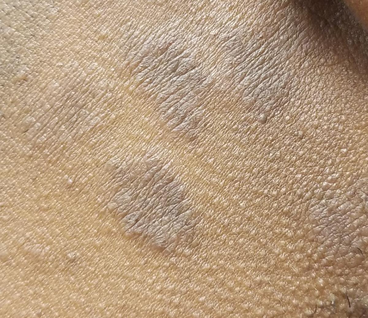
Lichen planus (LP) is a chronic inflammatory skin and mucous membrane condition characterized by itchy, polygonal, flat-topped papules, typically violaceous or reddish-purple in color. It is a non-contagious disease of unclear origin that may affect the skin, nails, scalp, oral cavity, and genitals. LP can occur at any age, but is most commonly seen in adults between 30 and 60 years old.
Despite its benign nature, LP can significantly impair quality of life due to persistent pruritus, mucosal involvement, or scarring alopecia in severe cases. In most patients, the disease resolves spontaneously within months to a few years, though relapses are possible.
Etiology and Possible Triggers
The exact cause of lichen planus is still unknown. However, it is believed to be a cell-mediated autoimmune reaction in which cytotoxic T-cells attack basal keratinocytes. The following factors have been associated with triggering or exacerbating the condition:
- Hepatitis C virus (HCV): An epidemiological association, strongest for oral and erosive disease, and one whose strength differs markedly between regions — it is well documented in some populations and barely detectable in others;
- Autoimmune diseases: LP may coexist with other autoimmune conditions (e.g., vitiligo, alopecia areata, thyroiditis);
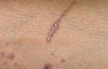
- Local trauma: New lesions may appear along a scratch or scar (the Koebner phenomenon).
A separate condition, lichenoid drug eruption, looks similar and must be distinguished from idiopathic lichen planus. It has been reported with a number of medicines — among them antihypertensives, antimalarials and NSAIDs — though for most of these the association rests on case reports rather than on established causation. What points to it is the timing: an eruption that began after a drug was started, and settles after it is stopped. A medication review is therefore part of assessing new lichen planus, but the drug list should not be read as a list of causes.
Cutaneous Lichen Planus: Clinical Manifestations
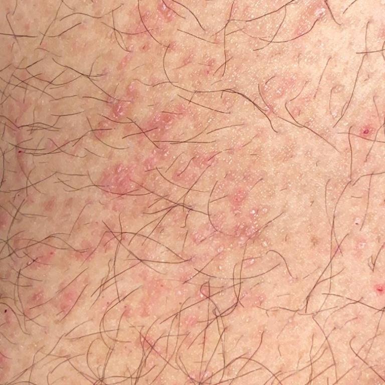
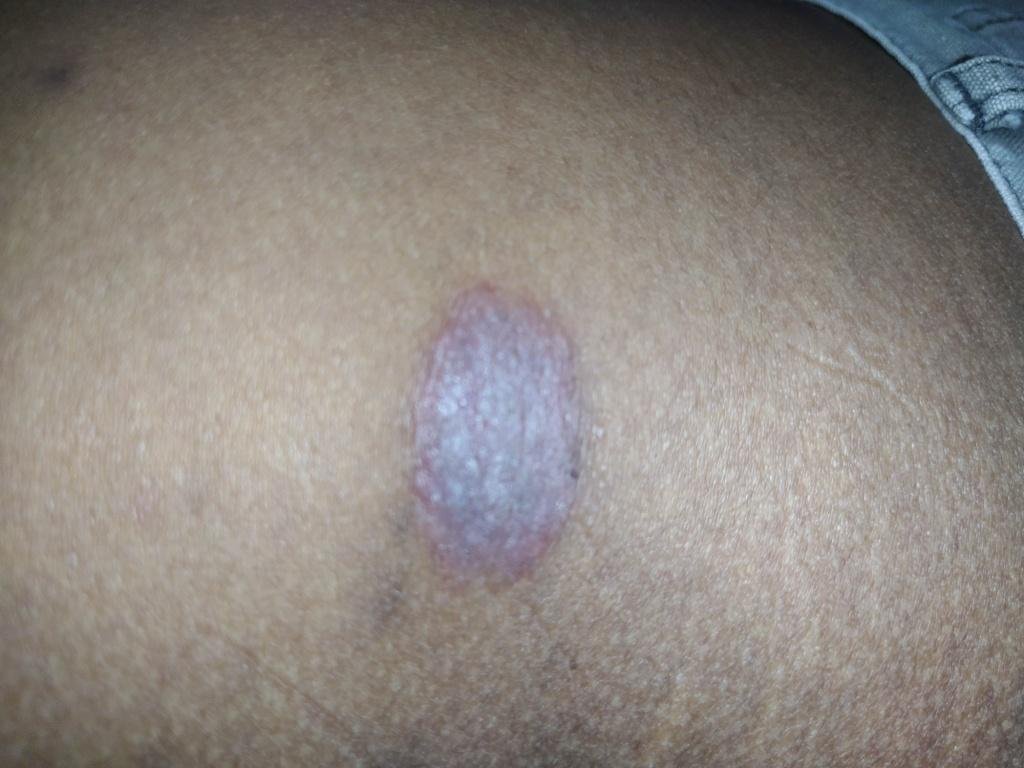
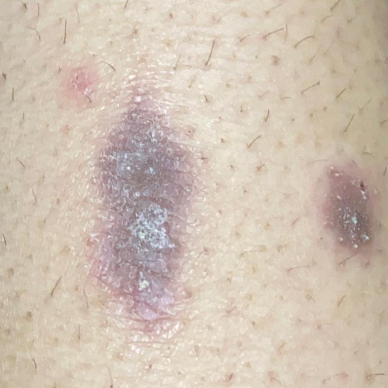
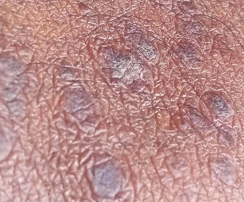
The cutaneous form of lichen planus typically presents as:
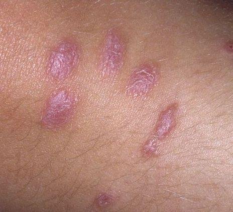
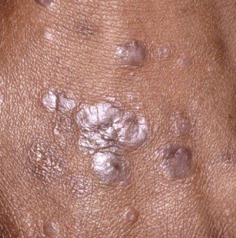
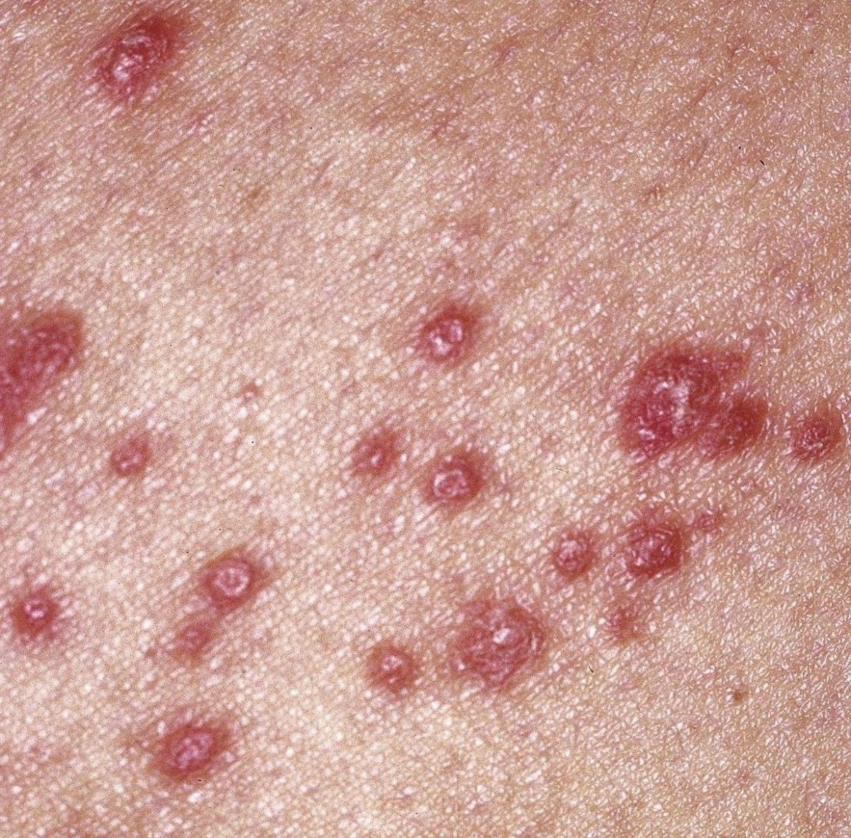
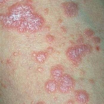
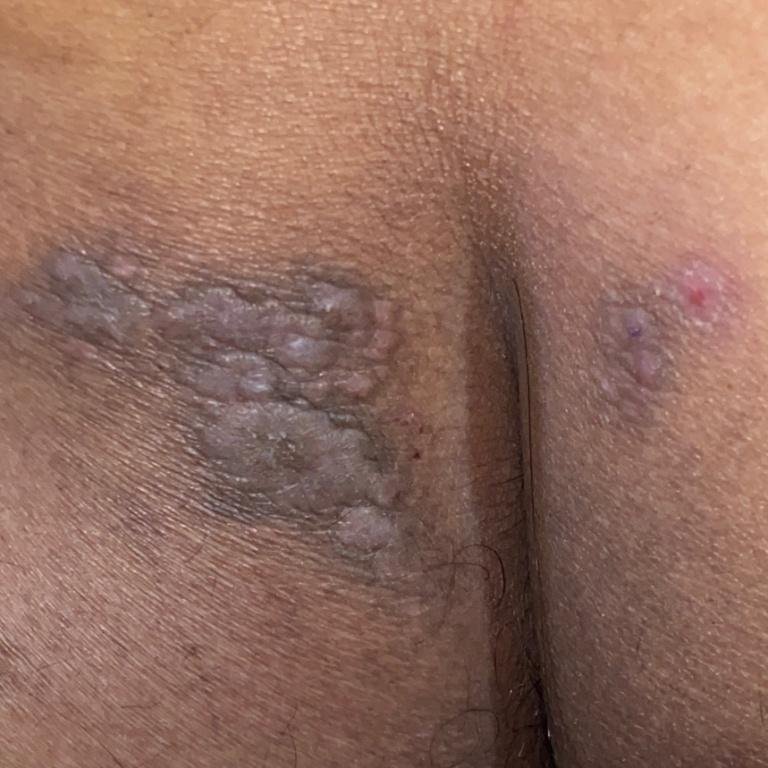
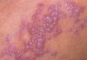
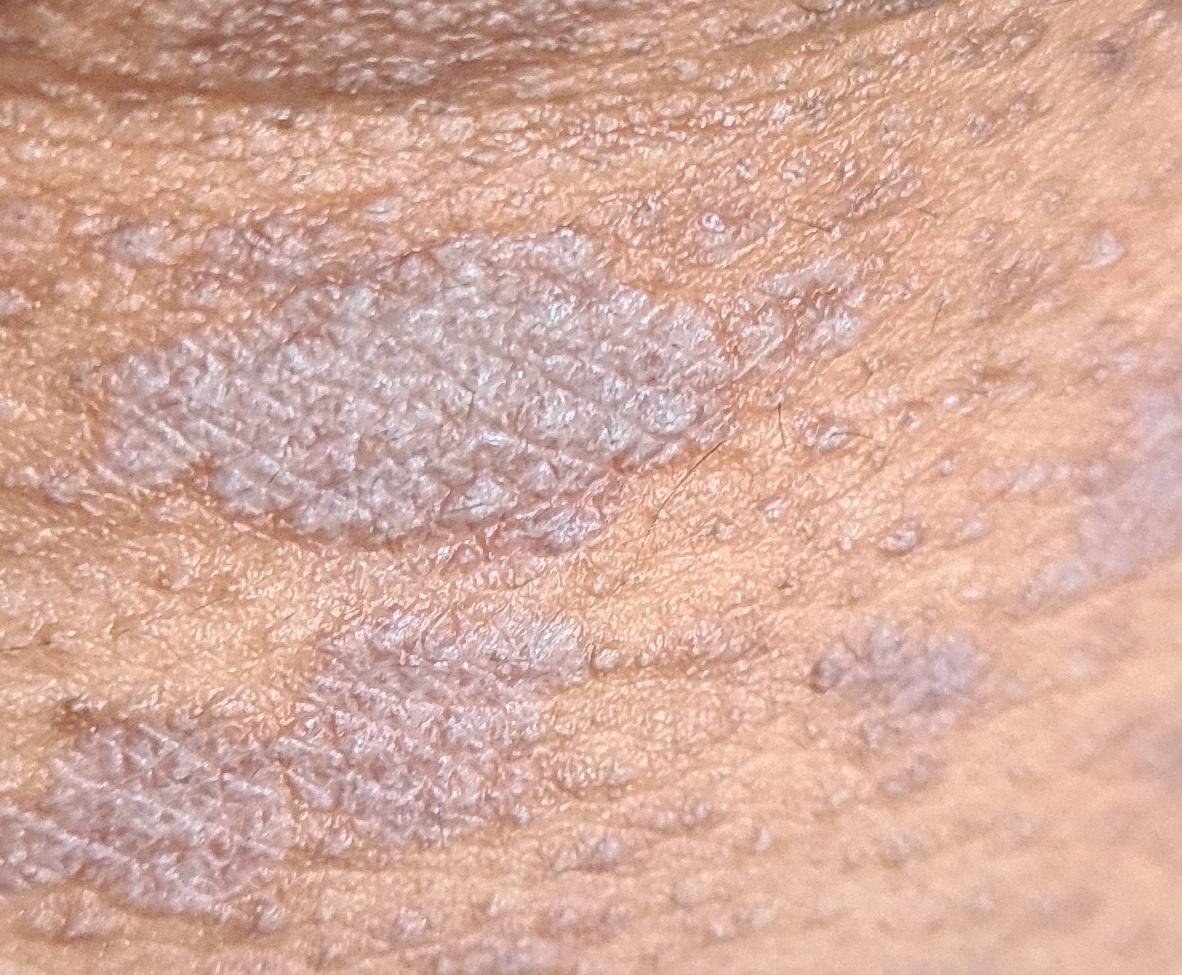
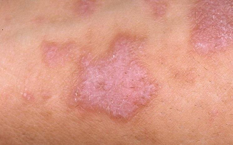
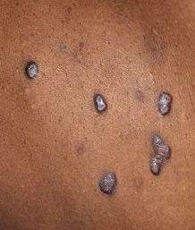
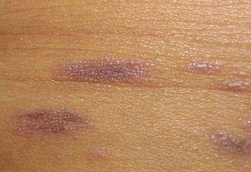
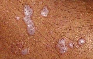
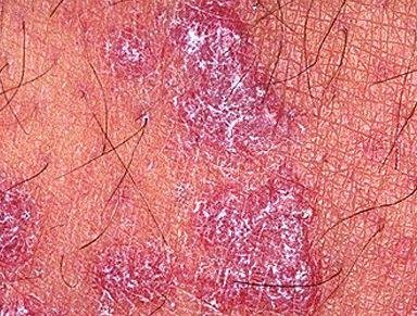
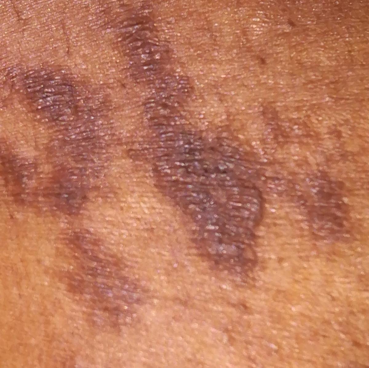
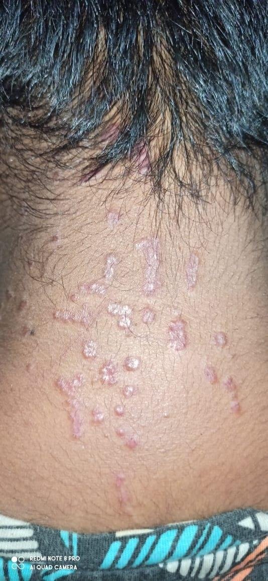
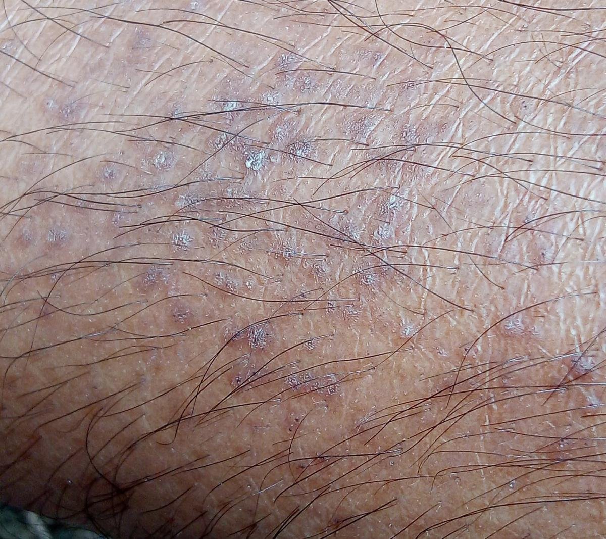
- Multiple polygonal, flat-topped papules 3–5 mm in size, often grouped symmetrically;
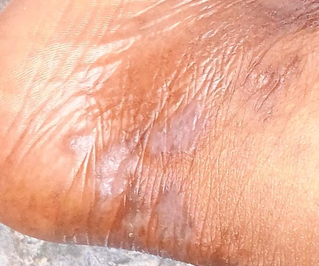
- Color: Pink, red, or violaceous; often shiny due to a smooth surface;
- Wickham striae: Fine, whitish reticulated lines visible on the surface of the papules;
- Itching: Varies from mild to intense, often worsened by heat or stress;
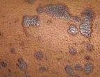
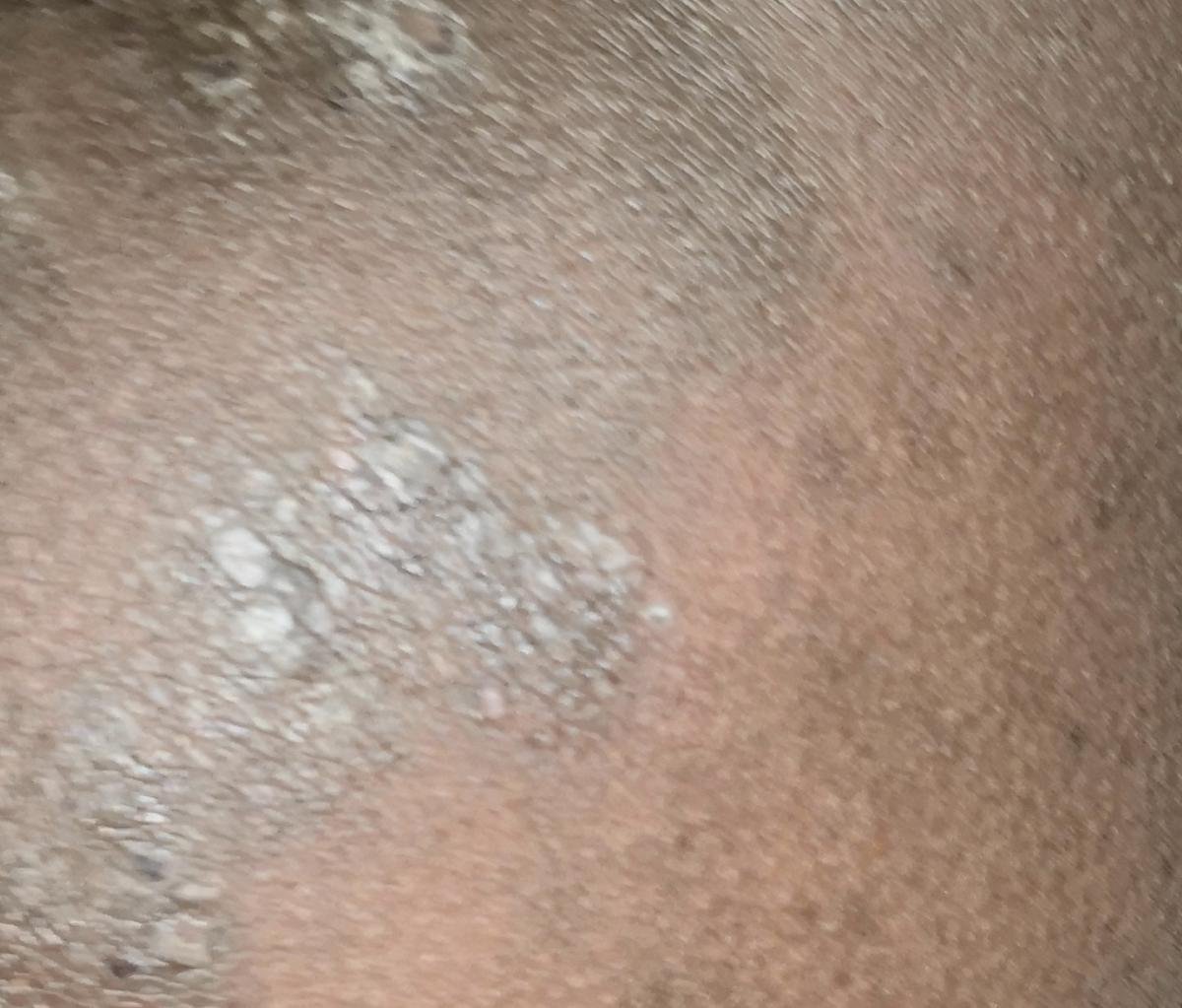
- Distribution: Common sites include wrists, forearms, lumbar area, ankles, and shins. Lesions may coalesce into larger plaques or form linear patterns due to the Koebner phenomenon (lesions developing at trauma sites);
- Thickened plaques: Seen over shins or ankles as hypertrophic LP variants.
In some patients, the initial eruption may be misdiagnosed due to its resemblance to eczema, psoriasis, or fungal infections. A thorough clinical examination is essential for proper identification.
Mucosal, Nail, Scalp, and Genital Involvement
Oral Lichen Planus
Oral lichen planus (OLP) often appears as reticular white patches with a lace-like pattern, known as Wickham striae. Lesions are typically found on the buccal mucosa, tongue, or gingiva. In erosive forms, ulcers, redness, burning, and pain may occur, especially when eating spicy or acidic foods. OLP may persist for years and carries a small but recognised risk of malignant transformation, highest in erosive and atrophic disease. This warrants regular clinical follow-up, and biopsy of any ulcer that persists, any area that becomes firm or indurated, and any new or changing suspicious patch. The same risk should not be read across to lichen planus on the skin, where it does not apply.
Nail Lichen Planus
LP may affect one or more nail plates and present as:
- Thinning and longitudinal ridging;
- Splitting and onychorrhexis;
- Pterygium formation: Adhesion of the nail fold to the nail bed leading to scarring;
- Total nail loss: In advanced, untreated cases.
Scalp (Lichen Planopilaris)
Scalp involvement can lead to follicular papules, redness, and scaling. In progressive disease, scarring alopecia may develop, resulting in permanent hair loss. Early treatment is crucial to preserve hair follicles.
Genital Lichen Planus
LP in the genital area typically manifests as red, erosive or atrophic plaques or papules that may cause burning or itching. It affects both sexes and can be mistaken for infections or other dermatologic conditions.
Diagnostics
A diagnosis of lichen planus is often based on clinical evaluation and characteristic morphology. However, in atypical cases or when mucosal or nail involvement is predominant, additional tests may be required:
- KOH test: To exclude fungal infection;
- Biopsy: Confirms diagnosis. Histopathology typically shows hypergranulosis, saw-tooth acanthosis, basal layer degeneration, and band-like lymphocytic infiltrate at the dermoepidermal junction;
- Hepatitis C testing: Considered according to the clinical history, the patient’s risk factors and local guidance, and more readily in oral or erosive disease. It is not a test that every patient with lichen planus needs;
- Allergy or drug history: In suspected lichenoid drug eruptions.
Differential Diagnosis
Conditions that may resemble LP include:
- Psoriasis: Thicker plaques, silvery scale, and typical nail pitting;
- Pityriasis rosea: Herald patch, “Christmas tree” pattern, and self-limiting course;
- Eczema or atopic dermatitis: More exudative with vesicles and intense pruritus;
- Cutaneous lupus erythematosus: Photosensitive with atrophic scarring and positive ANA;
- Drug-induced lichenoid reactions;
- Tinea corporis or versicolor: Confirmed by KOH microscopy.
Treatment
There is no universal cure for lichen planus, but most cases are self-limiting and resolve within months to a few years. Treatment is aimed at alleviating symptoms and preventing complications, especially in mucosal, scalp, and nail involvement.
Symptomatic and Pharmacologic Therapy:
- Topical corticosteroids: First-line therapy for skin and mucosal LP;
- Topical calcineurin inhibitors: Tacrolimus or pimecrolimus for sensitive areas (e.g., face, genitals);
- Oral antihistamines: For pruritus relief;
- Phototherapy: Narrowband UVB for widespread cutaneous LP;
- Systemic therapy: Reserved for disease that is severe, widespread, scarring or refractory to topical treatment — not an alternative to it. Oral corticosteroids are used for short courses to settle an acute flare; acitretin, methotrexate and ciclosporin are options for longer-term control; biologics have been used in refractory disease, where the evidence is limited and licensing varies by country. Each carries its own monitoring requirements.
Oral and Genital Lesions:
Mucosal disease is managed differently from skin disease, and in formulations made for the site rather than creams intended for the skin.
- Oral disease: Topical corticosteroids as a mouthwash, gel or paste that will stay in contact with the mucosa, or a calcineurin inhibitor where steroids are not enough. Good oral hygiene and dental care matter, and candidiasis is a common complication of topical steroid use in the mouth;
- Genital disease: Needs assessment in its own right, since erosive genital disease can scar and cause adhesions if it is left. It is usually managed jointly with a gynaecologist or urologist, and treatment aims to prevent scarring as much as to relieve symptoms;
- Avoidance of irritants such as spicy foods, alcohol and tobacco.
Prevention and Patient Guidance
- Avoid mechanical trauma, scratching, or friction in affected areas;
- Maintain hydration and use moisturizers to support skin barrier function;
- Identify and minimize exposure to potential triggers (e.g., medications, allergens);
- Eliminate stress where possible through behavioral or psychological support;
- Patients with oral LP should avoid smoking, alcohol, and spicy foods;
- Regular monitoring is important for mucosal LP due to a small risk of malignancy (especially in erosive oral LP).
Conclusion
Lichen planus is a multifaceted chronic inflammatory condition that may affect the skin, mucous membranes, nails, and scalp. While it is not life-threatening, its symptoms and cosmetic implications can cause significant distress. Early diagnosis, symptom management, and tailored therapy allow for improved patient outcomes and quality of life.
Most cases resolve over time, but some forms—especially mucosal or scalp variants—require long-term follow-up and specialist care.
Copyright concern about an image on this page? Write to mail@skinai.pro. Images may not be reused without permission unless an open licence is stated on this page.

Medical review
Board-Certified Dermatologist · Fellow of the American Academy of Dermatology (FAAD)