Atopic Dermatitis (ICD-10: L20) 🚨
Atopic Dermatitis (Atopic Eczema): Chronic Inflammatory Skin Disease
Overview
Atopic dermatitis (AD), also called atopic eczema, is the specific chronic, relapsing inflammatory disease coded ICD-10 L20—one form of “eczema”, which is a broader umbrella term. It is characterized by severe itching, skin dryness, and eczematous lesions, typically begins in early childhood, and is associated with a family or personal history of other atopic conditions such as allergic rhinitis or bronchial asthma. AD is not simply an allergic disease: it is multifactorial, arising from skin-barrier dysfunction, immune dysregulation, genetic susceptibility, and environmental factors.
In approximately 60% of cases, atopic dermatitis begins within the first year of life, most often by the age of 3 months. The disease affects males slightly more often in infancy, while females predominate in adolescence. Asthma and allergic rhinitis are frequent associated comorbidities—the “atopic triad”—but many patients never develop them; up to 70% of patients have a family history of atopic disease. Although it can persist into adulthood, onset in adulthood is rare.
Triggering Factors
Atopic dermatitis flares are often triggered by a combination of environmental, immunological, and lifestyle factors. Common exacerbating factors include:
- Allergens: Inhaled (dust mites, pollen) or contact allergens (nickel, fragrances) aggravate some patients; food is relevant in a minority;
- Dry skin: Due to over-washing, use of harsh soaps, or low humidity;
- Hormonal shifts: Puberty, menstruation, pregnancy, thyroid dysfunction;
- Emotional stress: Anxiety, fatigue, or psychological overload may initiate or aggravate symptoms;
- Infections: Secondary bacterial (e.g., Staphylococcus aureus), viral (herpes simplex), or fungal infections;
- Heat and sweating: Overheating, hot baths, or sweat trapped against the skin;
- Clothing irritants: Wool, synthetic fabrics, feather pillows, harsh detergents;
- Climatic conditions: In temperate climates, symptoms often worsen in winter and improve in summer.
Pathogenesis
Atopic dermatitis involves a complex interplay between genetic predisposition, immune dysregulation, and environmental exposure. Key mechanisms include:
- IgE-mediated hypersensitivity: Although the precise role is unclear, elevated IgE levels and sensitization to allergens are often present. Langerhans cells and mast cells play important roles in initiating inflammatory responses via IgE binding;
- Skin barrier dysfunction: Due to filaggrin gene mutations and lipid deficiencies, leading to increased transepidermal water loss and allergen penetration;
- Chronic inflammation: Sustained immune activation with Th2 cytokine dominance leads to persistent skin inflammation and pruritus;
- Neuroimmune pathways: Itching and scratching perpetuate the inflammatory cycle through neuronal and immune signaling pathways.
Clinical Presentation
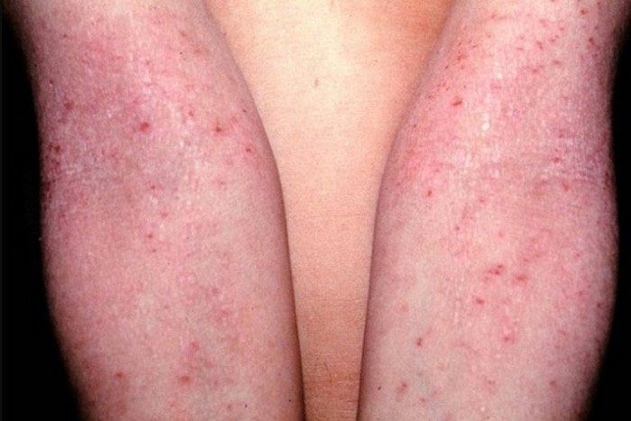
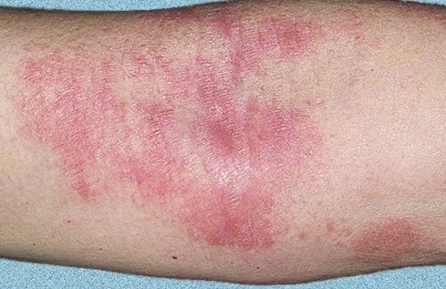
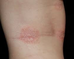
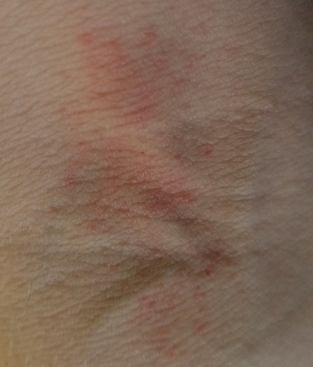
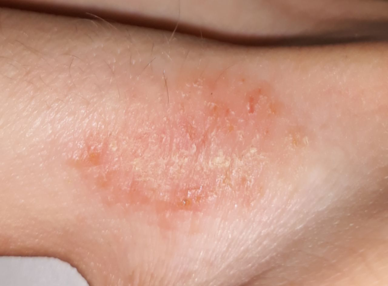
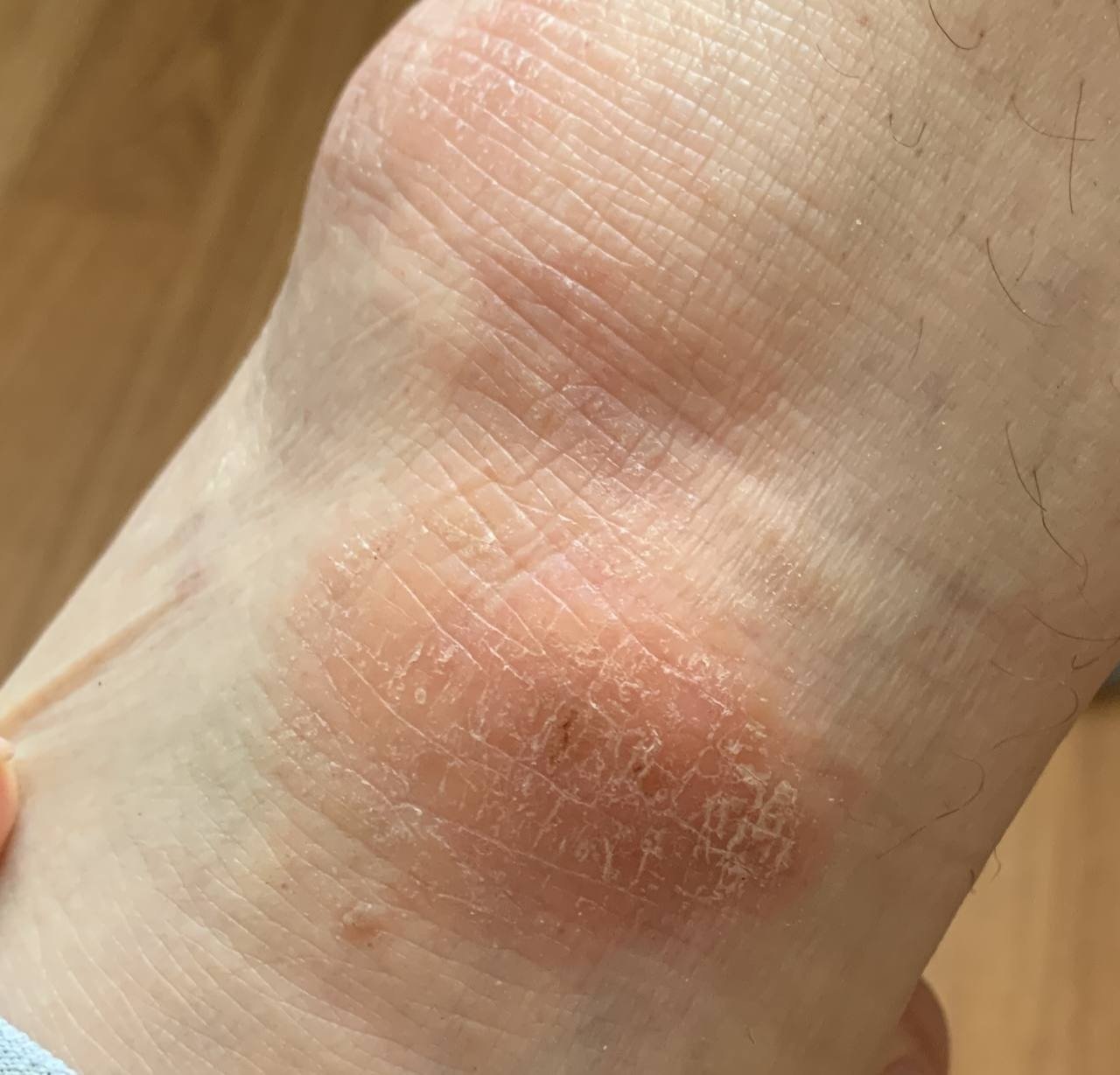
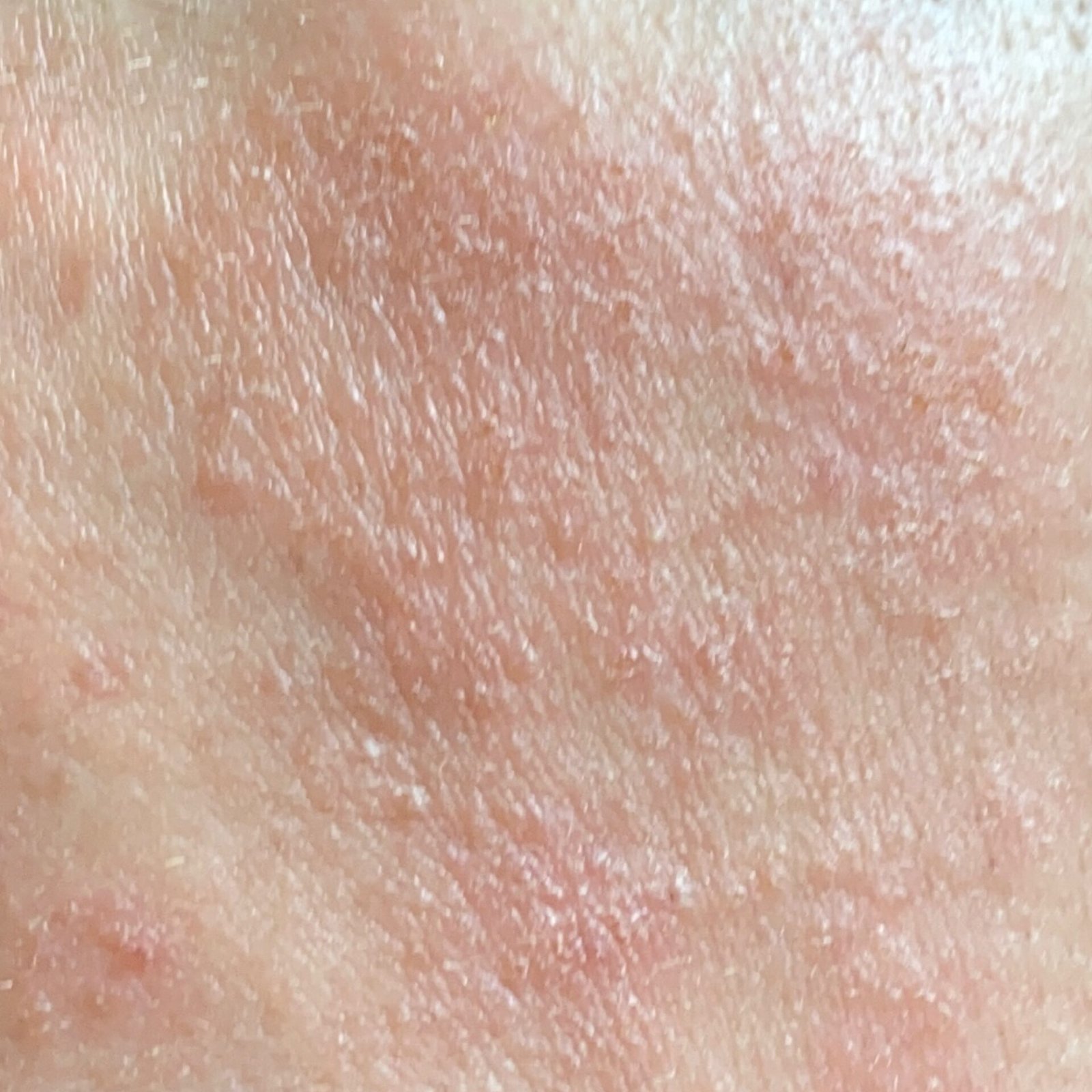
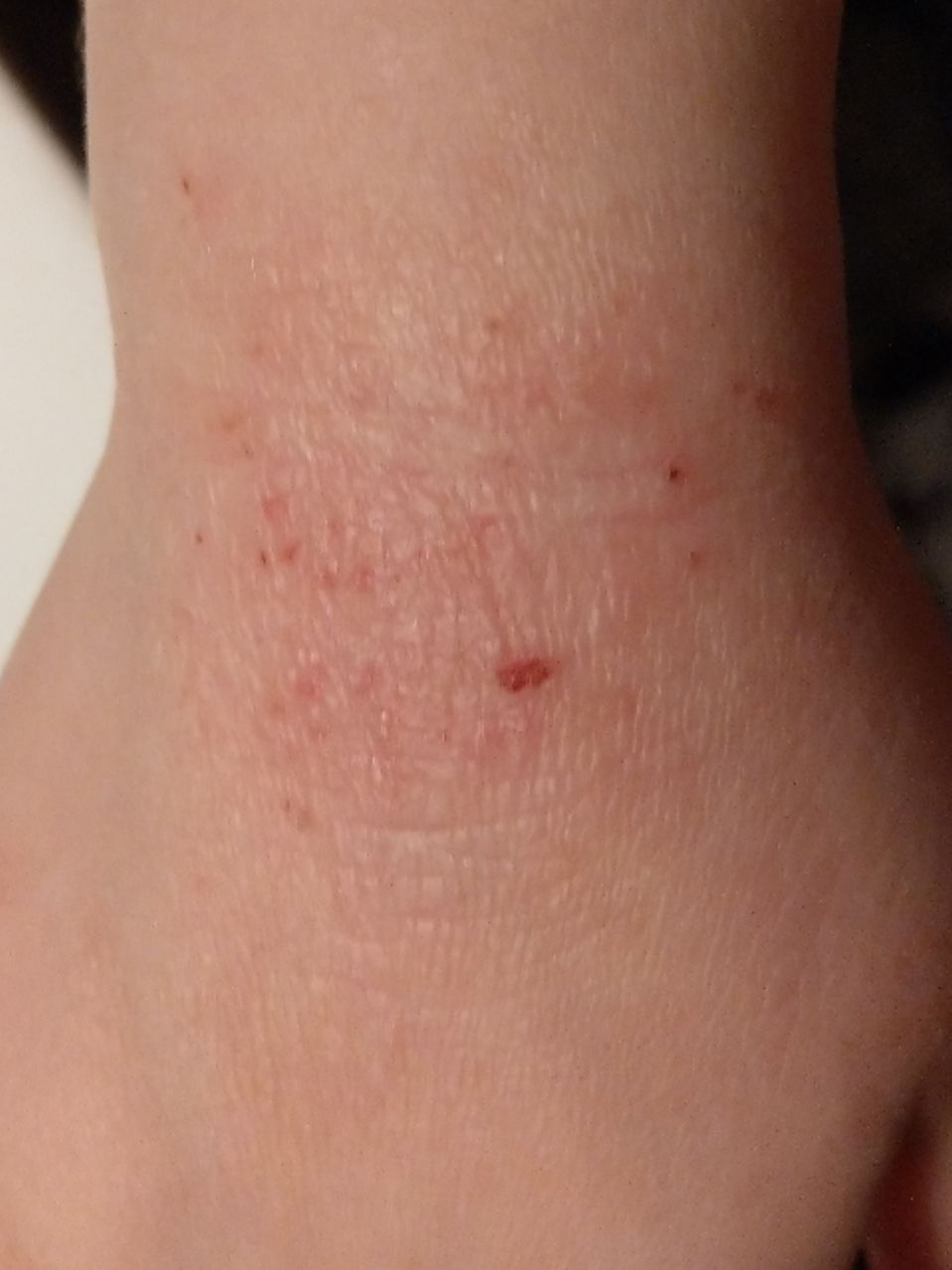
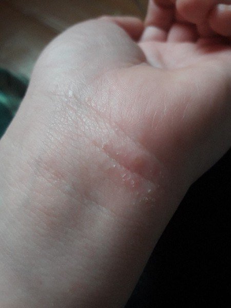
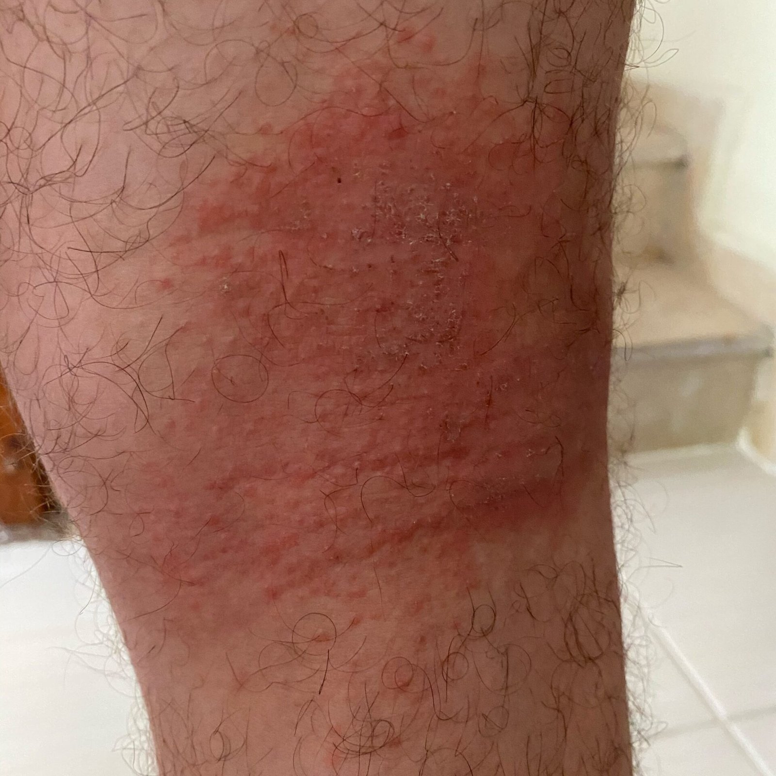
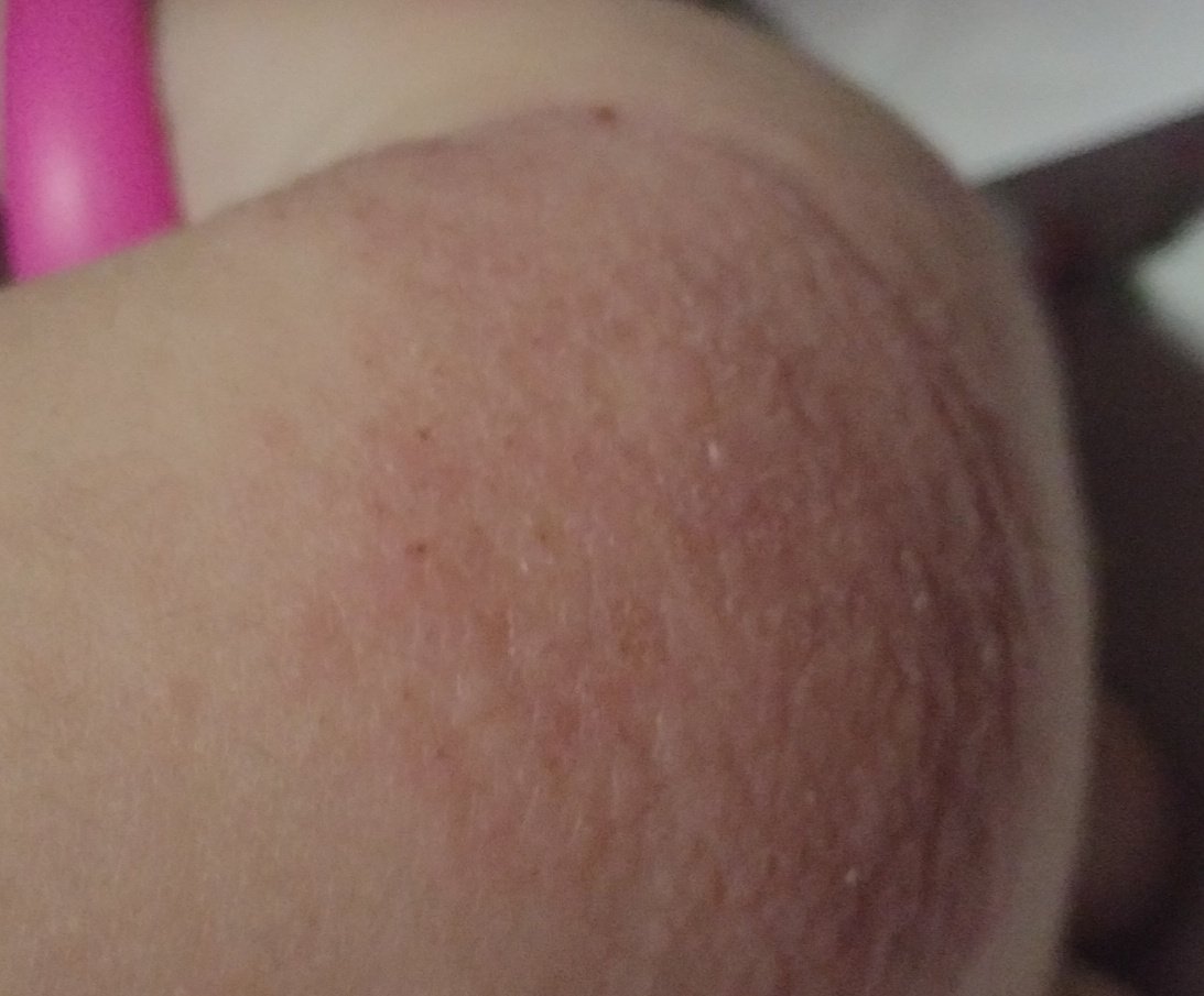
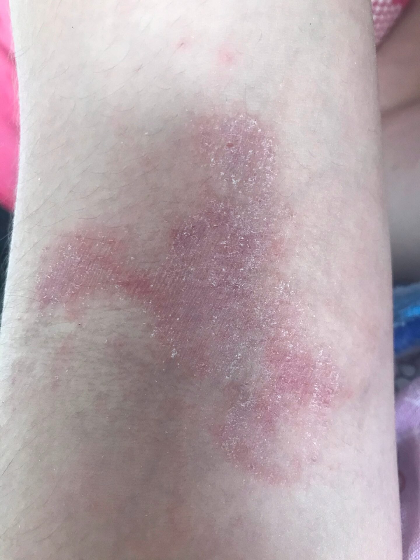
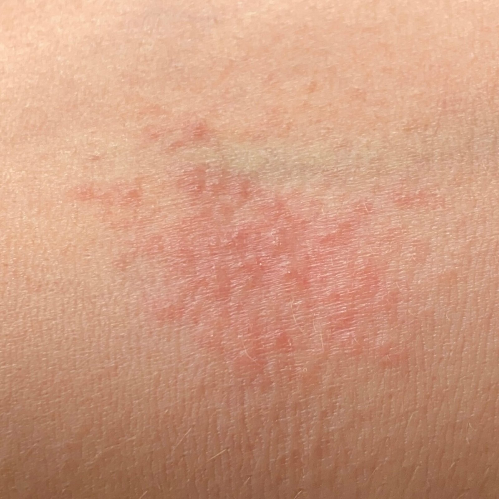
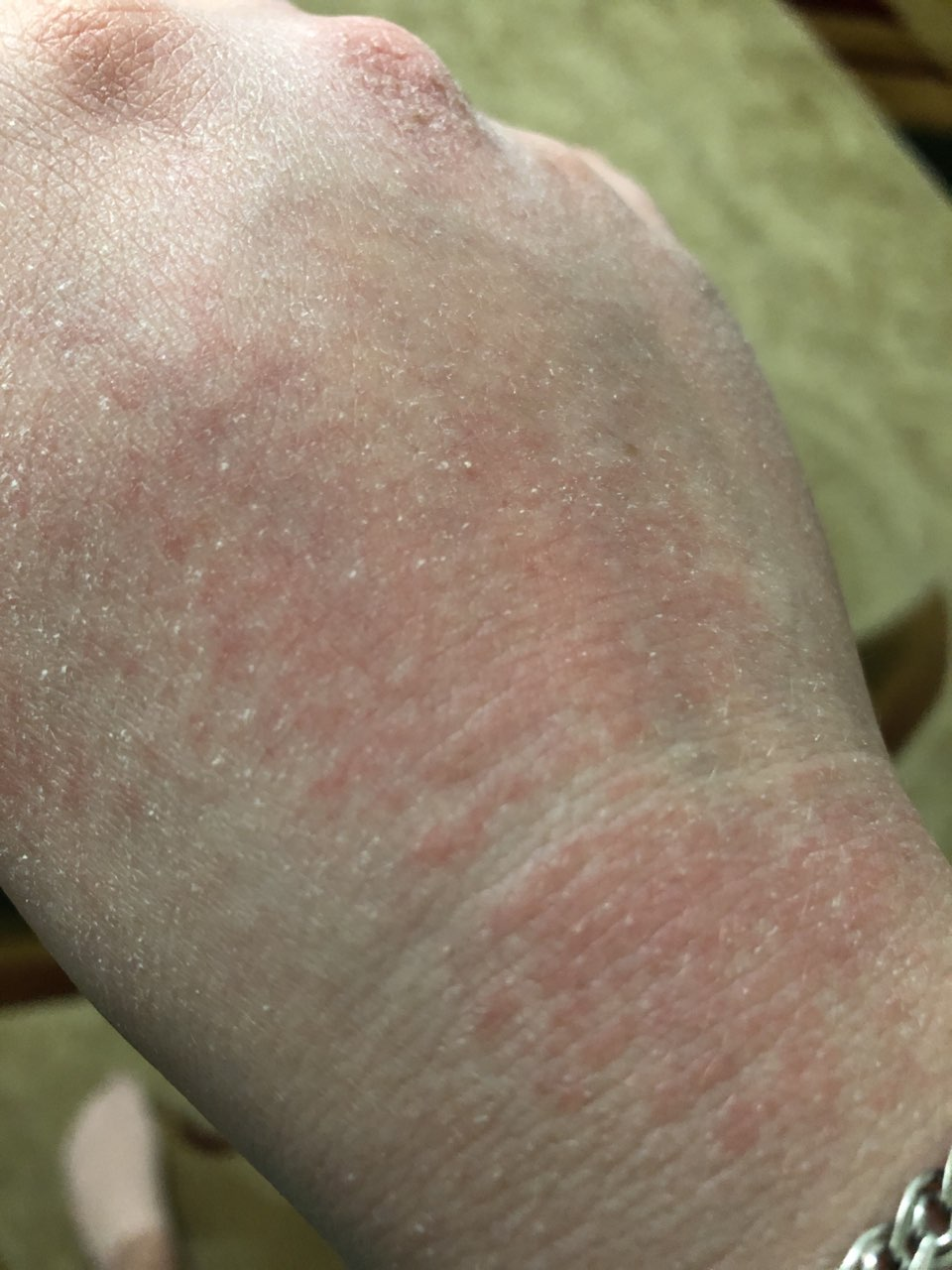
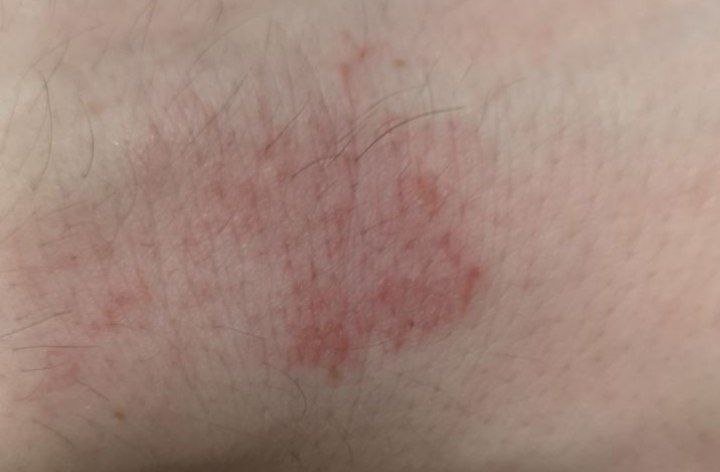
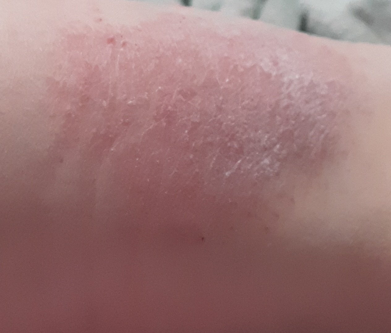
AD is marked by intense itching, xerosis (dry skin), eczematous eruptions, and lichenification. The disease evolves through acute, subacute, and chronic stages, with different morphological features.
Acute Stage:
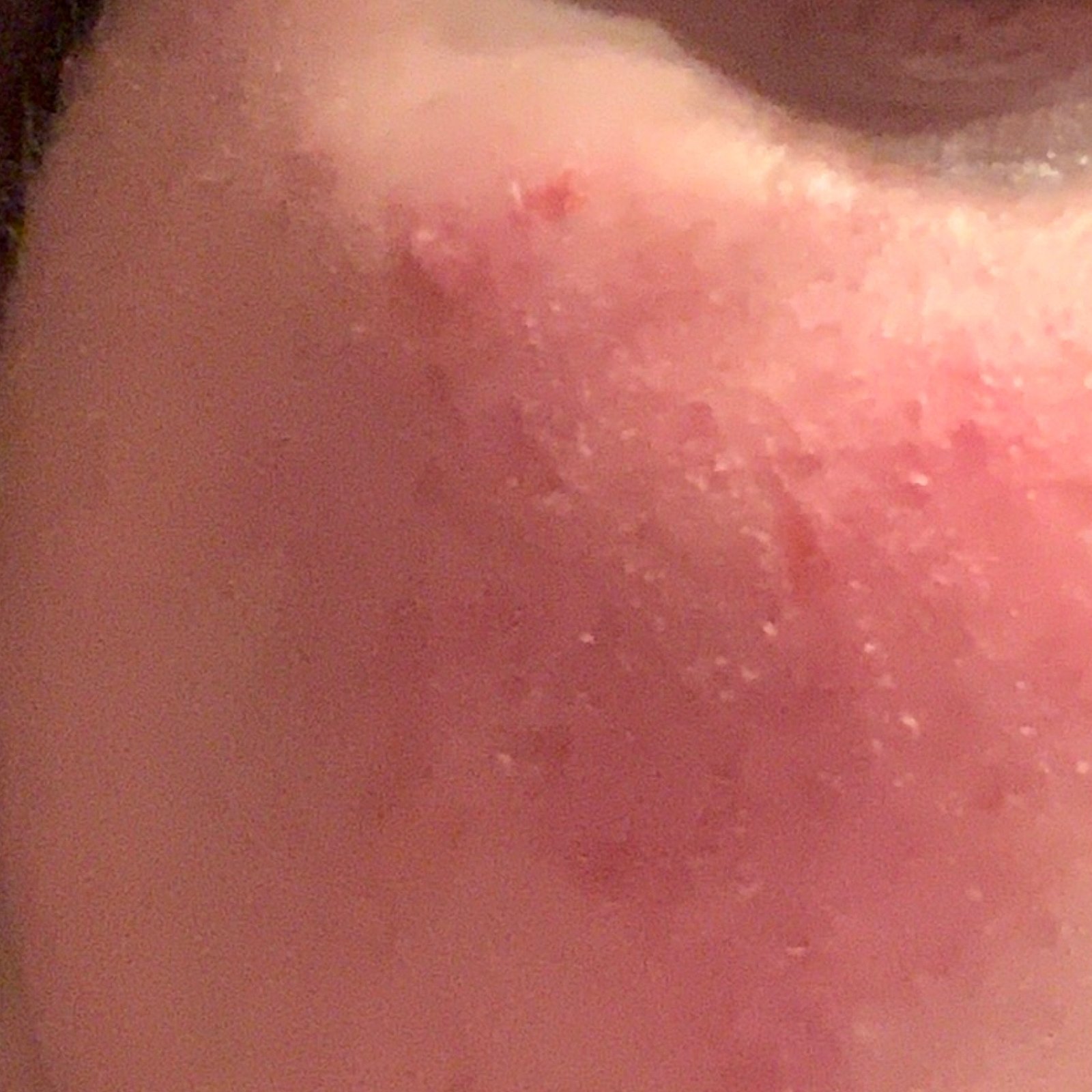
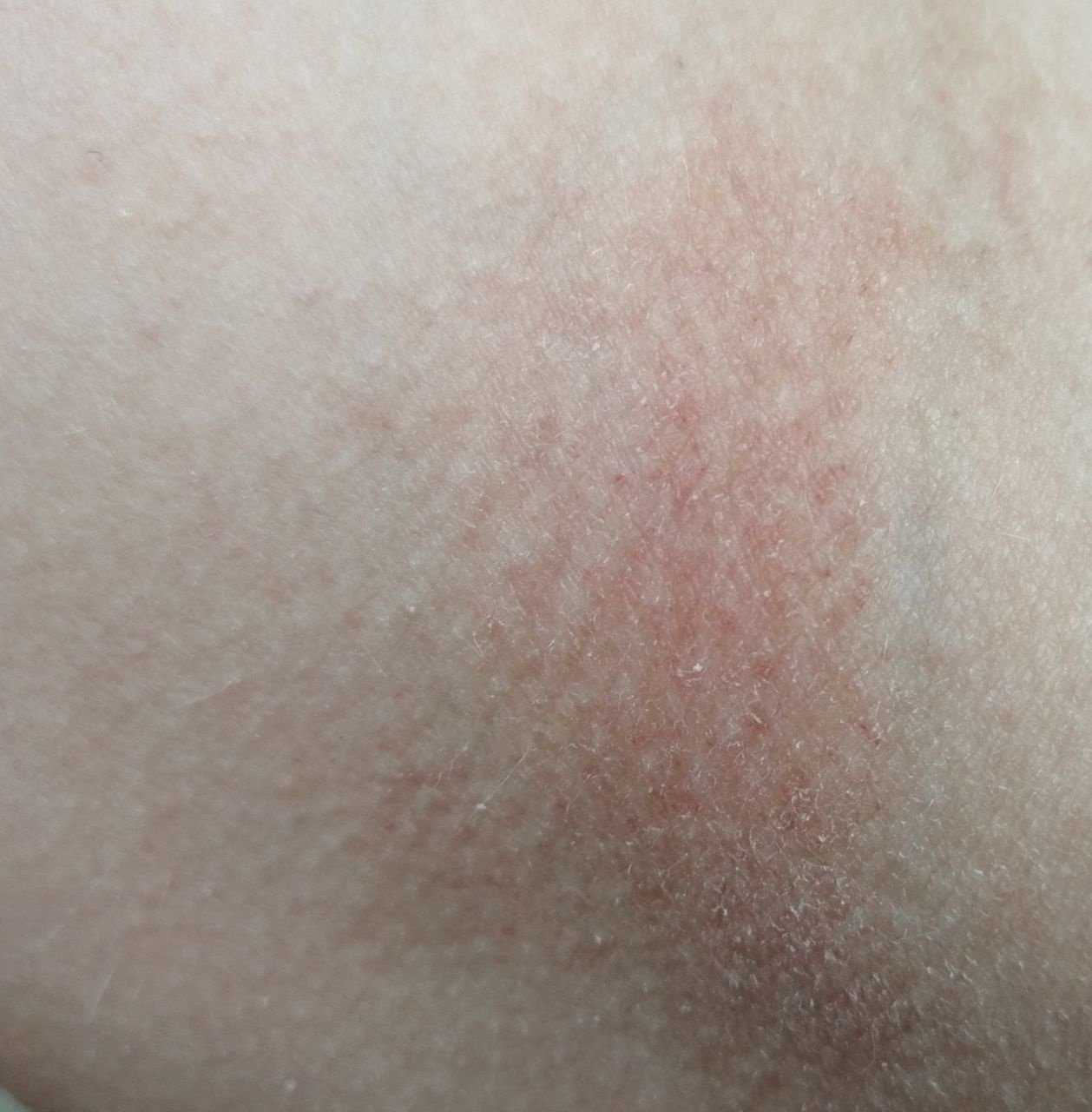
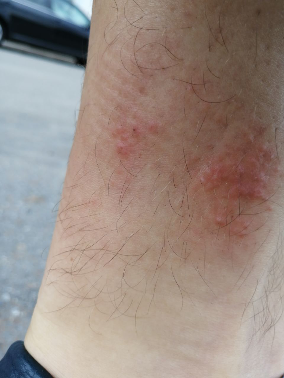
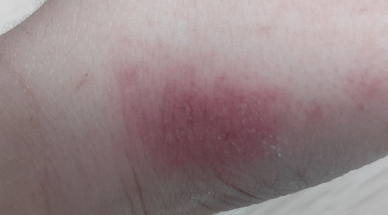
- Erythematous patches and plaques with ill-defined borders;
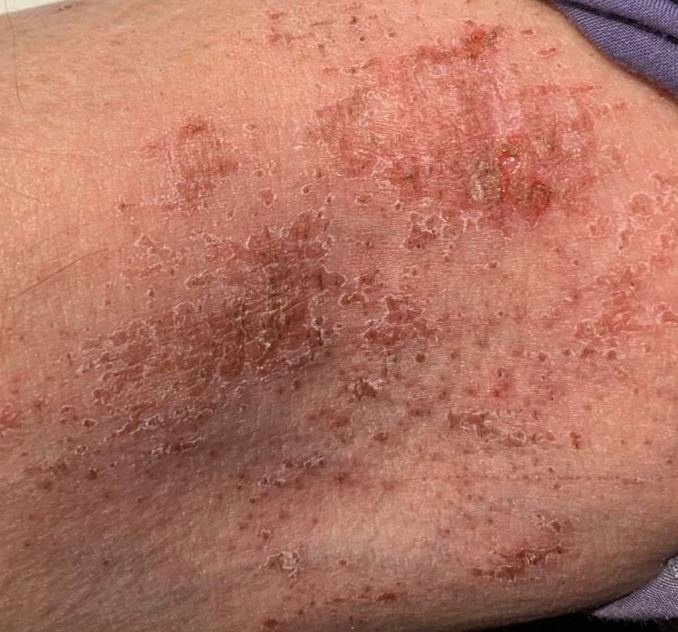
- Exudation, vesicles, and crusts;
- Swelling and edema of the affected skin;
- Excoriations and secondary infections with pustules (often S. aureus);
- Localized or generalized skin involvement.
Chronic Stage:
- Lichenification: Thickening of the skin with enhanced skin lines from repeated scratching;
- Hyperpigmentation and cracking: Especially on hands, feet, fingers, and palms;
- Small papules on hair follicles;
- Loss of lateral eyebrows, darkening of eyelids, and Denny-Morgan lines beneath eyes;
- White dermographism: A white line appears after stroking the skin, due to vasospasm.
Age-Specific Features of Atopic Dermatitis
Infants (0–2 years):
Often presents as a severe, early-onset skin condition with erythema, edema, vesicles, crusts, and fissures. Common locations include the face (excluding lips) and extensor surfaces of the limbs. Food allergy is relevant in a minority and should not be assumed.
Children (2–12 years):
Lesions become more chronic with lichenified plaques, excoriations, and erosions. Most commonly affects the flexural surfaces of elbows and knees, as well as the neck and wrists.
Adolescents and Adults:
The disease takes on a chronic, relapsing course, often triggered by stress or hormonal shifts. Lesions tend to be more generalized or affect typical flexural sites, the face, neck, and upper limbs. Exacerbations can present as papules, crusted plaques, fissures, and pustules with lichenification. Nodular variants may resemble prurigo nodularis.
Complications of Atopic Dermatitis
While atopic dermatitis is not life-threatening, it can lead to several complications that significantly impact the patient’s quality of life:
- Secondary bacterial infection: Often due to scratching, primarily caused by Staphylococcus aureus, resulting in impetiginization, crusts, and oozing erosions;
- Kaposi’s varicelliform eruption: A rare but severe complication caused by herpes simplex virus, characterized by widespread vesiculopustular rash, fever, and lymphadenopathy;
- Sleep disturbances: Due to persistent itching, especially at night;
- Psychosocial distress: Low self-esteem, anxiety, or depression from visible lesions and chronic symptoms;
- Progression to other atopic conditions: Up to 50% of children may develop allergic rhinitis or bronchial asthma (“atopic march”).
Diagnosis
The diagnosis of atopic dermatitis is primarily clinical, based on history and physical examination. Typical features include:
- Early onset in infancy or childhood;
- Chronic relapsing course with pruritic, eczematous lesions;
- Characteristic distribution by age;
- Family history of atopy;
- White dermographism and lichenification;
- Elevated total serum IgE (in many, but not all cases).
Additional diagnostic tools:
- Skin swabs: To detect Staphylococcus aureus in nasal or skin colonization;
- Viral culture: If eczema herpeticum is suspected (herpes simplex virus);
- Allergy testing: Not routine; skin prick or specific IgE tests only when the history suggests an allergen;
- Food challenge tests: Used in suspected food-triggered cases under medical supervision;
- Histology (rare): For unclear cases; findings include spongiosis, acanthosis, lymphocytic infiltrates, occasional mast cells;
- Serologic testing: Radioallergosorbent test (RAST) for allergen-specific IgE antibodies.
Treatment Strategy
Management of atopic dermatitis is multifactorial and individualized. The goals are to reduce inflammation and itching, restore the skin barrier, prevent flare-ups, and manage comorbidities.
Core treatment components:
- Diet: Elimination only for confirmed, relevant food allergy—not routine;
- Environmental control: Avoid known triggers (dust, pets, heat, fabrics, etc.);
- Topical therapy: Emollients as the foundation, plus corticosteroids and calcineurin inhibitors (tacrolimus, pimecrolimus), and newer agents such as roflumilast, ruxolitinib, or tapinarof;
- Systemic therapy: For moderate-to-severe disease uncontrolled by topical treatment—phototherapy, biologics (dupilumab, tralokinumab, lebrikizumab, nemolizumab), and selected oral JAK inhibitors or immunosuppressants under specialist supervision; oral corticosteroids and antihistamines are not routine;
- Secondary infection: Antibiotics only for clinically infected skin, not routine antiseptic use;
- Adjunctive therapy: Psychotherapy for stress-related flares, patient and family education, and support programs.
Prognosis
The long-term outlook varies by individual:
- In many children, symptoms significantly improve or remit by adolescence;
- Exacerbations in adolescents tend to be more severe but may be manageable with consistent care;
- In adults, the disease often presents chronically with periods of remission and relapse, and may coexist with other atopic conditions;
- Comorbid asthma or allergic rhinitis develops in 30–50% of patients.
Differential Diagnosis
Conditions that may resemble atopic dermatitis and must be ruled out include:
- Seborrheic dermatitis;
- Contact dermatitis (allergic or irritant);
- Psoriasis;
- Nummular eczema;
- Dermatophytosis (tinea);
- Cutaneous T-cell lymphoma (early stages);
- Genodermatoses (e.g., Wiskott-Aldrich syndrome, acrodermatitis enteropathica);
- Systemic diseases with skin manifestations (e.g., celiac disease, glucagonoma, histiocytosis X).
Preventive Measures
Prevention focuses on skin care, allergen avoidance, and health maintenance:
- Daily use of emollients to maintain hydration;
- Limit use of hot water and soap, use gentle cleansers only;
- Avoid confirmed personal triggers, not foods in general;
- Wear breathable, non-irritating clothing (preferably cotton);
- Manage comorbid conditions (asthma, rhinitis, GI disorders);
- Educate caregivers and patients to ensure compliance with treatment and reduce anxiety;
- Regular follow-up with dermatologists or allergists for early flare detection and long-term management planning.
Atopic dermatitis is chronic and relapsing and has no definitive cure, but with consistent care and education substantial disease control is achievable, with fewer flares and less impact on physical and emotional well-being.