
Venous Eczema (ICD-10: I83) 🚨
Venous Eczema: Inflamed Skin on the Lower Leg from Venous Disease
Venous eczema — also called stasis eczema, gravitational eczema or venous stasis dermatitis — is inflammation of the skin of the lower leg caused by long-standing high pressure in the leg veins. Unlike most forms of eczema, it is a sign of a circulatory problem rather than a primary skin disease, and treating the skin without addressing the veins produces disappointing results.
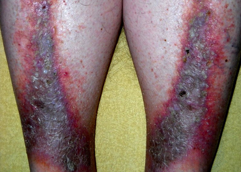
It affects the lower third of the leg, classically the skin over and just above the inner ankle, where venous pressure is highest. The skin becomes red or discoloured, itchy, scaly and sometimes weeping, and it sits on a leg that usually shows other signs of venous disease: swelling that is worse by evening, visible varicose veins, and a brownish staining from iron released by leaked red blood cells.
It is a condition of middle and later life, and it is common — venous insufficiency affects a substantial proportion of adults over sixty. Its importance is out of proportion to how it looks, because the same process that inflames the skin also leads to venous leg ulceration.
Predisposing Factors: Why Venous Eczema Develops
The underlying problem is venous hypertension: the valves in the leg veins fail, blood is not returned efficiently against gravity, and pressure in the small vessels of the skin rises. Fluid and red cells leak into the tissue, and the resulting chronic inflammation damages the skin barrier.
- Varicose veins and valve incompetence: The most common route to venous hypertension.
- Previous deep vein thrombosis: Damages the deep venous system and its valves, often years before the skin changes appear.
- Immobility and prolonged standing: The calf muscle pump is what returns blood from the leg; occupations that involve standing still, and reduced mobility, both impair it.
- Excess body weight: Increases venous pressure and makes compression harder to apply effectively.
- Previous pregnancy, which raises venous pressure and can leave lasting valve changes.
- Leg injury or surgery, which may disturb venous or lymphatic drainage.
- Age, through reduced vessel elasticity and thinner skin.
Diagnostics: How Venous Eczema Is Diagnosed
The diagnosis is made from the appearance of the skin together with the signs of venous disease in the same leg. The assessment does not stop at the skin, and two parts of it are important enough to name.
Assessment of the arterial circulation comes before compression. Compression is the central treatment, but applying it to a leg with significant arterial disease can cause harm, and it is the clinician who decides whether compression is safe at all and at what strength — reduced compression, or none, may be appropriate. Do not start therapeutic compression without a clinician assessing the arterial circulation first. Measuring the ankle–brachial pressure index is standard practice; where that index is unreliable — in diabetes, or where the arteries are calcified — toe pressures or another vascular test are used instead.
Patch testing is worth considering early here. Contact allergy is common in chronic venous disease, and the highest rates are reported in people with chronic ulcers and long exposure to topical products rather than in everyone with uncomplicated venous eczema. The mechanism is the same either way: inflamed, broken skin repeatedly exposed to creams, dressings and bandages. Reported rates vary widely, but the pattern is consistent enough to act on. A leg that is worsening despite treatment is often reacting to the treatment.
Duplex ultrasound maps the venous system where intervention is being considered. Swabs are taken when infection is suspected, and biopsy is reserved for an ulcer or plaque that behaves atypically or fails to heal.
Symptoms: What Venous Eczema Looks Like
- Itchy, scaly, discoloured skin on the lower leg, most marked over the inner ankle. In lighter skin this reads as redness; in deeper skin tones it may appear violet, grey or darker brown, and is easy to overlook.
- Swelling that is mild in the morning and worse by the end of the day.
- Brown staining (haemosiderin pigmentation): Discolouration from iron deposited by leaked red cells. It fades slowly at best and often persists.
- Varicose veins and small fans of dilated vessels around the ankle.
- Weeping and crusting when the barrier breaks down.
- Lipodermatosclerosis: Hardening and tightening of the skin and underlying tissue, which narrows the lower leg and produces the “inverted champagne bottle” shape in advanced disease.
- Atrophie blanche: Smooth white patches surrounded by dilated vessels, seen in chronic venous disease. They often sit where an ulcer has healed, but their presence does not prove that one did.
- Ulceration: A break in the skin, typically near the inner ankle, that does not heal.
Differential Diagnosis: Ruling Out Other Conditions
- Cellulitis: The most consequential confusion, and it runs in both directions. Redness of both lower legs is commonly misdiagnosed as cellulitis when the cause is venous eczema or another inflammatory mimic. Cellulitis is usually one-sided and usually comes with pain, fever and feeling unwell, but it can affect both legs and the systemic features may be absent early or in older people — so bilateral redness calls for assessment rather than for assuming infection either way. Repeated courses of antibiotics for a red leg that keeps coming back is a pattern worth questioning.
- Allergic contact dermatitis: Very often superimposed rather than an alternative; suspect it when the eczema spreads beyond the usual distribution or worsens with treatment.
- Tinea: Fungal infection of the leg or foot, distinguished by microscopy.
- Vasculitis: Palpable purpura rather than scaling, and usually with systemic features.
- Lymphoedema: Swelling that does not reduce overnight, with thicker skin and a positive Stemmer sign; the two frequently coexist.
- Pretibial myxoedema or drug-induced oedema, considered when the pattern does not fit.
Risks: Why Venous Eczema Should Be Treated
The reason to take venous eczema seriously is ulceration. The inflamed, fragile skin over a chronically congested lower leg breaks down easily — from a minor knock, from scratching, or on its own — and venous leg ulcers are slow to heal, prone to recurrence and a substantial burden to live with. Treating the eczema and the venous pressure behind it is the most effective ulcer prevention available.
Chronic inflammation also causes lasting change. Lipodermatosclerosis is difficult to reverse once established, and haemosiderin staining may never fully clear. Broken skin on the leg is a common entry point for cellulitis, and a leg that has had cellulitis is more likely to have it again.
Contact sensitisation is a further risk that is specific to this condition: each new topical product applied to chronically inflamed leg skin carries a real chance of creating an allergy that then complicates treatment for years.
Tactics: When and How to Seek Treatment
See a clinician if the skin of the lower leg becomes itchy, scaly or discoloured, particularly if the leg also swells by the end of the day or has visible varicose veins. Seek care promptly if the skin breaks, if an area becomes painful, hot and spreading, or if the leg is suddenly swollen, since that requires a different assessment.
Two common self-management mistakes are worth naming. Buying compression stockings without having the arterial circulation checked is not safe for everyone. And applying a succession of different creams, ointments and antiseptics to a leg that is not improving raises the chance of becoming allergic to one of them, which then obscures why the leg is deteriorating — if treatment is not working, the answer is a review, not another product.
Treatment: Modern Strategies for Venous Eczema
- Compression therapy: The central treatment, once arterial supply has been assessed. Graduated compression counteracts venous hypertension, reduces swelling and allows the skin to recover. It has to be strong enough to work and comfortable enough to be worn every day; the level is set after arterial assessment rather than chosen by the patient.
- Leg elevation and calf exercise: Raising the legs above hip level during the day, and regular ankle movement and walking, both assist venous return.
- Emollients: Applied generously and frequently, using a plain preparation with few potential allergens. A soap substitute replaces soap.
- Topical corticosteroids: Used in courses for active inflammation, with strength matched to severity, then stepped down rather than continued indefinitely.
- Treating infection when it is present, guided by assessment rather than by the appearance of redness alone.
- Avoiding sensitisers: Fragranced products, and topical antibiotics and antihistamines applied to the leg, are best avoided. Where patch testing has identified allergens, a written avoidance list is part of the treatment.
- Treating the veins: Endovenous ablation, sclerotherapy or surgery for suitable patients reduces recurrence and improves ulcer healing. This is assessed by a vascular service.
- Weight management and mobility, both of which improve the function of the calf muscle pump.
Prevention: Protecting the Leg
- Wear prescribed compression consistently, including after the skin has settled; stopping is a frequent reason the problem returns, though recurrence can also reflect progressing venous disease or another cause.
- Move regularly and avoid long periods of standing still or sitting with the legs down.
- Elevate the legs when resting, and raise the foot of the bed slightly if advised.
- Moisturise daily, even when the skin looks well.
- Protect the leg from knocks, which is how many ulcers begin.
- Do not scratch; treat the itch instead, since scratching is a direct route to ulceration.
- Have the leg reviewed rather than adding new products when it deteriorates.
Venous eczema is manageable, but it is managed through the circulation rather than through the skin alone. Compression, movement and attention to the veins do more for the skin of the lower leg than any cream applied to it.
Image: Cardiologist61, public domain, via Wikimedia Commons.

Medical review
Board-Certified Dermatologist · Fellow of the American Academy of Dermatology (FAAD)