
What does skin cancer look like? Pictures and warning signs
This tool is for informational purposes only and does not constitute medical advice. Always consult a qualified dermatologist.
The pictures and answers on this page are free. SkinAI Lab is a paid AI analysis; pricing is shown before you scan. A photo check cannot identify or rule out skin cancer, and it does not cover every type on this page.
What does skin cancer look like?
Skin cancer does not have one look. The three main types are basal cell carcinoma, squamous cell carcinoma and melanoma. Basal cell carcinoma often looks like a round, pearly or shiny bump or a raised, scaly patch. Squamous cell carcinoma often looks like a rough, scaly patch, a firm, dome-shaped growth or a sore that does not heal. Melanoma can appear within a mole you already have, or as a new dark spot that looks different from the rest of your moles. Pictures can give you an idea of what skin cancer can look like, but they cannot identify it or rule it out. First, should you see a doctor?
- See a doctor about a new mole, or a mole that has changed size, shape or colour; a sore that does not heal, or heals and comes back; a growth that is getting bigger or has changed colour or texture; any spot that is itching or bleeding; or a dark area under a nail that was not caused by an injury. All the signs
- Often harmless: moles are usually nothing to worry about unless they change size, shape or colour, and a seborrheic keratosis is a harmless warty spot. But skin cancers can be hard to tell apart from harmless spots, so check the signs first, and see a doctor if you are not sure what a spot is.
Sources: Cleveland Clinic — Skin cancer · AAD — Types of skin cancer · AAD — Basal cell carcinoma · NHS — melanoma symptoms · NHS — non-melanoma skin cancer symptoms · AAD — ABCDEs of melanoma · NHS — Moles · DermNet — Seborrhoeic keratosis
See a doctor — don’t rely on a photo check — if you have:
- A new mole, or a mole that has changed size, shape or colour
- A mole or spot with an uneven shape or edges, or a mix of 2 or more colours
- A mole that is painful or itchy, or inflamed, bleeding or crusty
- A spot that looks different from your other spots or moles
- A sore that does not heal, or heals and then comes back
- A growth that is getting bigger or has changed colour or texture
- Any spot that is itching or bleeding; or a growth or area of skin that hurts, crusts or scabs for more than 4 weeks
- A new spot or lump that is growing quickly — see a doctor promptly
- A red or scaly patch that does not go away, when you do not know the cause
- A new or unusual mark on your skin that has not gone away after a few weeks, including on the palms of your hands or soles of your feet
- A dark area or a new dark streak under a nail that has not been caused by an injury
A symptom outweighs a reassuring result. Our atlas article on healthy skin puts it this way: “If something itches, bleeds, hurts or will not heal, that symptom carries more weight than a reassuring screening result.”
Do not wait for it to hurt. You can have melanoma without feeling any pain or discomfort, and basal cell carcinoma might not cause bothersome symptoms until it has grown quite large. Finding skin cancer early can mean it is easier to treat.
Protect your skin from the sun. The World Health Organization says skin cancer is highly preventable: limit time in the midday sun, seek shade, wear protective clothing, use a broad-spectrum sunscreen on skin that clothes cannot cover, and avoid sunbeds.
Sources: NHS — melanoma symptoms · NHS — non-melanoma skin cancer symptoms · AAD — ABCDEs of melanoma · AAD — Melanoma signs · AAD — Merkel cell carcinoma signs · NHS — Bowen’s disease · AAD — Nail melanoma · Atlas — Healthy skin · American Cancer Society — Signs of basal cell carcinoma · WHO — Ultraviolet radiation
ABCDE: the warning signs of melanoma
-
Asymmetry
“One half of the lesion does not match the other half in shape or size.”
-
Border
“The edges of the lesion are irregular, notched, or blurred, especially in more advanced stages.”
-
Colour
“Uneven pigmentation with multiple shades of brown, black, or other colors like blue, pink, or white.”
-
Diameter
“Melanomas are usually larger than 5–6 mm, although smaller lesions can still be dangerous.”
-
Evolving
“The lesion changes over time in size, shape, surface texture, or symptoms such as itching or bleeding.”
-
The “ugly duckling”
“Because a person’s moles usually look alike, one that does not belong deserves attention.”
ABCDE does not catch every melanoma. Some melanomas have little or no dark colour: they may be pink, red or the colour of your skin, and may not show the ABCDE signs. Others look like a firm, dome-shaped growth. Have a mole checked if it looks very different from your others, even if it has none of the ABCDE signs.
The six descriptions are quoted from our atlas articles on melanoma and healthy skin. Sources: Atlas — Melanoma · Atlas — Healthy skin · AAD — ABCDEs of melanoma · DermNet — Amelanotic melanoma · AAD — Melanoma signs · Cancer Research UK — melanoma symptoms
Normal mole or melanoma?
Normal mole
Normal moles are usually round with smooth edges, one colour, and about the size of the end of a pencil or smaller.
Melanoma
Melanomas are often an uneven shape with uneven edges, a mix of 2 or more colours, and often more than 6 mm wide, though they can be smaller when found early. A mole that changes size, shape or colour may be a melanoma.
Melanoma does not have to start in a mole. Our atlas article on melanoma says: “A minority of melanomas arise from a pre-existing pigmented lesion such as a mole; most arise de novo on previously normal skin.” So look for new spots as well as for moles that change. These two descriptions are common patterns, not a test: it is sometimes hard to tell the difference between melanoma and an ordinary mole, so show a doctor any mole you are unsure of.
Sources: NHS — melanoma symptoms · Atlas — Melanoma · American Cancer Society — Signs of melanoma
Signs of basal cell and squamous cell skin cancer
-
Basal cell carcinoma
The most common type of skin cancer. It can look like a firm, round, raised growth; a round area that dips in the centre; a scaly spot or patch; a sore that does not heal, or heals and returns; a new mark that looks like a scar; or a cluster of shiny bumps. It tends to grow slowly and rarely spreads, but it can grow deep.
-
Squamous cell carcinoma
It can look like a rough, scaly patch; a firm, dome-shaped growth; a sore that does not heal, or heals and comes back; or a wart-like or horn-like growth. It tends to grow slowly, but sometimes growth is quick, and left untreated it may grow deep and spread.
-
Where they appear
Any part of your skin can be affected, but these cancers are most common in areas exposed to the sun, such as the head, face and ears, the neck and shoulders, the back, the hands and the lower legs.
-
How they feel
For many people the only sign is a slow-growing bump, a sore-like growth or a rough-feeling area of skin. Some people also notice pain, itching, numbness, scabbing or bleeding.
A sore that does not heal. Our atlas article on healthy skin lists “A sore that has not healed within about four weeks, or that heals and reopens.” among the changes that need an in-person assessment. Do not wait 4 weeks if a spot is growing, bleeding or changing.
Sources: AAD — Basal cell carcinoma · AAD — Squamous cell carcinoma · AAD — Types of skin cancer · NHS — non-melanoma skin cancer symptoms · Atlas — Healthy skin
Skin cancer pictures: examples by type
Photos from our dermatology atlas, grouped by type. Each description is quoted word for word from the atlas article; open a card for the full article and all its photos. Skin cancers can look quite different from one person to another, so these pictures cannot identify or rule out skin cancer. Look through all four groups.
Source: Skin Cancer Foundation — Skin cancer information
Melanoma
Melanoma is known as the most serious skin cancer: it can grow rapidly and spread beyond the skin (AAD). When melanoma is suspected, see a dermatologist without delay (atlas).
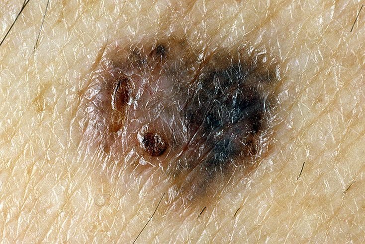 32 photos
Melanoma
“A melanoma lesion may appear as a flat or raised spot, or a combination of both, on the surface of the skin.”
“These lesions often exhibit a multiform appearance and tend to differ in texture, color, and shape from normal skin or benign moles.”
Read the article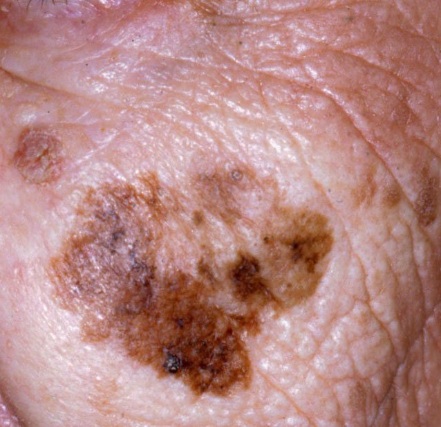 24 photos
Lentigo maligna melanoma
“Gradual enlargement of a previously stable pigmented patch.”
“Over time, however, the lesion can darken, develop uneven borders, and change in texture.”
Read the articleBasal cell and squamous cell skin cancers
Bowen’s disease is squamous cell carcinoma in situ, an early form of skin cancer confined to the top layer of the skin. A keratoacanthoma cannot be reliably told apart from a squamous cell carcinoma on examination, so it is generally managed as one (atlas).
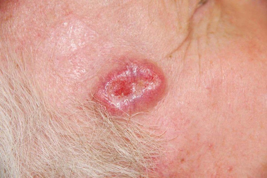 32 photos
Basal cell carcinoma
“Basal cell carcinoma typically presents as a slowly enlarging, raised lesion or plaque that may appear pink, flesh-colored, or red.”
“In its classic form, BCC may resemble a small crater-like ulcer with rolled, pearly edges and a depressed central area.”
Read the article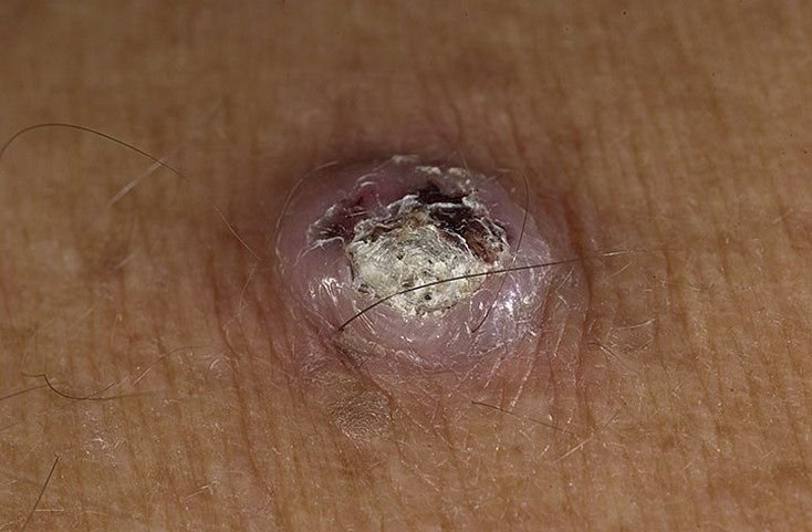 16 photos
Squamous cell carcinoma
“The tumor often appears as a persistent, thickened plaque or a nodule that protrudes from the surface of the skin.”
“The lesion may have a rough, scaly, or wart-like texture and is commonly covered by crusts or horny (keratinized) masses.”
Read the article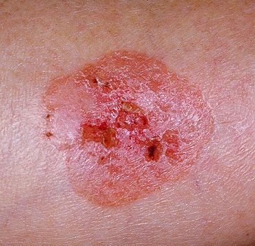 12 photos
Bowen’s disease (squamous cell carcinoma in situ)
“It presents as a persistent red or pink patch or plaque with a rough, scaly surface.”
Read the article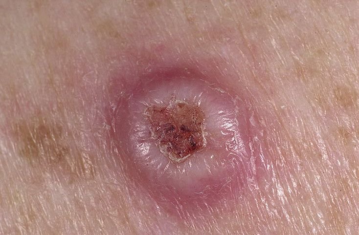 12 photos
Keratoacanthoma
“Keratoacanthoma often appears as a raised, dome-shaped lesion with a central crater or keratin-filled core.”
“The tumor may initially grow rapidly over weeks, reaching a size of 10–20 mm, after which growth typically slows.”
Read the articlePrecancerous and borderline growths
Most individual actinic keratoses do not progress, but one can need assessment and treatment even when it is not changing and does not hurt (atlas). An atypical mole is benign and rarely transforms, but people with several have a higher melanoma risk and need regular skin checks (atlas).
 20 photos
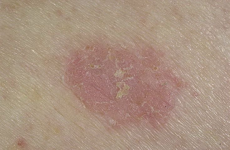
Actinic keratosis (solar keratosis)
“On visual examination, actinic keratosis presents as single or multiple flat or raised lesions with a rough, dry surface.”
“Actinic keratosis is considered a precancerous condition, as it carries a significant risk of transforming into squamous cell carcinoma of the skin.”
Read the article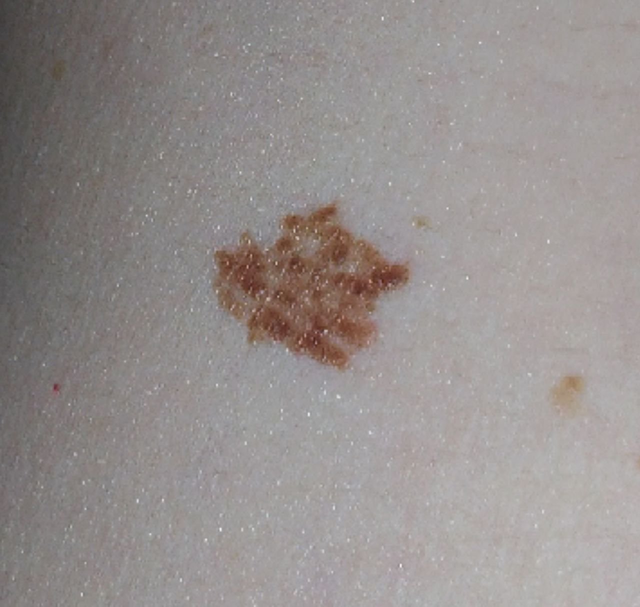 24 photos
Atypical mole (dysplastic nevus)
“The borders of dysplastic nevi are usually irregular, with uneven edges that can be considered an unfavorable sign.”
“Rapid growth is a reason to have the lesion assessed for melanoma without delay.”
Read the articleNon-cancerous growths that can resemble skin cancer
These growths are benign, yet skin cancers can look like them: pigmented basal cell carcinoma, squamous cell carcinoma and melanoma “can look like a seborrheic keratosis, and a photograph cannot settle which it is” (atlas). Show a doctor any mole or growth that has not been checked before, even if it looks like these pictures. If a spot that was found to be harmless later changes, itches or bleeds, have it checked again.
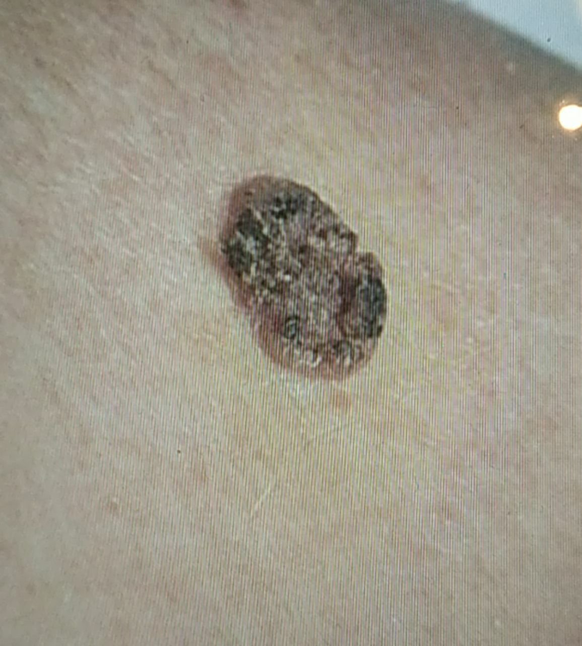 30 photos
Seborrheic keratosis
“The surface texture of seborrheic keratosis differs from that of normal skin: it is rough, dry, and may show signs of peeling.”
Read the article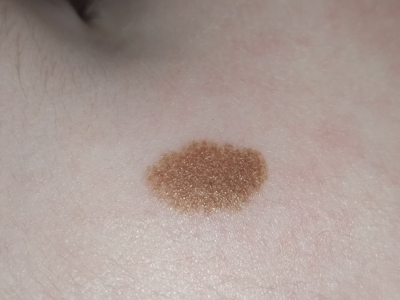 24 photos
Mole (benign nevus)
“The color of a simple nevus ranges from light brown to dark brown, with uniform pigment distribution.”
“Any one typical nevus has a very low chance of becoming a melanoma.”
Read the article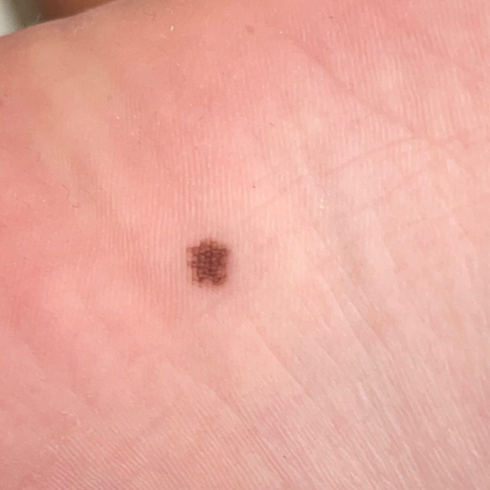 24 photos
Mole on a palm or sole (acral nevus)
“On visual examination, a benign acral nevus appears as a pigmented growth that is typically symmetrical, oval or round, with a regular outline.”
“Most acral nevi are benign, but acral melanoma can resemble them clinically, so a newly detected or changing acral lesion should be evaluated by a dermatologist rather than watched.”
Read the articleSee every condition in the dermatology atlas →

Medical review of the atlas articles above
Board-Certified Dermatologist · Fellow of the American Academy of Dermatology (FAAD)
How the main types differ
A summary of features from our atlas articles and the sources on this page. Skin cancers and harmless spots overlap, so this table cannot tell you which one you have. Tap a name to see its picture.
| Condition | What it often looks like | How it changes or feels | Where it often appears |
|---|---|---|---|
| Melanoma | Often an uneven shape or edges and a mix of 2 or more colours; often more than 6 mm across, but can be smaller | Changes size, shape or colour; can be swollen and sore, bleeding, itchy or crusty, but may cause no pain | Anywhere; more common on sun-exposed skin |
| Lentigo maligna melanoma | A flat coloured patch that can darken and develop uneven borders | Enlarges gradually; the texture can change | Face, neck, ears and scalp, most often after the age of 50 |
| Basal cell carcinoma | A firm, round, raised growth, shiny and pink or red on lighter skin, brown or black on darker skin; a round area with a dip in the centre; a scaly patch; or a scar-like mark | Grows slowly; may bleed, crust over and break open several times | Head and neck, including the ears, nose, eyelids and scalp |
| Squamous cell carcinoma | A rough, scaly patch, a firm, dome-shaped growth, a wart-like growth, or a sore surrounded by raised skin | Usually grows slowly, sometimes quickly; may scab and bleed; can be painful or tender | Face, scalp, neck, arms, hands, lower legs |
| Bowen’s disease | A scaly or crusty patch with clear edges, flat or raised, up to a few centimetres across; red or pink on white skin, harder to see on brown and black skin | Usually very slow growing; does not heal; sometimes itchy | Lower legs, neck and head |
| Keratoacanthoma | A firm, dome-shaped growth, red or skin-coloured, with a plug or crater in the centre | Grows rapidly within a few weeks, up to 1–2 cm | Sun-exposed skin of the face and arms |
| Actinic keratosis | Dry, scaly patches, usually 1–2 cm, skin-coloured or pink to red to brown | Rough, like sandpaper; may itch | Face, hands and arms, ears, scalp, legs |
| Atypical mole | Usually 10–15 mm, with irregular borders; light to dark brown, sometimes with red, pink or blue | Slightly denser than normal skin; usually no symptoms | Anywhere; often sun-exposed skin |
| Seborrheic keratosis | Raised growths with a rough, dry surface, round or oval, 5–20 mm, skin-coloured or grey to grey-brown | Usually no symptoms; older ones may itch mildly | Trunk, arms, face |
| Mole | Small round or oval spot or bump with a smooth edge, light to dark brown, usually one colour | Usually does not change over time; flat or raised, smooth or rough | Trunk and limbs; fewer on the head and neck |
Sources: the atlas articles linked in the cards · NHS — melanoma symptoms · AAD — Melanoma signs · AAD — Basal cell carcinoma · AAD — Squamous cell carcinoma · NHS — Bowen’s disease · DermNet — Keratoacanthoma · NHS — Actinic keratoses · NHS — Moles
Skin cancer on brown and black skin
-
Often found late
People with darker skin tones can get skin cancer. When they do, it is often advanced by the time it is diagnosed, which can make treatment difficult.
-
Places that get little sun
On darker skin, skin cancer may show up in areas that get little, if any, sun: the palms, soles, fingers and toes, nails, inside the mouth or on a lip, the buttocks and around the genitals. It can also appear on the face and neck.
-
Colour
On darker skin tones a basal cell carcinoma is usually brown, black, bluish or grey, and a patch of non-melanoma skin cancer may be darker than the skin around it rather than red.
-
What to look for
A bump, spot or mole that is growing, bleeding, itching or changing in any way; a patch of skin that is getting darker; a sore that does not heal, or heals and returns; a dark line or streak in a nail; and a scaly or rough patch that does not go away.
Soles, palms and nails. Our atlas notes that acral melanoma, more common in people with darker skin tones, often develops on the soles, the palms and under the nails. Have a dermatologist look at a new or changing mark on a palm or sole, or a new dark streak under a nail, rather than waiting for it to go away. In a nail it may look like a brown or black band, often on the thumb or big toe.
Sources: AAD — Skin cancer in darker skin tones · AAD — Basal cell carcinoma · NHS — non-melanoma skin cancer symptoms · Cancer Research UK — non-melanoma skin cancer symptoms · Atlas — Melanoma · AAD — Nail melanoma
Rarer types of skin cancer
There are no pictures of these types on this page. They are described using the linked sources.
Melanoma that is not a dark mole
Any skin, nails, soles
Amelanotic melanoma has little or no dark pigment: it may be pinkish, reddish, white, the colour of your skin or even colourless, which makes it difficult to recognise. Melanoma can also look like a firm, dome-shaped growth that may bleed, like a bruise on the bottom of the foot, or like a dark brown or black line beneath a fingernail or toenail.
See a dermatologist promptly about a new dark band in a nail, or any new, changing or unusual spot, including one that is pink, red or skin-coloured.
Skin Cancer Foundation — Melanoma warning signs · DermNet — Amelanotic melanoma · AAD — Melanoma signs · AAD — Nail melanoma
Merkel cell carcinoma
Head or neck
A rare, aggressive skin cancer that often looks harmless: people can mistake it for an insect bite, a sore, a cyst, a stye or a pimple. It tends to grow quickly, usually becoming noticeably bigger in a few weeks to months, to be pink, red or purple, and to feel painless. It usually appears after the age of 50 on skin that has had a lot of sun over the years.
See a dermatologist promptly if you notice a new spot on your skin that is growing rapidly.
Cutaneous T-cell lymphoma
Skin that gets little sun
A rare cancer that starts in white blood cells called T-lymphocytes and shows on the skin. Its most common type, mycosis fungoides, often begins as a rash that can last for months or years, usually on the upper thighs, buttocks, belly or groin. It tends to be red or pink on lighter skin and brown or purple on darker skin. It can turn into flat, scaly patches that may be mistaken for eczema or psoriasis, and it can be itchy.
AAD — Cutaneous T-cell lymphoma signs · Cancer Research UK — Skin lymphoma · Skin Cancer Foundation — Rare skin cancers
Kaposi’s sarcoma
Legs, face, groin, mouth
A rare cancer that develops from the cells lining blood or lymph vessels. The first symptom is usually a coloured spot on the skin, which may be pink, brown, brown-red or reddish-purple and flat or raised. The spots can develop anywhere on the skin, including inside the mouth. It is caused by a virus (human herpesvirus 8) and is more likely in people with a weakened immune system, for example from HIV or after an organ transplant. A slow-growing type is seen in older men of Mediterranean or Middle Eastern descent.
Macmillan Cancer Support — Kaposi’s sarcoma · Skin Cancer Foundation — Rare skin cancers · DermNet — Kaposi sarcoma
Dermatofibrosarcoma protuberans
Trunk, arms, legs
A rare cancer that starts in the deeper layer of the skin. It usually appears as a painless, thickened area of skin or a lump that feels rubbery or firm, red-brown or skin-coloured, and it grows very slowly over months to years. It can look like an ordinary scar, cyst or bruise. It rarely spreads to other parts of the body, but it tends to come back after it is removed.
DermNet — Dermatofibrosarcoma protuberans · Skin Cancer Foundation — Rare skin cancers
Sebaceous carcinoma
Eyelids, face
A rare, aggressive cancer of the skin’s oil glands. On an eyelid it is a small, firm, slowly enlarging lump, reddish or yellowish and often painless, which is often mistaken for a chalazion, a benign inflammation of an eyelid gland. On other parts of the body it may look like a yellowish lump that sometimes bleeds.
DermNet — Sebaceous carcinoma · Skin Cancer Foundation — Rare skin cancers
Terms used in the quotes
- Lesion
- An area of abnormal or damaged tissue caused by injury, infection, or disease.
- Plaque
- In medicine, a small, abnormal patch of tissue on a body part or an organ.
- Nodule
- A growth or lump that may be malignant (cancer) or benign (not cancer).
- Ulcer
- A break on the skin, in the lining of an organ, or on the surface of a tissue.
- Tumor
- An abnormal mass of tissue that forms when cells grow and divide more than they should or do not die when they should. Tumors may be benign (not cancer) or malignant (cancer).
- Benign
- Not cancer. Benign tumors may grow larger but do not spread to other parts of the body.
- Precancerous
- A term used to describe a condition that may (or is likely to) become cancer.
- Nevus
- A benign (not cancer) growth on the skin that is formed by a cluster of melanocytes (cells that make a substance called melanin, which gives color to skin and eyes).
- Pigment
- A substance that gives color to tissue. Pigments are responsible for the color of skin, eyes, and hair.
- Keratin
- A type of protein found on epithelial cells, which line the inside and outside surfaces of the body. Keratins help form the tissues of the hair, nails, and the outer layer of the skin.
Source: NCI Dictionary of Cancer Terms.
What skin cancer looks like: FAQ
What does skin cancer look like when it starts?
There is no single early look. A small basal cell carcinoma can be a shiny, pearly bump or a flat, pale area that looks like a scar. The earliest form of squamous cell carcinoma can look like scaly, dry-looking, rough skin. An early melanoma could be mistaken for a mole. Many early melanomas do not feel like anything, and basal cell carcinoma might not cause bothersome symptoms until it has grown quite large. That is why a spot that is new, changing or not healing matters more than how it feels. Pictures cannot identify or rule out skin cancer, so have such a spot checked by a doctor. Sources: American Cancer Society — Signs of basal cell carcinoma, AAD — Squamous cell carcinoma, AAD — Melanoma signs, Skin Cancer Foundation — Melanoma warning signs.
How can you tell if a spot is skin cancer?
You cannot tell for certain by looking, and neither can a picture. A doctor examines the spot and, if it could be skin cancer, takes a sample of it (a skin biopsy) to be examined under a microscope. Our atlas article on melanoma says that melanoma “cannot be reliably diagnosed by a fixed symptom count and requires clinical and histological assessment.” What you can do is notice the warning signs: a spot that is new, growing or changing, one that looks different from your others, a sore that does not heal, or a spot that itches or bleeds. Sources: AAD — Squamous cell carcinoma, NCI Dictionary of Cancer Terms, Atlas — Melanoma, AAD — ABCDEs of melanoma.
What are the warning signs of skin cancer?
The American Cancer Society lists a new mole, spot or bump that is growing or changing in shape or colour; a sore that bleeds and does not heal after several weeks; a rough or scaly red patch, which might crust or bleed; a wart-like growth; and a mole with an odd shape, irregular borders or areas of different colours. Two more signs are a spot that looks different from all the other spots on your skin, known as the ugly duckling sign, and a dark line or streak in a nail. Not all skin cancers look the same, so show a doctor any area of skin that concerns you. Sources: American Cancer Society — Signs of basal cell carcinoma, American Cancer Society — Signs of melanoma.
What does skin cancer look like on the face?
Most basal cell carcinomas develop on the head and neck, including the ears, nose, eyelids and scalp; on lighter skin they are often a firm, round, shiny, pink or red growth, and on darker skin brown or black. Squamous cell carcinoma, which can look like a rough, scaly patch, a firm, dome-shaped growth or a sore that does not heal, is also common on the face and scalp. Lentigo maligna melanoma is a flat, unevenly coloured patch that slowly enlarges, usually on the face, neck, ears or scalp of people over 50. Actinic keratoses, which are rough, scaly patches of sun-damaged skin, often appear on the face, ears and scalp too. A doctor should look at any of these. Sources: AAD — Basal cell carcinoma, AAD — Squamous cell carcinoma, Atlas — Lentigo maligna melanoma, NHS — Actinic keratoses.
What is the difference between a mole and melanoma?
Normal moles are usually round with smooth edges, only one colour, about the size of the end of a pencil or smaller, and they usually do not change over time. Melanomas often have an uneven shape, uneven edges or a mix of 2 or more colours. They are often more than 6 mm wide, but can be smaller. A new spot, or a change in size, shape or colour, can be a warning sign. Our atlas adds that “Any one typical nevus has a very low chance of becoming a melanoma.” It is sometimes hard to tell the difference between melanoma and an ordinary mole, so show a doctor any mole you are unsure of. Sources: NHS — melanoma symptoms, Atlas — Mole, American Cancer Society — Signs of melanoma.
Does skin cancer itch or hurt?
Often it does not. You can have melanoma without feeling any pain or discomfort, and for many people the only sign of basal cell carcinoma is a slow-growing bump, a sore-like growth or a rough-feeling area of skin. Skin cancers can also itch, hurt, feel tender or bleed. Itching alone does not diagnose or rule out melanoma: many melanomas never itch, and plenty of harmless spots do. Do not wait for a spot to hurt before you have it checked. Sources: AAD — Melanoma signs, AAD — Basal cell carcinoma, Skin Cancer Foundation — Melanoma warning signs.
Can skin cancer look like a pimple, a scar or a wart?
Yes. Merkel cell carcinoma can be mistaken for a pimple, an insect bite, a cyst or a stye. Basal cell carcinoma can look like a sore that does not heal or a new mark that looks like a scar. Squamous cell carcinoma can be a wart-like growth. Melanoma can look like an age spot, a bruise on the bottom of the foot or a cyst. A “pimple” or sore that does not heal, keeps coming back or keeps growing should be looked at by a doctor. Sources: AAD — Merkel cell carcinoma signs, AAD — Basal cell carcinoma, AAD — Squamous cell carcinoma, AAD — Melanoma signs.
Can a photo or an app tell me if I have skin cancer?
No. Pictures can give you an idea of what skin cancer can look like, but you cannot know or rule out that you have it by looking at pictures, and the output of an AI skin check is not a medical diagnosis. A reassuring result does not cancel a warning sign: our atlas says “If something itches, bleeds, hurts or will not heal, that symptom carries more weight than a reassuring screening result.” Photos are useful in another way. Cancer Research UK suggests taking a photo of anything that does not look quite right, with a ruler next to it, so that you can tell if it is changing and show the photos to your doctor. Sources: AAD — Basal cell carcinoma, Atlas — Healthy skin, Cancer Research UK — non-melanoma skin cancer symptoms.
How serious is skin cancer?
It depends on the type and on how early it is found. The three main types are basal cell carcinoma, squamous cell carcinoma and melanoma. Basal cell carcinoma grows slowly and rarely spreads, but it can grow deep. Squamous cell carcinoma can spread or return after treatment, though it is highly treatable when caught early. Melanoma is known as the most serious skin cancer because it can grow rapidly and spread beyond the skin; the outcome can be good when it is diagnosed early and properly treated. That is the reason not to wait with a spot that worries you. Sources: Cleveland Clinic — Skin cancer, AAD — Types of skin cancer, AAD — Basal cell carcinoma.
When should I see a doctor about a spot?
See a doctor about a new mole, or a mole that has changed size, shape or colour; a mole that is painful, itchy, inflamed, bleeding or crusty; a spot that looks different from your other spots or moles; a sore that does not heal, or heals and comes back; a growth that is getting bigger or has changed colour or texture; any spot that is itching or bleeding; a new spot or lump that is growing quickly; a new or unusual mark that has not gone away after a few weeks, including on the palms or soles; or a dark area under a nail that was not caused by an injury. Sources: NHS — melanoma symptoms, NHS — non-melanoma skin cancer symptoms, AAD — ABCDEs of melanoma, AAD — Melanoma signs, AAD — Merkel cell carcinoma signs.
For a clear photo: daylight, close and in focus, no filters. Upload a photo for an AI skin check. A photo check cannot identify or rule out skin cancer, and it does not cover every type on this page. AI output is not a medical diagnosis. If you have any of the signs above, see a doctor instead.
Check my skin ↑SkinAI Lab is a paid AI analysis; pricing is shown before you scan.


Prepared by the SkinAI Lab editorial team · Updated . Apart from the quoted atlas text, this page has not been reviewed by a doctor. It is general information, not medical advice.