Actinic keratosis (ICD-10: L57) ⚠️
Actinic Keratosis (AK, Solar Keratosis)
Actinic Keratosis (also known as solar keratosis) is a premalignant skin lesion that appears as a rough, hyperemic spot with keratinization. This condition is most commonly observed in individuals over the age of 40, particularly in areas of the skin frequently exposed to solar ultraviolet (UV) radiation. Actinic keratosis is considered a precancerous condition, as it carries a significant risk of transforming into squamous cell carcinoma of the skin. The incidence of actinic keratosis increases with age, and it is characterized by its multiplicity, with new lesions appearing over time. Both men and women are equally affected by this condition.
Predisposing Factors
Although the exact cause of actinic keratosis is not fully understood, several factors are known to increase the risk of developing this condition. These factors primarily involve environmental and genetic influences that make the skin more susceptible to damage and subsequent lesion formation:
- Age-Related Skin Changes: As individuals age, their skin undergoes changes at both the histological and functional levels, leading to aging of the epidermal keratinocytes and a decline in cellular function.
- Excessive UV Radiation: Prolonged and repeated exposure to UV radiation, whether from the sun or artificial sources like tanning beds, is the primary cause of actinic keratosis. The damaging effects of UV radiation accumulate over time, increasing the risk of skin lesions.
- Genetic Factors: A genetic predisposition to developing actinic keratosis may exist, and certain individuals may be more vulnerable due to their skin type or family history.
- Ionizing Radiation: Exposure to ionizing radiation, whether occupational or medical, can trigger the development of actinic keratosis in some cases.
- Chemical Compounds and Chronic Skin Injuries: Exposure to arsenic or to ionizing radiation is also associated with keratotic lesions of this kind, though ultraviolet light is by far the usual cause of actinic keratosis.
Diagnostics
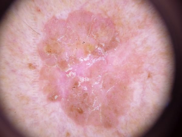
The diagnosis of actinic keratosis is based on a clinical examination, which includes a visual inspection of the lesions and dermatoscopic analysis to assess the characteristics of the skin lesions. Biopsy is considered when a lesion is thick, indurated, ulcerated, bleeding, rapidly enlarging or painful, or otherwise suspicious for squamous cell carcinoma. If there is suspicion that the lesion may be malignant or atypical, a biopsy may be performed for further evaluation.
Symptoms
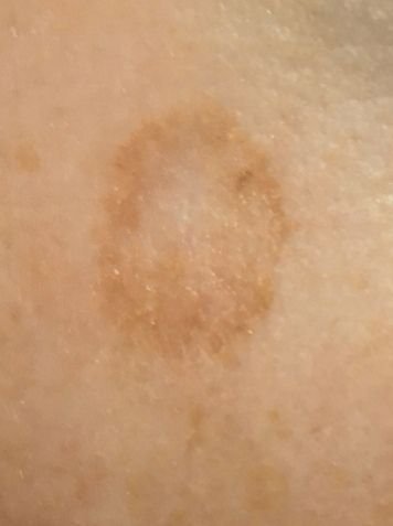
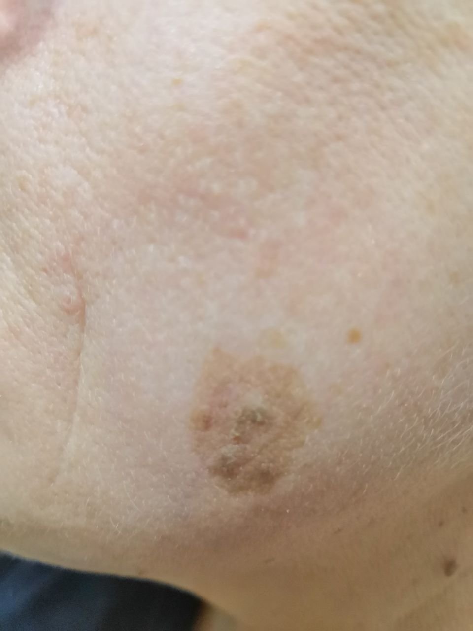
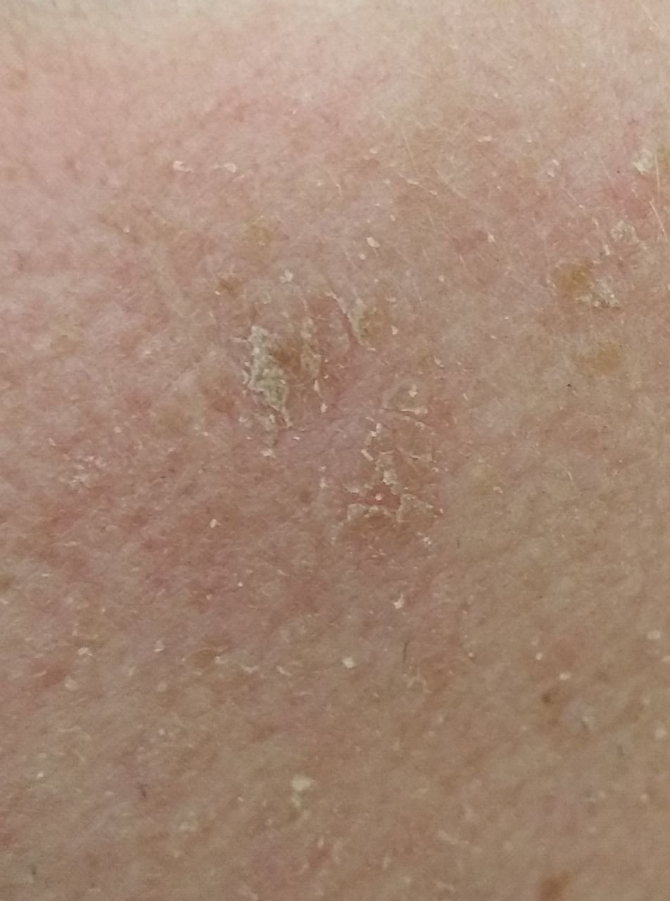
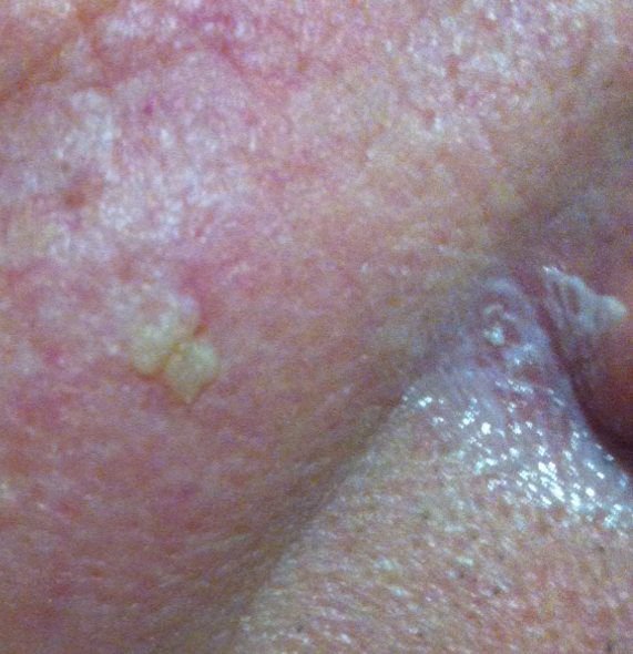
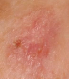
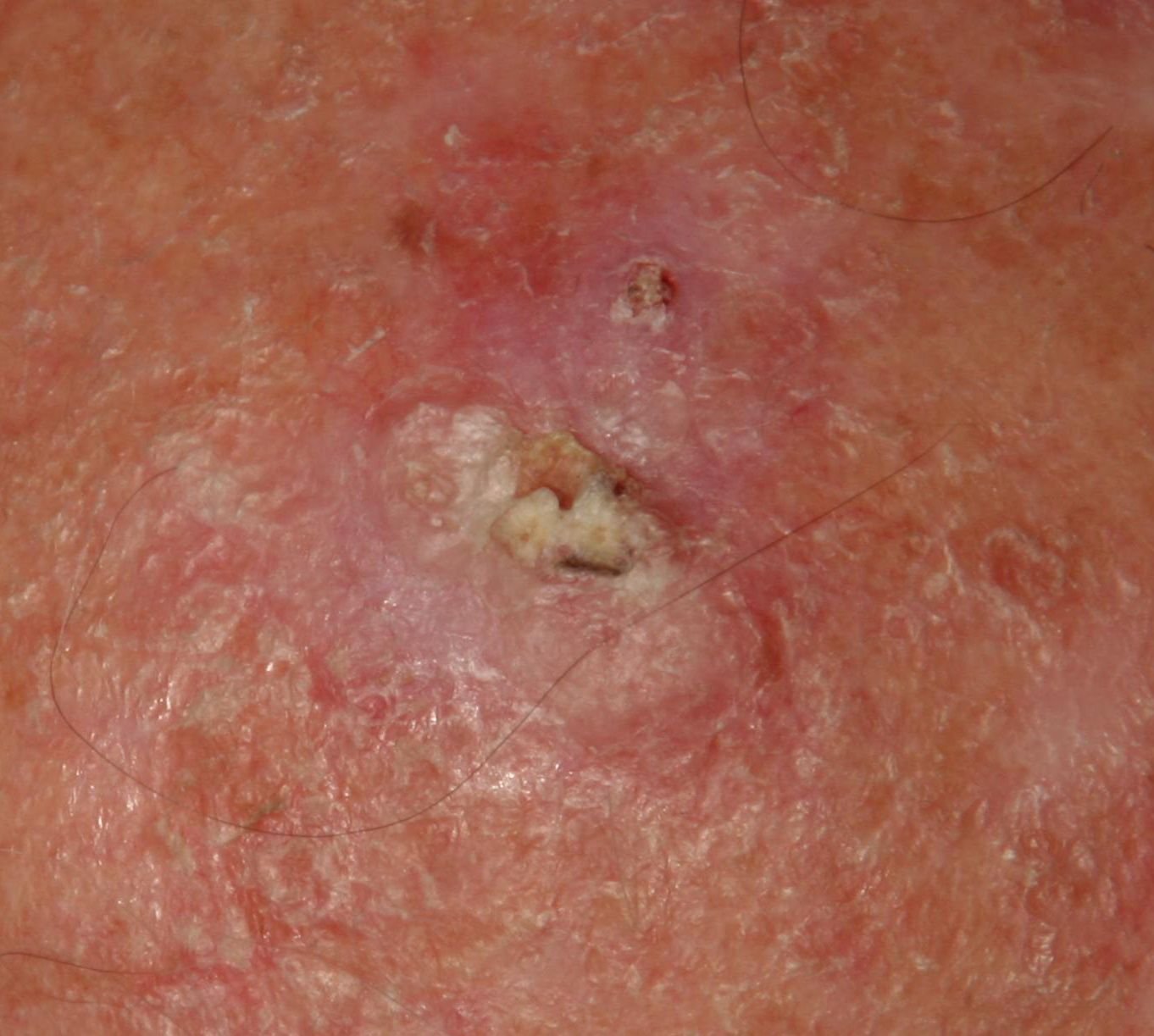
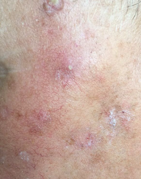
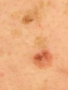
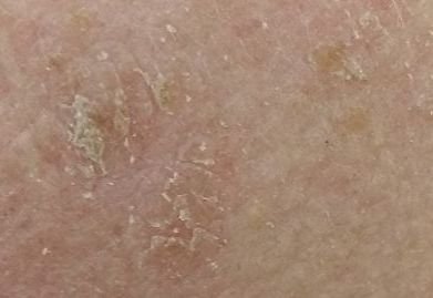
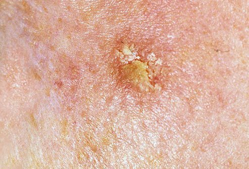
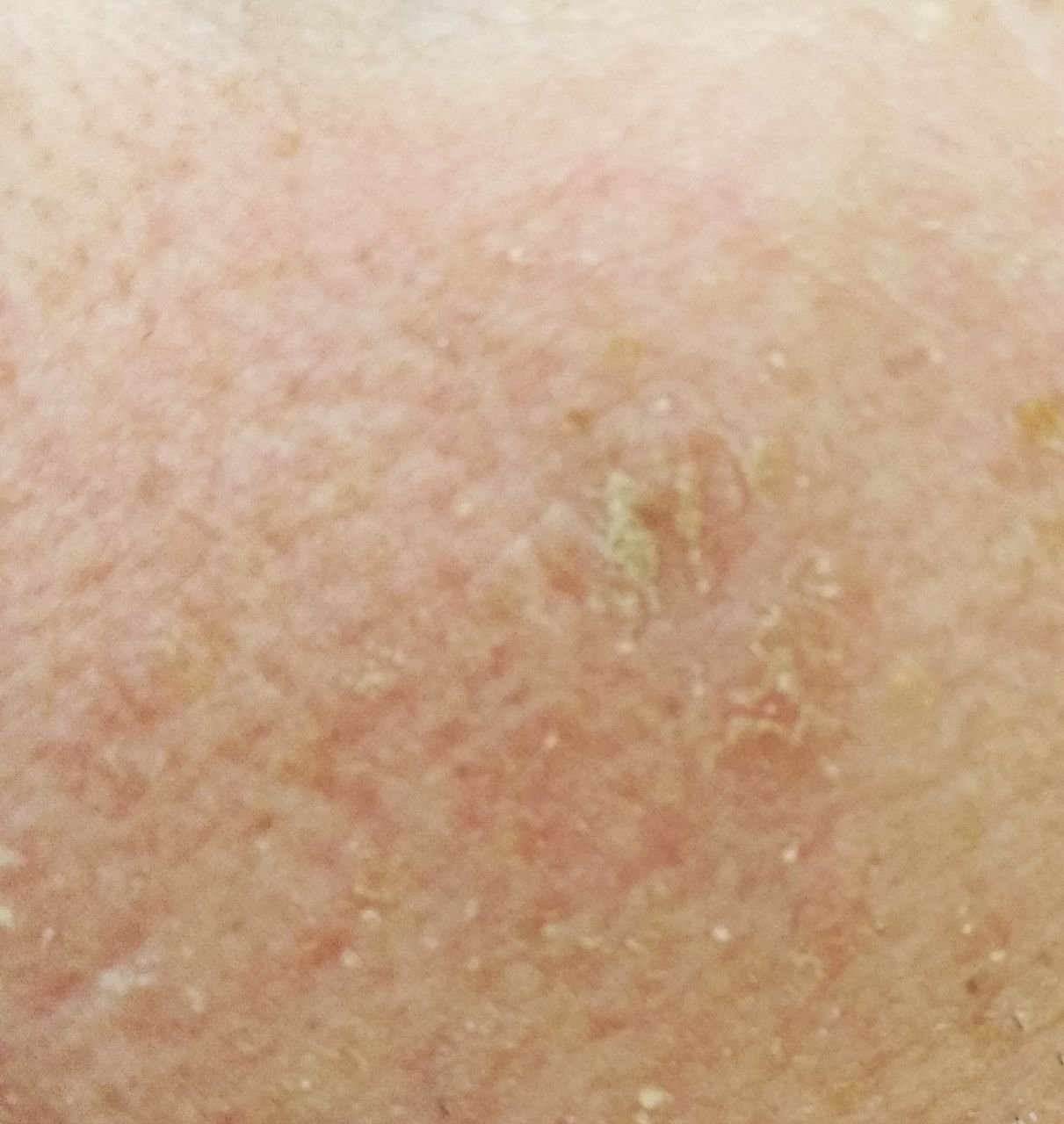
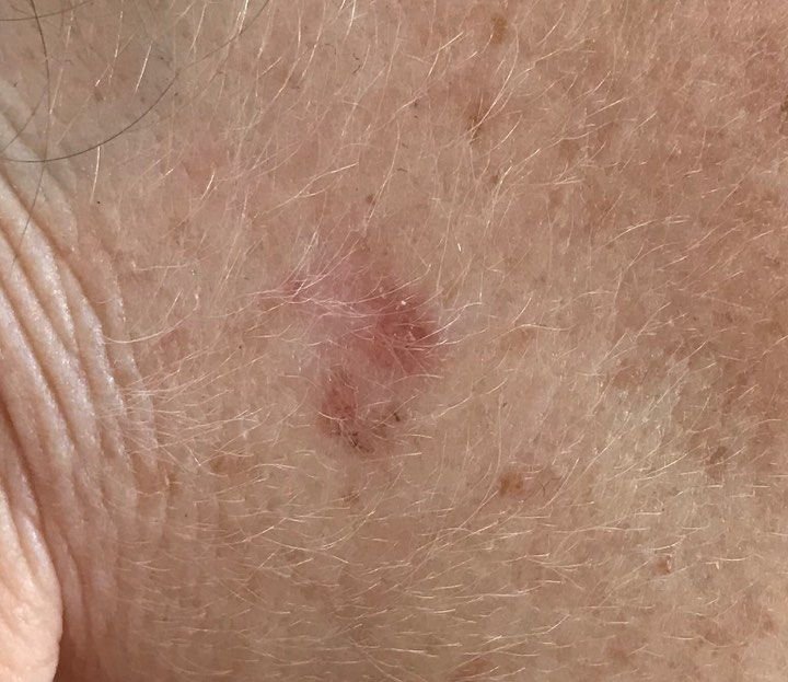
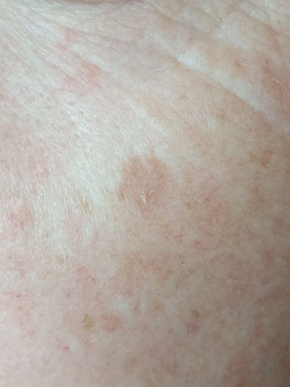
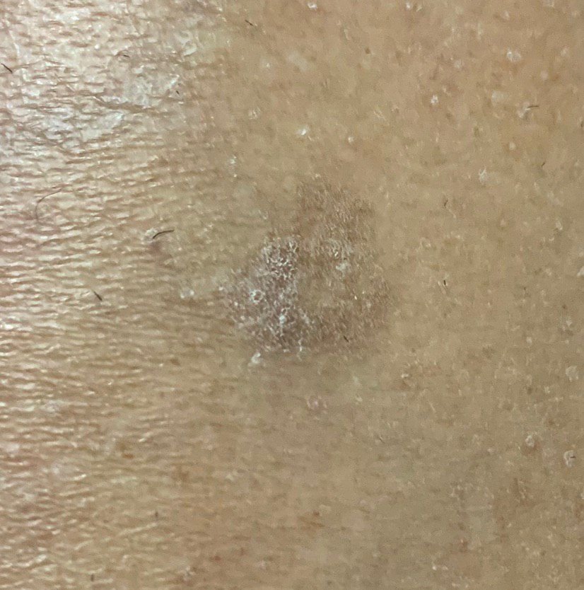
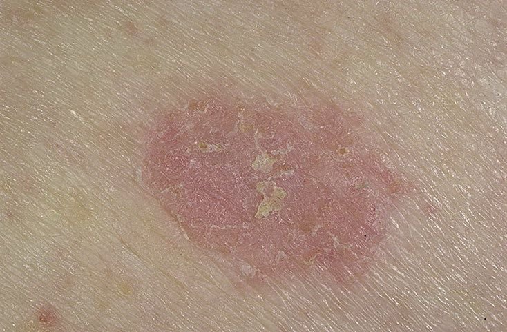
On visual examination, actinic keratosis presents as single or multiple flat or raised lesions with a rough, dry surface. These spots are typically covered with crusts and may show signs of erosion or bleeding. The lesions often appear asymmetrical, with uneven, poorly defined borders. The color of the lesions varies, ranging from flesh-colored to gray, gray-brown, or pink. In some cases, the lesions may show redness surrounding the lesion, which is a common feature.
The size of actinic keratosis can vary from 5 mm to 20 mm, and grouped lesions may cover an area of 3-4 cm or more. The height of the lesions above the skin surface usually does not exceed 5-7 mm. These lesions typically do not affect hair growth, and in some cases, they can become itchy or uncomfortable, although subjective sensations are usually minimal.
Actinic keratosis most commonly affects sun-exposed areas of the skin, such as the face, ears, scalp, neck, upper limbs (particularly the shoulder girdle and wrists), and chest. The lesions are less commonly found on the palms and soles of the feet.
Dermatoscopic Description
Dermatoscopy of actinic keratosis reveals several characteristic features that can help in the diagnosis:
- Erythema: The presence of redness or flushing around the lesions is a common feature.
- Red Pseudo-Network: A reticular structure formed by blood vessels beneath the surface of the skin.
- Superficial Keratin Flakes: The presence of dry, flaky skin on the surface of the lesion.
- White Halo Around Hair Follicles: A white ring of tissue around hair follicles within the lesion.
- Yellowish Keratotic Plugs: Horny masses or plugs of keratin can be seen in the expanded hair follicles.
- Vascular Structures: The lesion often contains convoluted and linear vascular structures, indicating blood vessel formation.
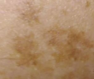
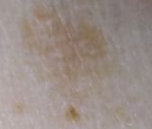
- Dark Brown Dots and Veins: In pigmented forms of actinic keratosis, dark brown veins, dots, and globules may be visible under dermatoscopy.
Differential Diagnosis
Actinic keratosis must be differentiated from other skin lesions and conditions, including:
- Psoriasis, eczema, and dermatitis
- Seborrheic keratosis
- Lentigo
- Post-inflammatory hyperpigmentation
- Melanosis
- Dysplastic nevus
- Bowen’s disease
- lentigo maligna melanoma
- Basal cell carcinoma
- Squamous cell carcinoma
- Melanoma
Risks
Actinic keratosis is a keratinocytic lesion caused by ultraviolet damage, with the potential to progress to cutaneous squamous cell carcinoma. Most individual lesions do not progress, and the risk varies with the lesion and with the patient — it is higher in people with extensive sun damage or with suppressed immunity. Actinic keratosis can progress to invasive squamous cell carcinoma; Bowen’s disease is an in situ form of SCC and is a related but distinct entity. Due to the cumulative effect of UV radiation, actinic keratosis lesions can increase in number and size over time, further raising the risk of malignant transformation.
Patients with many actinic keratoses have substantial cumulative UV damage and warrant regular skin surveillance, since other UV-related skin malignancies (such as BCC or SCC) may develop on the same exposed areas.
Tactics
Self-monitoring supplements clinical care rather than replacing it: an actinic keratosis can need assessment and treatment even when it is not changing and does not hurt, because it can progress to squamous cell carcinoma. This should include regular checks, at least once a year, especially for lesions in hard-to-see areas. If the lesion experiences mechanical injury, is exposed to UV radiation, or if any changes are observed, a consultation with a dermatologist is necessary.
A healthcare provider will determine whether further monitoring or removal of the lesions is necessary. Actinic keratosis lesions that are subject to chronic trauma, such as from clothing, jewelry, or occupational activities, should be evaluated by a dermatologist. It is also recommended to document any changes using photographs for dynamic observation.
Someone with multiple actinic keratoses is not cured by having individual lesions removed: new ones appear, and dermatological follow-up watches for signs of squamous cell carcinoma. How often depends on the extent of the sun damage and on whether the immune system is suppressed. Creating a map of skin neoplasms can help with ongoing monitoring and identification of any new or altered lesions.
Treatment
Treatment of actinic keratosis is necessary due to the risk of malignant transformation. Actinic keratoses generally require direct treatment and can persist or recur if untreated. Symptomatic treatments may include:
- Sun Protection: Sun protection is a core part of management, not an adjunct to it: continuing ultraviolet exposure drives new actinic keratoses and raises the risk of skin cancer generally. Broad-spectrum sunscreen used regularly, protective clothing, hats and limiting exposure all matter, and they matter after treatment as much as before it.
- Local Removal: Removal of keratotic plaques can be achieved through various methods, including laser treatment, cryodestruction (liquid nitrogen), and diathermocoagulation.
- Surgical Excision: In rare cases, surgical excision is used to remove large or problematic actinic keratoses. However, this method is less frequently used due to the high number of lesions and potential cosmetic consequences.
Where lesions are multiple or the surrounding skin is diffusely sun-damaged — field cancerisation — treatment is directed at the whole area rather than at single lesions. Which agent is chosen depends on the patient and on the clinician’s assessment, and availability differs by country. Options include:
- 5-fluorouracil, alone or with salicylic acid
- Imiquimod, photodynamic therapy, diclofenac gel or tirbanibulin
It is important that these treatments be administered under the supervision of a healthcare professional, as side effects and relapse are possible.
Prevention
Prevention of actinic keratosis and its potential for malignancy involves careful management of sun exposure and skin health:
- Limiting ultraviolet radiation, including avoiding tanning beds and excessive sun exposure.
- Using protective creams during periods of active sun exposure.
- Treating existing lesions rather than waiting, since new ones continue to appear on sun-damaged skin.
- Limiting or excluding ionizing radiation and occupational hazards.
- Following safety measures when handling skin-damaging substances.
- Examining your own skin regularly and reporting new or changing rough patches.
Regularly checking the skin for actinic keratosis, seeking timely consultation with a healthcare professional if changes occur, and removing potentially dangerous lesions are essential for maintaining skin health and preventing complications.