
Brown and dark spots on skin: pictures and possible causes
This tool is for informational purposes only and does not constitute medical advice. Always consult a qualified dermatologist.
The pictures and answers on this page are free. SkinAI Lab is a paid AI analysis; pricing is shown before you scan. The check does not cover every cause on this page, so a result cannot rule one out.
What causes brown or dark spots on the skin?
Brown or dark spots have several possible causes, including sun spots, freckles, moles, seborrheic keratoses and marks left after acne or a rash. Many of these are harmless, but some skin cancers, including melanoma, can also look brown, so a new or changing spot needs checking. Pictures can help you describe what you see, but they cannot tell you which condition you have. First, should you see a doctor?
- See a doctor about a new mole, a mole that has changed size, shape or colour, a spot with uneven edges or several colours, or a mark that has not gone away after a few weeks. All the signs
- Often harmless: freckles, sun spots (lentigo) and seborrheic keratoses are harmless, and moles are usually nothing to worry about unless they change. Check the signs first, and see a doctor if you are not sure what a spot is.
See a doctor — don’t rely on a photo check — if you have:
- A new mole, or a mole that has changed size, shape or colour
- A mole that is painful or itchy, or inflamed, bleeding or crusty
- A spot with an uneven shape or edges, or a mix of 2 or more colours
- A new or unusual mark on your skin that has not gone away after a few weeks, including on the palms of your hands or soles of your feet
- A spot that looks different from your other spots or moles
- A dark area under a nail that has not been caused by an injury
- A large mole that has been there since birth, in you or your child
- A growth that is getting bigger or has changed colour or texture; any spot that is itching or bleeding; or a growth or area of skin that hurts, crusts or scabs for more than 4 weeks
- A sore that does not heal, or heals and then comes back
- An age spot that darkens or changes
- New dark patches on your skin, such as dry, velvety patches in the armpits, neck or groin
- Round or oval brown spots on your shins: they can be a sign of diabetes or pre-diabetes
- Areas of darker skin together with extreme tiredness, weight loss, or feeling dizzy when standing up
- A child with 6 or more café-au-lait spots
Age spots are not dangerous, but they can look like some skin cancers, says Cleveland Clinic, so have a spot diagnosed by a doctor rather than by its colour.
Protect your skin from the sun. Sunlight makes many brown spots darker and helps new ones form; use a broad-spectrum sunscreen with SPF 30 or higher.
Sources: NHS — melanoma symptoms · AAD — Melanoma signs · NHS — skin cancer symptoms · AAD — Basal cell carcinoma signs · Atlas — Mole · Cleveland Clinic — Age spots · NHS — Acanthosis nigricans · AAD — Diabetes warning signs · NHS — Addison’s disease symptoms · NHS — Birthmarks · AAD — Dark spots in darker skin tones · DermNet — Postinflammatory hyperpigmentation
Dark spots on brown and black skin
-
Marks after acne or a rash
Dark marks left after acne, a rash or an injury are more common on darker skin, and they tend to be more intense and to last longer. Dark spots and patches are one of the most common reasons people with darker skin tones see a dermatologist.
-
Melasma
Melasma is most common in people who tan easily or have naturally brown skin.
-
Small dark bumps on the face
Dermatosis papulosa nigra, small dark bumps on the face, is common on darker skin.
-
Moles and growths
Moles are usually darker on brown and black skin, and on darker skin a basal cell carcinoma can be a firm, raised, round brown or black growth.
Soles, palms and nails. Our atlas notes that acral melanoma, more common in people with darker skin tones, often develops on the soles, the palms and under the nails, so a new dark mark there that does not go away needs checking.
Sources: DermNet — Postinflammatory hyperpigmentation · AAD — Dark spots in darker skin tones · DermNet — Melasma · DermNet — Dermatosis papulosa nigra · NHS — Moles · AAD — Basal cell carcinoma signs · Atlas — Melanoma · NHS — melanoma symptoms
Possible causes: examples with pictures
Grouped by features described in the atlas articles, not by every way a condition can look. Groups overlap and cannot identify or rule out a condition, so look through all three; pictures of skin cancers are in a separate section further down. Each description is quoted word for word from the atlas article; open a card for the full article and all its photos.
Flat brown spots or patches
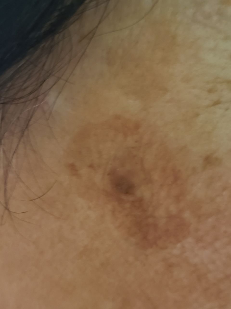
Sun spots are solar lentigines; other types of lentigo are not necessarily caused by sun exposure (atlas). Sun spots can be harder to see on darker skin (DermNet). On brown or black skin, tinea versicolor patches tend to be paler than the skin around them (NHS).
 20 photos
Lentigo (sun spots, age spots)
“The coloration ranges from light brown to dark brown, and the pigment is usually evenly distributed throughout the lesion.”
Read the article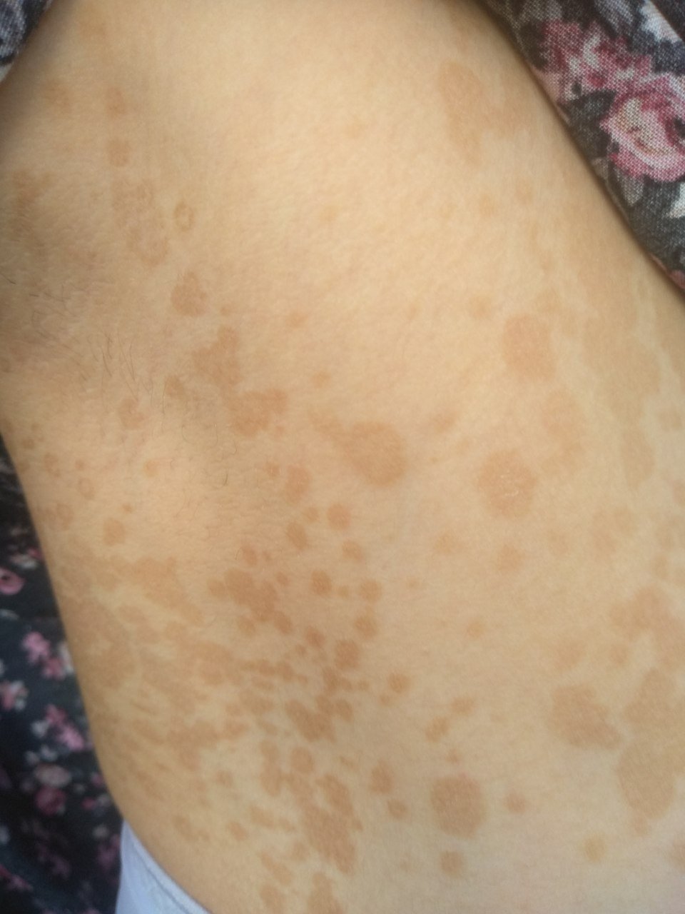 25 photos
Tinea versicolor (pityriasis versicolor)
“The hallmark of pityriasis versicolor is the development of well-demarcated, scaly, discolored patches that vary in hue—from pink, tan, and light brown to hypopigmented or chalky white, especially in darker-skinned individuals.”
Read the articleRaised, rough or firm brown spots
Actinic keratosis can develop into squamous cell carcinoma, a type of skin cancer, so have it assessed by a doctor even if it is not changing or painful (atlas).
 30 photos
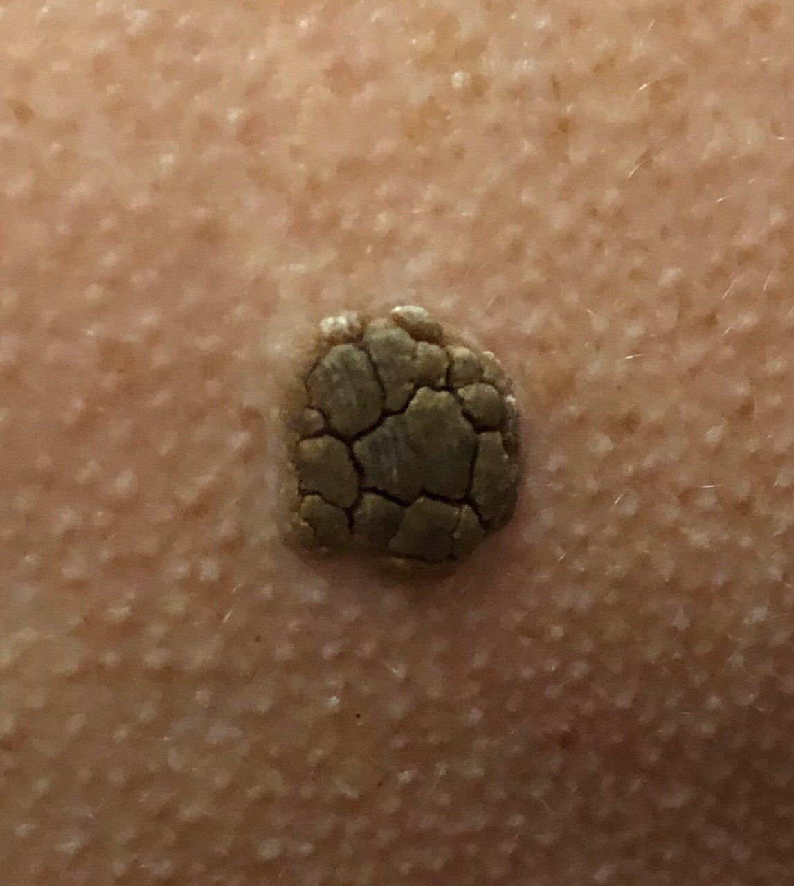
Seborrheic keratosis
“The surface texture of seborrheic keratosis differs from that of normal skin: it is rough, dry, and may show signs of peeling.”
“The color of seborrheic keratosis varies from nude or gray to gray-brown.”
Read the article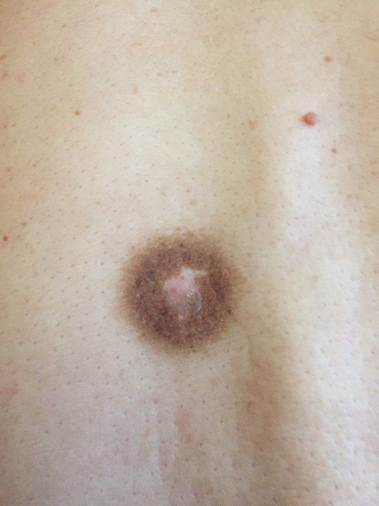 24 photos
Dermatofibroma
“The color of the lesion ranges from flesh-colored to gray or brown shades, with the pigment distribution being uneven.”
Read the article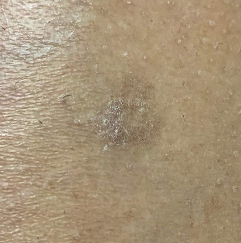 20 photos
Actinic keratosis (solar keratosis)
“The color of the lesions varies, ranging from flesh-colored to gray, gray-brown, or pink.”
Read the articleMoles
Moles are usually darker on brown and black skin (NHS). Atypical moles can look like melanoma; people with several have a higher melanoma risk and need regular skin checks by a dermatologist (atlas).
 24 photos
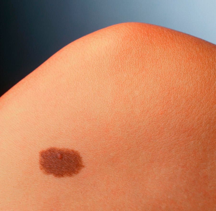
Mole (benign nevus)
“The color of a simple nevus ranges from light brown to dark brown, with uniform pigment distribution.”
Read the article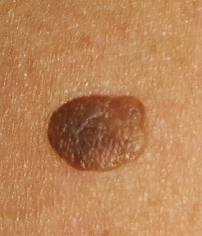 30 photos
Raised mole (papillomatous nevus)
“The color of the nevus can range from flesh-colored to tan and dark brown, and the pigment distribution is typically uniform.”
Read the article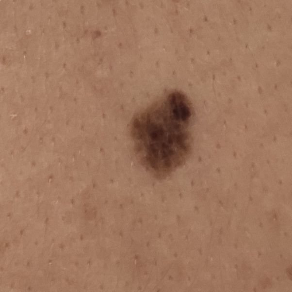 24 photos
Atypical mole (dysplastic nevus)
“The color of the nevus can vary widely, ranging from light brown to dark brown, and may also include shades of red, pink, and even blue.”
Read the articleBrown spots that appear suddenly
Brown spots can become noticeable for several reasons.
- Freckles multiply after time in the sun and darken in summer, and sun spots can appear on sun-damaged skin after a sunburn.
- Dark marks can appear where a rash, acne or an injury has healed.
- Melasma often first shows during pregnancy or after starting birth control pills.
- Some medicines darken the skin, tinea versicolor makes patches of skin change colour, and capillaritis can cause groups of new red-brown patches on the lower legs for no apparent reason.
- It is normal for new moles to appear in children and teenagers.
See a doctor
- See a doctor about a new mole or one that changes size, shape or colour.
- Have a new or unusual mark checked if it has not gone away after a few weeks, including on the palms or soles.
- A dark area under a nail that was not caused by an injury should also be checked.
Sources: DermNet — Freckles · DermNet — Solar lentigo · DermNet — Postinflammatory hyperpigmentation · AAD — Melasma · DermNet — Drug-induced pigmentation · NHS — Pityriasis versicolor · DermNet — Capillaritis · NHS — Moles · NHS — melanoma symptoms
Other possible causes
More possible causes, described using the linked sources; where relevant, we also quote related atlas articles. For pale patches, see our page on white spots on skin.
Melasma
Forehead, cheeks, nose, upper lip
Blotchy, light to dark brown patches with irregular borders on both sides of the face, most often the forehead, cheeks, nose and upper lip. It is more common in women, usually starting between 20 and 40, and in people who tan easily or have naturally brown skin. Sun exposure, pregnancy and hormonal birth control can play a part; it is so common in pregnancy that it is sometimes called the “mask of pregnancy”. For some women it goes away after the baby is born or after stopping the pill. It is not contagious.
Freckles
Nose, cheeks
Small, flat, light to dark brown spots, 3 to 10 mm across, on sun-exposed skin, particularly the nose and cheeks. They are inherited, not present at birth, multiply after sun exposure, darken in summer and fade in winter. They need no treatment; sun protection reduces the summer darkening.
Dermatosis papulosa nigra
Cheeks, forehead
Many small, smooth, firm, black or dark brown bumps, 1 to 5 mm across, mainly on the cheeks and forehead. It is common in people with darker skin and usually begins in adolescence. The bumps are the same as small seborrheic keratoses and are generally best left untreated.
Dark marks after inflammation (post-inflammatory hyperpigmentation)
Where the skin has healed
Light brown to black patches left where a rash, acne, a burn or another injury has healed. They can occur in anyone, but are more common in people with darker skin, in whom the colour tends to be more intense and to last longer. Sunlight can make them darker. Our atlas article on acne describes “Post-inflammatory brown spots that remain after the resolution of acne lesions.”
DermNet — Postinflammatory hyperpigmentation · AAD — Dark spots in darker skin tones
Café-au-lait spots
Anywhere on the body
Flat, light or dark brown patches that can be anywhere on the body. They are usually present at birth or appear in early childhood, and many children have 1 or 2. They look darker on brown or black skin.
A child with 6 or more may have neurofibromatosis type 1, so they should be seen by a doctor.
Acanthosis nigricans
Armpits, neck, groin
Dry, dark patches of skin that feel like velvet, usually in the armpits, neck or groin. The most common cause is obesity; others include type 2 diabetes, hormone conditions and some medicines, and rarely cancer. Sometimes healthy people get it, which is more common in people with black or brown skin.
It is usually harmless, but it needs to be checked by a primary care doctor (GP).
Erythrasma
Skin folds, between the toes
Well-defined pink or brown patches with fine scale in skin folds: the armpits, groin, under the breasts and between the toes. It is caused by bacteria and can itch mildly.
Shin spots (diabetic dermopathy)
Shins
Round or oval, brown or reddish-brown spots on the shins, scaly at first and later leaving small dents. They are common in people with diabetes and are often mistaken for age spots. They often fade once diabetes is well controlled.
The American Academy of Dermatology lists them among skin signs that can mean undiagnosed diabetes or pre-diabetes.
AAD — Diabetes warning signs · DermNet — Skin problems in diabetes
Brown staining on the lower legs
Lower legs
Long-standing high pressure in the leg veins can cause venous (varicose) eczema: itchy, scaly, discoloured skin on the lower leg, with swelling that is worse by evening, visible varicose veins and “a brownish staining from iron released by leaked red blood cells”, as our atlas article on venous eczema puts it. On black or brown skin it tends to look dark brown, purple or grey.
See a doctor if you have these symptoms, and seek care promptly if the skin breaks, an area becomes painful, hot and spreading, or the leg suddenly swells.
Capillaritis (pigmented purpura)
Lower legs
A harmless condition in which tiny blood vessels leak, causing small red and brown “cayenne pepper” dots that can group into flat red-brown patches and slowly fade over weeks to months. It is most common on the lower legs and usually causes no symptoms.
Medicines
Some medicines can darken the skin, including antimalarials, some chemotherapy drugs, anti-inflammatory painkillers (NSAIDs), antipsychotics, amiodarone, some epilepsy medicines and minocycline. A doctor diagnoses it from your medicine history and the pattern of the colour change.
Do not stop a prescribed medicine on your own: ask the doctor who prescribed it whether it could be causing the change.
DermNet — Drug-induced pigmentation · AAD — Dark spots in darker skin tones
Addison’s disease
A rare hormone condition. Its symptoms include areas of skin that are darker than the skin around them, which may be harder to see on brown or black skin, along with extreme tiredness, weight loss and feeling dizzy when standing up.
How selected causes differ
A summary of selected features from our atlas articles and the sources on this page. Many conditions overlap, so this table cannot tell you which one you have. Tap a condition to see its picture or description.
| Condition | What it usually looks like | How it feels | Where it often appears |
|---|---|---|---|
| Lentigo (sun spots) | Flat light to dark brown spots, evenly coloured, from 2–3 mm to 3–4 cm | Like normal skin; older spots can feel rough | Face, neck, hands |
| Freckles | Light to dark brown flat spots, 3–10 mm, poorly defined; darker in summer, fading in winter | Flat | Sun-exposed skin, especially the nose and cheeks |
| Melasma | Blotchy light to dark brown patches with irregular borders, on both sides of the face | No symptoms | Forehead, cheeks, nose, upper lip, jawline; sometimes forearms |
| Marks after inflammation | Light brown to black patches where a rash, acne or an injury has healed | Not stated in our sources | Where the skin was inflamed or injured |
| Tinea versicolor | Flat, round patches that can join up: pink, red or pale brown on white skin; paler than the skin around them on brown or black skin | May feel scaly, sometimes itchy | Chest, upper back, upper arms, neck, tummy |
| Seborrheic keratosis | Raised growths with a rough, dry surface, round or oval, 5–20 mm, skin-coloured or grey to grey-brown | Usually no symptoms; older ones may itch mildly | Trunk, arms, face |
| Mole | Small round or oval spot or bump, light to dark brown, usually one colour and up to 10 mm | Flat or raised; smooth or rough | Trunk and limbs; fewer on the head and neck |
| Atypical mole | Usually 10–15 mm, with irregular borders; light to dark brown, sometimes with red, pink or blue | Slightly denser than normal skin; usually no symptoms | Anywhere; often sun-exposed skin |
| Dermatofibroma | Slightly sunken or dome-shaped growth, flesh-coloured to grey or brown, usually under 10 mm | Firm and dense; mild itching is rare | Legs and shoulder area |
| Actinic keratosis | Patches 1–2 cm with a rough, dry surface: skin-coloured, pink, red or brown | Rough, like sandpaper; may itch | Face, ears, scalp, hands, arms, legs |
| Café-au-lait spots | Flat, light or dark brown patches with a sharp border, usually from birth or early childhood | Flat | Anywhere on the body |
| Acanthosis nigricans | Patches of skin that are darker and thicker than usual | Dry, feels like velvet; sometimes itchy | Armpits, neck, groin |
| Shin spots (diabetic dermopathy) | Round or oval brown or reddish-brown spots that can leave small dents | Scaly at first; no itch or pain | Shins |
| Melanoma | Often an uneven shape or edges, a mix of 2 or more colours, and changes over time; often more than 6 mm across, but can be smaller | Can be swollen and sore, bleeding, itchy or crusty | Anywhere; more common on sun-exposed skin |
Sources: the atlas articles linked in the cards · DermNet — Freckles · DermNet — Melasma · DermNet — Postinflammatory hyperpigmentation · NHS — Pityriasis versicolor · NHS — Moles · NHS — Actinic keratoses · NHS — Birthmarks · DermNet — Café-au-lait macule · NHS — Acanthosis nigricans · AAD — Diabetes warning signs · NHS — melanoma symptoms
Can a brown spot be skin cancer?
Normal mole
Normal moles are usually round with smooth edges, one colour, and about the size of the end of a pencil or smaller.
Melanoma
Melanomas are often an uneven shape with uneven edges, a mix of 2 or more colours, and often more than 6 mm wide, though they can be smaller when found early. A mole that changes size, shape or colour may be a melanoma.
Non-melanoma skin cancers can also be brown: on darker skin, a firm, raised, round brown or black growth can be a sign of basal cell carcinoma (AAD).
Sources: NHS — Symptoms of melanoma skin cancer · AAD — Basal cell carcinoma signs
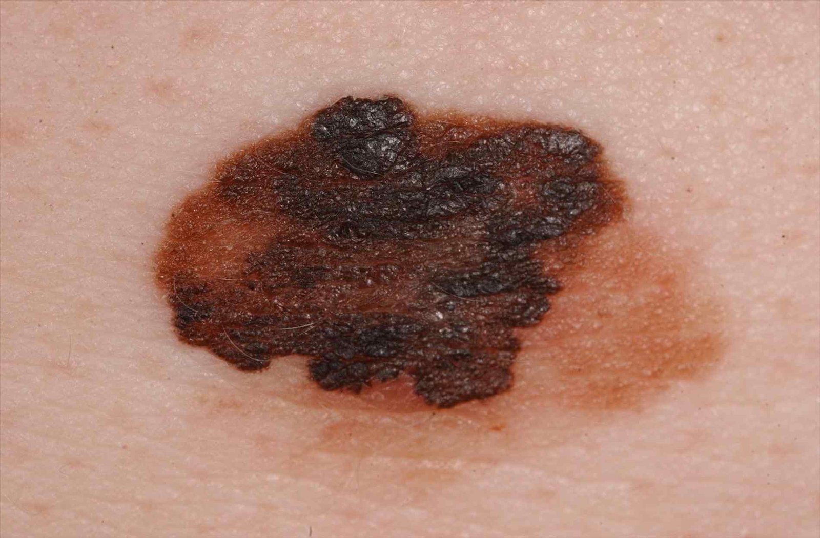 32 photos
Melanoma
“C – Color: Uneven pigmentation with multiple shades of brown, black, or other colors like blue, pink, or white.”
Read the article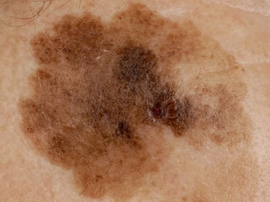 24 photos
Lentigo maligna melanoma
“Gradual enlargement of a previously stable pigmented patch.”
“This tumor most often arises in individuals over the age of 50 and typically affects areas chronically exposed to ultraviolet radiation, such as the face, neck, ears, and scalp.”
Read the article
Medical review of the atlas articles above
Board-Certified Dermatologist · Fellow of the American Academy of Dermatology (FAAD)
Terms used in the quotes
- Pigment
- A substance that gives color to tissue. Pigments are responsible for the color of skin, eyes, and hair.
- Hyperpigmentation
- A common, usually harmless condition in which patches of skin are darker than the surrounding skin.
- Hypopigmented
- Lighter in colour than normal skin.
- Lesion
- An area of abnormal or damaged tissue caused by injury, infection, or disease.
- Nevus
- A benign (not cancer) growth on the skin that is formed by a cluster of melanocytes (cells that make a substance called melanin, which gives color to skin and eyes).
- Tumor
- An abnormal mass of tissue that forms when cells grow and divide more than they should or do not die when they should. Tumors may be benign (not cancer) or malignant (cancer).
Source: NCI Dictionary of Cancer Terms.
Brown and dark spots on skin: FAQ
Why do I have brown spots on my skin?
Extra melanin, the pigment that gives skin its colour, can cause brown or dark spots. Common causes include sun spots (solar lentigo), freckles, moles, seborrheic keratoses, marks left after acne or a rash, and melasma. Sun exposure plays a part in many of them. Pictures and descriptions cannot tell you which one you have, so have a new or changing spot checked by a doctor. Sources: DermNet — Solar lentigo, DermNet — Freckles, NHS — Moles, DermNet — Postinflammatory hyperpigmentation, DermNet — Melasma, the atlas articles in the cards above.
Can a brown spot be skin cancer?
Yes, some can. Melanoma is often an uneven shape with uneven edges and a mix of 2 or more colours, and often more than 6 mm wide, though it can be smaller if found early. Lentigo maligna melanoma can start as a slowly growing, unevenly coloured flat patch on sun-damaged skin, most often on the face, neck, ears or scalp of people over 50. On darker skin, a firm, raised, round brown or black growth can be a sign of basal cell carcinoma. See a doctor about a new mole, a mole that has changed size, shape or colour, or a mole that is painful, itchy, inflamed, bleeding or crusty. Sources: NHS — melanoma symptoms, Atlas — Lentigo maligna melanoma, AAD — Basal cell carcinoma signs.
What is the difference between an age spot and melanoma?
You cannot be sure by looking. Our atlas describes lentigo (sun spots, age spots) as flat and evenly coloured, light to dark brown. Melanomas are often an uneven shape with a mix of colours, and they change over time. Age spots can look similar to some skin cancers, so Cleveland Clinic advises seeing a dermatologist for a diagnosis, and seeing one right away if an age spot darkens or changes. Sources: Atlas — Lentigo, NHS — melanoma symptoms, Cleveland Clinic — Age spots.
Are liver spots caused by the liver?
No. Liver spots is another name for age spots or sun spots (solar lentigines). They have nothing to do with your liver except a similar colour, tan to dark brown. They are caused by years of sun exposure and are more common in adults over 50. Sources: Cleveland Clinic — Age spots, DermNet — Solar lentigo.
What causes dark spots on the face?
Common causes are melasma, sun spots, freckles and dark marks left after acne or other inflammation. Small dark bumps on the cheeks and forehead can be dermatosis papulosa nigra, which is common on darker skin. In people over 50, a slowly growing, unevenly coloured patch on the face can be lentigo maligna melanoma, so a patch that changes should be checked by a doctor. Sources: DermNet — Melasma, DermNet — Solar lentigo, DermNet — Freckles, DermNet — Postinflammatory hyperpigmentation, DermNet — Dermatosis papulosa nigra, Atlas — Lentigo maligna melanoma.
What causes brown spots on the legs?
Possible causes include sun spots on the shins, shin spots linked to diabetes, brown staining from venous eczema, capillaritis, and dermatofibroma, a firm bump most common on the legs. Melanoma can also appear on the legs. See a doctor about round brown spots on your shins: if you have not been diagnosed with diabetes, ask whether you need testing; if you have diabetes, tell the doctor who manages it. Also have any new or changing spot checked. Sources: DermNet — Solar lentigo, AAD — Diabetes warning signs, NHS — Varicose eczema, DermNet — Capillaritis, Atlas — Dermatofibroma, NHS — melanoma symptoms.
Can a fungal infection cause brown spots?
Yes. Tinea versicolor (pityriasis versicolor) is a common fungal infection that causes patches of skin to change colour: on white skin they are usually pink, red or pale brown, and on brown or black skin they tend to be paler than the skin around them. It does not spread from person to person. The NHS advises not to diagnose it yourself and to see a GP or pharmacist if you are worried. Erythrasma, which causes pink or brown patches in skin folds, is caused by bacteria rather than a fungus. Sources: NHS — Pityriasis versicolor, DermNet — Erythrasma.
Can dark patches be a sign of an illness?
Sometimes. Dry, velvety dark patches in the armpits, neck or groin (acanthosis nigricans) can be linked to obesity, type 2 diabetes or hormone conditions. Brown spots on the shins can be a sign of diabetes. Darker skin together with extreme tiredness, weight loss and dizziness can be a symptom of Addison’s disease, which is rare. Some medicines darken the skin. See a GP about new dark patches or skin changes you are unsure about. Sources: NHS — Acanthosis nigricans, AAD — Diabetes warning signs, NHS — Addison’s disease symptoms, DermNet — Drug-induced pigmentation.
Why does my child have brown spots?
Many children have 1 or 2 café-au-lait spots, and it is normal for new moles to appear in children and teenagers. Freckles appear on sun-exposed skin and are most noticeable in childhood. A child with 6 or more café-au-lait spots should see a doctor, because this can be a sign of neurofibromatosis type 1. Blue-grey birthmarks that look like a bruise are common on babies with brown or black skin and usually go away by the age of 4. Sources: NHS — Birthmarks, NHS — Moles, DermNet — Freckles.
How do I get rid of brown or dark spots?
Find out what the spots are first: treatment depends on the cause, and a spot that could be melanoma needs a doctor, not a cream. Many harmless spots, such as freckles and sun spots, need no treatment. Sun protection helps prevent new spots and stop existing ones getting darker; the American Academy of Dermatology recommends a broad-spectrum, water-resistant sunscreen with SPF 30 or higher, and for dark marks left after acne or a rash, a tinted sunscreen containing iron oxide, which also protects against visible light. If the cause can be removed, many dark marks clear on their own, but do not stop a prescribed medicine on your own: ask the doctor who prescribed it. A dermatologist can explain options such as prescription creams, chemical peels, freezing and laser. Avoid skin-lightening creams that contain mercury, which can damage the kidneys and nerves. Sources: AAD — Dark spots in darker skin tones, Cleveland Clinic — Age spots, DermNet — Freckles, NHS — melanoma symptoms.
When should I worry about a brown spot?
See a doctor about a new mole, or a mole that has changed size, shape or colour; a spot that looks different from your other spots or moles; a mole that is painful, itchy, inflamed, bleeding or crusty; a new or unusual mark that has not gone away after a few weeks, including on the palms or soles; or a dark area under a nail that was not caused by an injury. Also see a doctor about any spot that is itching or bleeding; a growth that is getting bigger or has changed colour or texture; a growth or area of skin that hurts, crusts or scabs for more than 4 weeks; or new dark patches you are unsure about. Sources: NHS — melanoma symptoms, AAD — Melanoma signs, NHS — skin cancer symptoms, NHS — Acanthosis nigricans.
For a clear photo: daylight, close and in focus, no filters. Upload a photo for an AI skin check. The check does not cover every cause on this page, so a result cannot rule one out. AI output is not a medical diagnosis. If you have any of the signs above, see a doctor instead.
Check my skin ↑SkinAI Lab is a paid AI analysis; pricing is shown before you scan.


Prepared by the SkinAI Lab editorial team · Updated . Apart from the quoted atlas text, this page has not been reviewed by a doctor. It is general information, not medical advice.