
Café-au-Lait Spots in Children: A Sign of Neurofibromatosis?
Café-au-lait spots in children: When might they need a closer look?
Café-au-lait spots are flat patches of skin that are darker than the surrounding area. They are common in children, and a few isolated spots usually do not point to a wider health problem.
Parents may worry because these spots can also be an early sign of neurofibromatosis type 1 (NF1), a genetic condition that can affect the skin, nerves, bones, and other parts of the body. The number and size of the spots, along with a child’s age and any other signs, can help doctors decide whether further evaluation is needed.
In an episode of Don’t Be Rash, the Society for Pediatric Dermatology’s official podcast, host Andrew Krakowski, MD, spoke with Joseph Lam, MD, FRCPC, of BC Children’s Hospital in Vancouver. They discussed how doctors distinguish ordinary café-au-lait spots from early NF1 and other conditions that can look similar. (Source: Don’t Be Rash, Society for Pediatric Dermatology)
What makes a café-au-lait spot different from a mole?
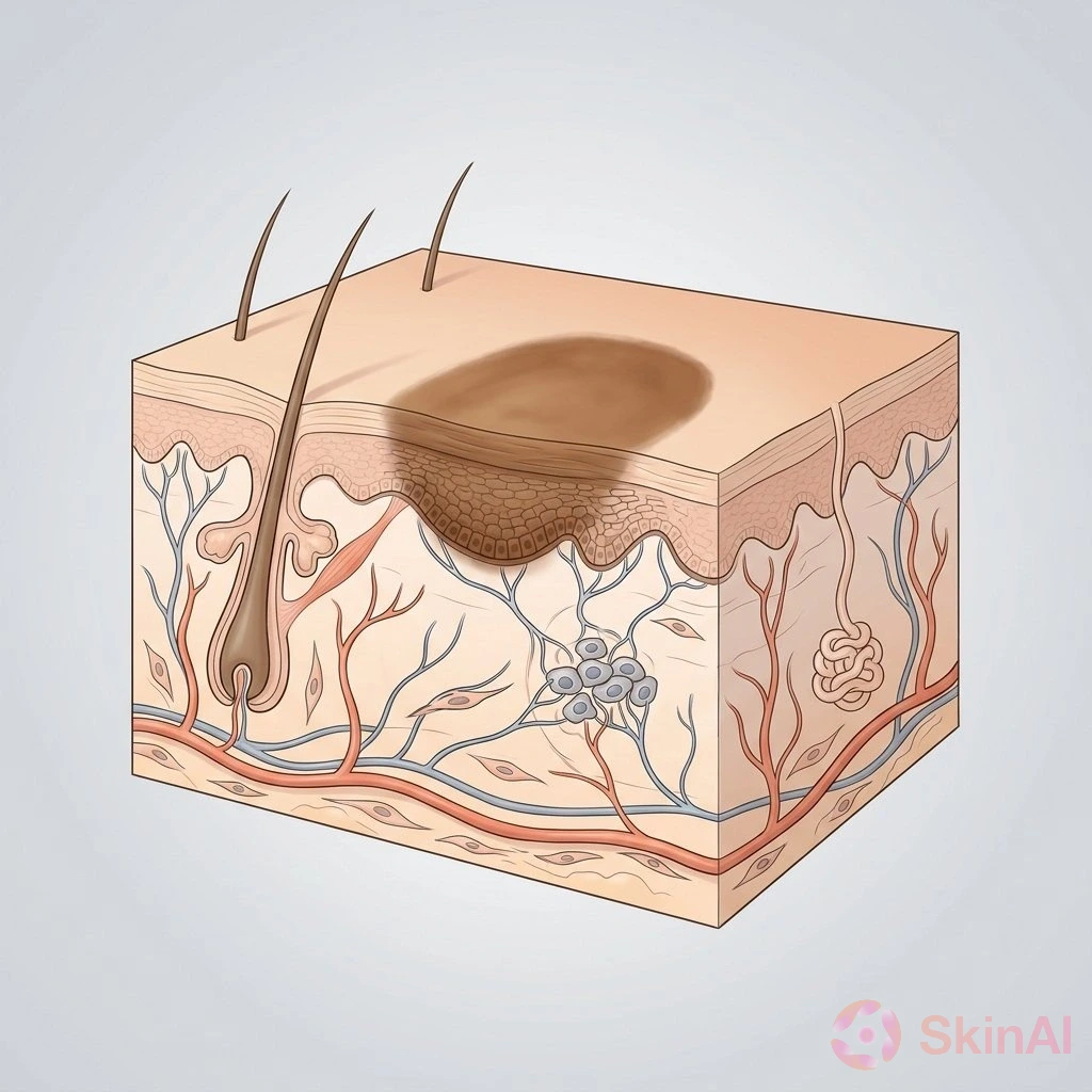
Café-au-lait spots, also called café-au-lait macules, get their color from an increase in skin pigment. Unlike many moles, they are not caused by an increased number of pigment-producing cells.
One or a few spots on their own are common. A larger number may prompt a doctor to look for other signs of NF1 or a related genetic condition.
How doctors assess the possibility of NF1
The 2021 international diagnostic criteria for NF1 include several features. For someone without an affected parent, a diagnosis generally requires at least two qualifying features. (Source: Legius et al., Genetics in Medicine, 2021)
Café-au-lait spots count as one feature when there are at least six of them, and each qualifying spot is more than 5 millimeters across before puberty or more than 15 millimeters across after puberty. Having six spots alone does not establish a diagnosis of NF1.
Other signs doctors consider include:
- Freckles in skin folds, such as the armpits or groin.
- Neurofibromas, which are usually noncancerous growths involving nerve tissue.
- Lisch nodules, which are small growths on the colored part of the eye, or certain other eye findings.
- An optic pathway glioma, a tumor affecting the pathway that carries visual information from the eyes.
- Certain distinctive bone changes.
- A disease-causing change in the NF1 gene.
An affected parent is also important to the assessment. Different diagnostic criteria apply when a parent has NF1. (Source: Legius et al., Genetics in Medicine, 2021)
Why a child’s age matters
Signs of NF1 can appear over time, so an examination at one age may not tell the whole story.
In one study of children with isolated café-au-lait spots, researchers reported that children younger than 29 months who had at least six spots had an 80.4% estimated risk of having constitutional NF1. “Constitutional” means the genetic change is present throughout the body, rather than only in some cells. For children older than 29 months who had fewer than six spots or spots with an atypical appearance, the reported risk was 0.9%. These figures come from a study group and do not predict what will happen for an individual child. (Source: Ben-Shachar et al., Journal of the American Academy of Dermatology, 2017)
A separate study from St. Louis found that most children with at least six typical café-au-lait spots met the diagnostic criteria for NF1 by age 6. (Source: Nunley et al., Archives of Dermatology, 2009)
Children who have fewer than six spots may still have periodic full-skin examinations, because new spots or freckles can appear as they grow. Lisch nodules also become more likely with age, so their absence in an adolescent can be reassuring.
Other conditions that can look like NF1
NF1 is not the only condition associated with café-au-lait spots. Some conditions share certain features but have different causes and health effects.
Legius syndrome
Legius syndrome is caused by changes in the SPRED1 gene, which is involved in the same cell-signaling pathway as the gene associated with NF1. It can cause café-au-lait spots and freckles in skin folds, but it does not have the full range of features seen in NF1. (Source: Legius et al., Genetics in Medicine, 2021)
Cutaneous mastocytosis
Maculopapular cutaneous mastocytosis is a skin condition that can cause brown spots or bumps. Compared with café-au-lait spots, these areas may be raised and have less sharply defined edges.
A doctor may look for Darier sign, a reaction in which a spot becomes swollen or develops a hive-like appearance after it is rubbed. This finding can support the diagnosis.
McCune-Albright syndrome and mosaic conditions
McCune-Albright syndrome is a condition caused by a genetic change that occurs after an egg is fertilized. It can cause café-au-lait spots in a limited area of the body, along with changes in several bones and overactive hormone production. Hormone-related problems can include early puberty or Cushing syndrome in a newborn.
Segmental pigmentary mosaicism, a pattern of differently colored skin affecting a limited area, is much more common. Another condition, mosaic NF1, occurs when the NF1-related genetic change is present in only some cells. Its appearance can resemble a speckled birthmark called a speckled lentiginous nevus.
When genetic testing may help
Genetic testing is most useful when the result could change a person’s care. A test that looks at a group of genes involved in related conditions can include SPRED1 and may help distinguish NF1 from Legius syndrome. A disease-causing NF1 gene change is also included in the updated diagnostic criteria. (Source: Legius et al., Genetics in Medicine, 2021)
If siblings have concerning findings but neither parent is affected, doctors may also consider a rare inherited condition called constitutional mismatch repair deficiency. This condition affects the body’s ability to repair certain mistakes in DNA.
Care and follow-up
For a child with confirmed NF1, health supervision guidance emphasizes regular monitoring for possible health concerns. Routine blood pressure checks are one part of that follow-up. The care plan and any decisions about testing or treatment should be discussed with the child’s doctor or dermatologist. (Source: Miller et al., Pediatrics, 2019)
For many families, careful counting and follow-up provide reassurance. For others, a referral can help identify whether more structured monitoring is needed.
When to speak with a doctor
A doctor or dermatologist can help assess café-au-lait spots if you are unsure what they are, notice several spots, or have concerns about a child’s skin or development. Seek medical advice for a spot or other skin change that is growing quickly, bleeding, painful, changing, or showing signs of infection.
Keeping a simple record of visible changes over time may help you prepare for an appointment, but it cannot establish a diagnosis.
This article is for information only and is not a substitute for care from a qualified health professional.
Sources
- Society for Pediatric Dermatology. Don’t Be Rash podcast interview with Andrew Krakowski, MD, and Joseph Lam, MD, FRCPC, on café-au-lait spots, NF1, and similar conditions.
- Legius E, Messiaen L, Wolkenstein P, et al. Revised diagnostic criteria for neurofibromatosis type 1 and Legius syndrome: an international consensus recommendation. Genetics in Medicine. 2021;23(8):1506–1513. doi:10.1038/s41436-021-01170-5.
- Ben-Shachar S, Dubov T, Toledano-Alhadef H, et al. Predicting neurofibromatosis type 1 risk among children with isolated café-au-lait macules. Journal of the American Academy of Dermatology. 2017;76(6):1077–1083.e3. doi:10.1016/j.jaad.2017.02.027.
- Nunley KS, Gao F, Albers AC, Bayliss SJ, Gutmann DH. Predictive value of café au lait macules at initial consultation in the diagnosis of neurofibromatosis type 1. Archives of Dermatology. 2009;145(8):883–887. doi:10.1001/archdermatol.2009.169.
- Miller DT, Freedenberg D, Schorry E, et al. Health supervision for children with neurofibromatosis type 1. Pediatrics. 2019;143(5):e20190660. doi:10.1542/peds.2019-0660.