
Erythema Annulare Centrifugum (ICD-10: L53.1) ⚠️
Erythema Annulare Centrifugum (EAC)
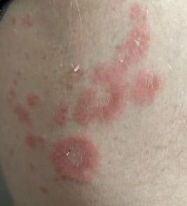
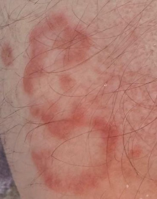
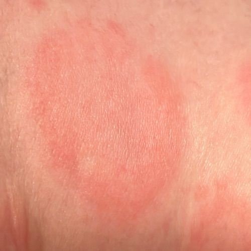
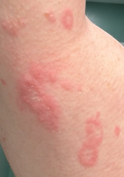
Erythema annulare centrifugum (EAC) is an uncommon reactive skin condition in which erythematous lesions enlarge slowly outwards, taking an annular or arciform shape. It belongs to the group of figurate or gyrate erythemas. In some cases an associated infection, medicine or underlying disorder can be identified; in many, no trigger is ever found.
Historical Background
EAC was first described in 1916 by French dermatologist Ferdinand-Jean Darier, who recognized its characteristic circular lesions and distinct clinical features.
Causes and Pathogenesis
EAC is a reactive erythema: the eruption reflects the skin responding to something else. Where no trigger can be found — which is common — it is termed idiopathic.
On histology, EAC shows a lymphohistiocytic infiltrate arranged tightly around the dermal vessels — the “coat-sleeve” pattern. The epidermis is spared in the deep form; in the superficial form it shows mild change, which is what produces the trailing scale.
Epidemiology
EAC is uncommon, and reliable incidence figures are not available: it is reported from case series rather than from population studies, so no rate, sex ratio or ethnic distribution can be stated with confidence. It is described at any age, including in children, and most often in adults. Rare familial cases with autosomal dominant inheritance have been reported.
Associated Conditions and Triggers
EAC is a reactive eruption: the skin is responding to something rather than being diseased in its own right. The important qualification is that in a large share of cases no trigger is ever identified, and the lists of specific organisms, drugs and foods that circulate in the literature come from individual case reports rather than from established causation.
The categories worth considering, guided by the patient’s history and examination rather than by a checklist, are:
- Infection — a dermatophyte infection elsewhere on the skin is the association most often worth looking for, and other bacterial, viral and fungal infections have been reported;
- Medicines — a number have been implicated in single reports. What points to a drug is the timing, and lesions settling after it is stopped;
- Underlying systemic disease, including autoimmune conditions;
- Malignancy — reported with lymphoproliferative disorders in particular. This association exists but is uncommon, and it does not justify investigating every patient for cancer.
Clinical Manifestations
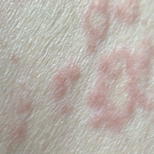
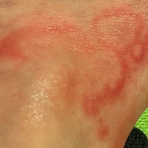
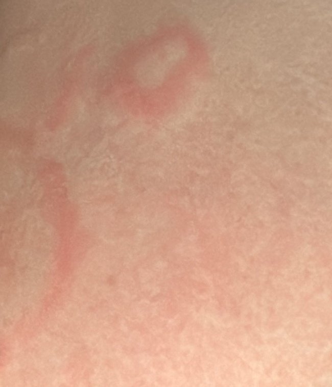
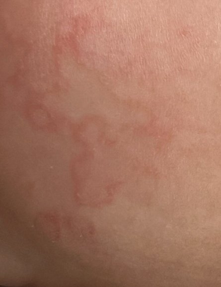
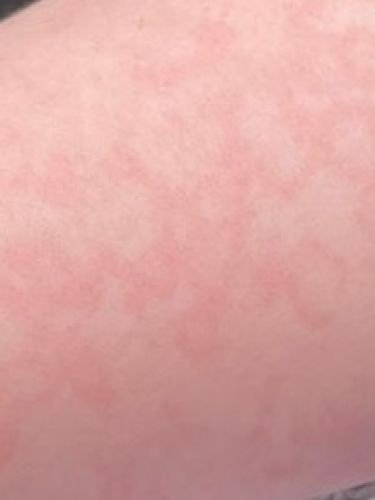
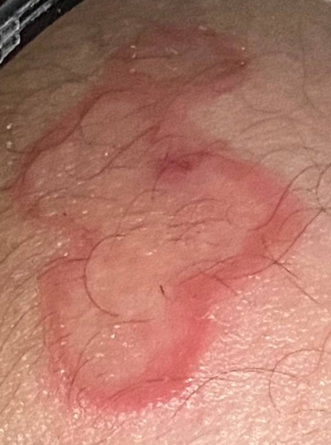
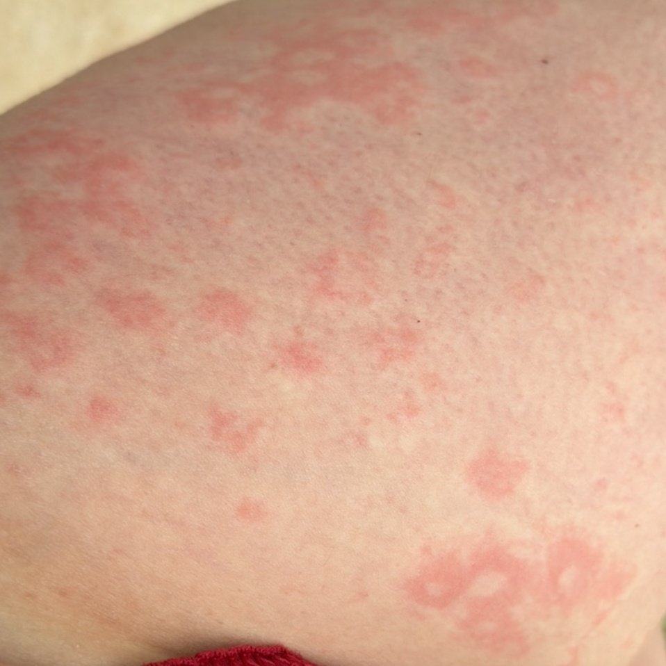
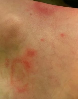
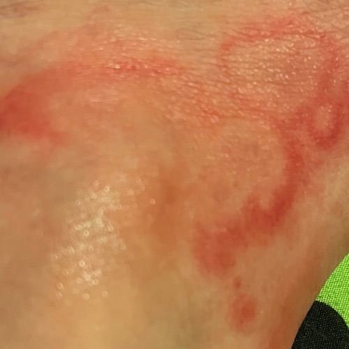
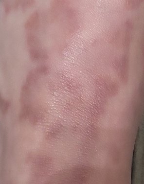
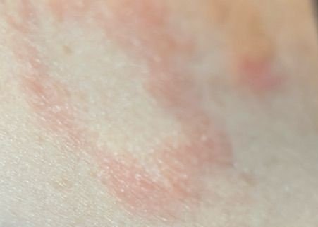
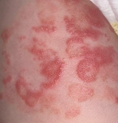
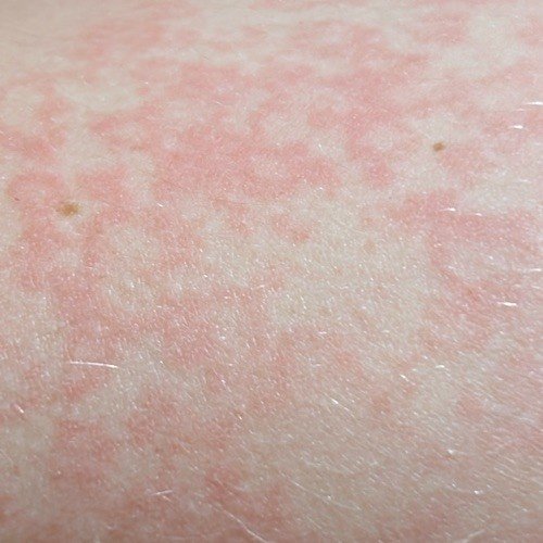
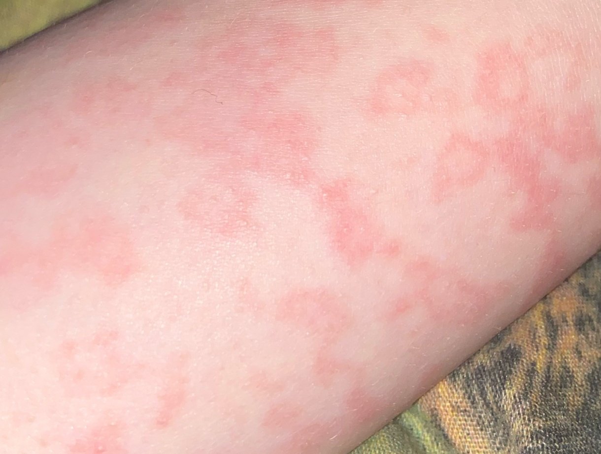
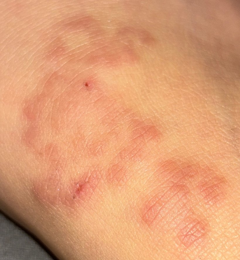
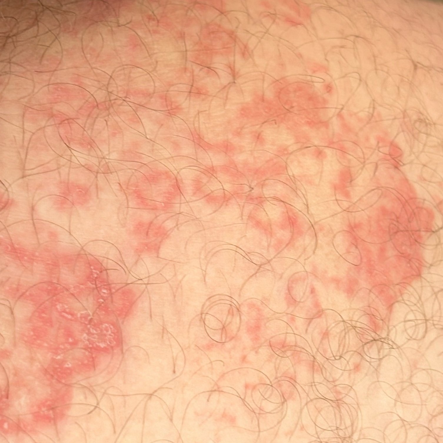
Patients with EAC typically present with annular erythematous lesions that begin as small, red spots in the center and expand outward. These lesions often appear on the thighs, shins, and occasionally on the trunk and face. Systemic symptoms, if present, usually reflect an underlying cause rather than the skin eruption itself.
The characteristic finding is an enlarging annular or arciform erythematous lesion that spreads outwards over weeks while clearing in the centre, and which may grow to several centimetres or more. In the superficial form the scale is trailing scale: it sits on the inner side of the advancing edge, just behind the border rather than on it. That is the feature which distinguishes EAC from tinea, where the scale lies on the leading edge. Vesicles and telangiectasias are not defining features. Resolving lesions may leave post-inflammatory pigment change, but no scarring.
Patients may experience either asymptomatic or itchy rashes, depending on the allergen or trigger. Some lesions may be painful, especially in cases involving systemic diseases or infections.
Prognosis
EAC generally has a favorable prognosis, especially when the underlying cause is identified and treated promptly. However, the prognosis may worsen in patients with concurrent systemic or malignant conditions. The duration of the lesions can vary, with some cases resolving within weeks, while others may persist for several years.
Classification
According to Ackerman, and later Bressler and Jones, EAC has two clinical forms:
- Superficial form: This type involves itching and flaking of the skin.
- Deep form: This form is characterized by the absence of itching and scaling, making it harder to distinguish from other conditions.
Diagnosis
The diagnosis is often made clinically, from the appearance and the slow centrifugal spread. Investigation is directed by what the history and examination suggest, not performed as a standard panel.
Diagnostic methods include:
- Medical History Review: The doctor will ask about symptoms, recent exposures, and underlying health conditions.
- Physical Examination: A thorough visual and tactile examination of the lesions will be performed.
- Skin biopsy: For uncertain cases, and the way EAC is distinguished from the conditions that mimic it;
- Testing for fungal infection: A KOH preparation or culture where the appearance or distribution raises tinea, which is the main clinical mimic;
- Investigation for systemic disease: Guided by the history and examination. There is no indication for routine allergy testing, and no basis for a blanket search for malignancy in a patient with no other features to suggest it.
Differential Diagnosis
EAC must be distinguished from various other dermatological conditions that present with similar symptoms, including:
- Autoimmune diseases (such as lupus or Sjögren’s syndrome)
- Infectious dermatoses (like tinea) and inflammatory dermatoses (like lichen planus)
- Oncodermatoses (cancer-related skin lesions)
- Pseudolymphomas and lymphomas of the skin
It is also important to differentiate EAC from other similar conditions like:
- Erythema gyratum repens
- Necrolytic migratory erythema
- Erythema marginatum
- Erythema migrans
- Erythema multiforme
- Erythema papulatum centrifugum
Treatment
Treatment for EAC focuses on addressing the underlying cause of the condition. Once the trigger is identified and treated, the skin lesions often resolve on their own. For idiopathic cases or when the cause remains unclear, symptomatic treatments may include:
- Topical Corticosteroids: These can reduce inflammation and alleviate itching associated with the lesions.
- Antihistamines: These medications help manage allergic reactions and can relieve itching.
Treating an identified underlying cause is the main approach, and where a trigger is found and dealt with the eruption usually settles. Topical corticosteroids help symptoms but do not shorten the course, and lesions commonly return when they are stopped. Systemic immunosuppressants are not a routine treatment for EAC. Lesions may come and go over months or years, which is in itself not a reason to escalate treatment; a dermatologist should guide care in persistent or recurrent disease.
Prevention
To prevent flare-ups of EAC, individuals should avoid known triggers and take proactive steps to maintain skin health:
- Where a specific trigger was identified in your case, avoid it.
- Wear loose, breathable clothing to minimize skin irritation.
- Limit exposure to extreme temperatures and always apply sunscreen in direct sunlight.
- Avoid scratching the affected areas to prevent worsening symptoms and secondary infections.
- Maintain good sleep hygiene and manage stress effectively to support immune function.
- Have any new or recurring rash looked at, since a change in the pattern may point to a cause that was not apparent before.
Copyright concern about an image on this page? Write to mail@skinai.pro. Images may not be reused without permission unless an open licence is stated on this page.

Medical review
Board-Certified Dermatologist · Fellow of the American Academy of Dermatology (FAAD)