Seborrheic keratosis (ICD-10: L82) 💚
Seborrheic Keratosis (Seborrheic Wart, Senile Wart, Senile Keratosis, Senile Keratoma)
Seborrheic Keratosis is a benign skin tumor characterized by keratinization, which rises above the skin surface. These lesions typically appear after the age of 50, with their prevalence increasing with age. Seborrheic keratosis is commonly found in multiples, and the number of lesions tends to grow over time. This type of neoplasm is equally common in both men and women, although the frequency may vary slightly based on age and other factors.
Predisposing Factors
The exact cause of seborrheic keratosis remains unclear, but several factors have been identified that may increase the risk of developing these benign skin lesions. These factors contribute to the appearance and growth of seborrheic keratosis:
- Involutive (Age-Related) Skin Changes: The aging of epidermal keratinocytes, coupled with late-stage apoptosis (cell death), is a significant factor in the development of seborrheic keratosis.
- Excessive Insolation: Prolonged exposure to sunlight and ultraviolet radiation increases the risk of developing seborrheic keratosis due to the effect of UV light on skin cells.
- Genetic Factors: Family history and genetic predisposition can play a role in the development of seborrheic keratosis, with some individuals being more susceptible due to their genetic makeup.
Diagnostics
The diagnosis of seborrheic keratosis is based on a clinical examination, which includes a routine visual inspection of the lesion and dermatoscopic evaluation to assess the characteristics of the growth. If there are concerns that the lesion may be malignant or atypical, a biopsy may be performed to confirm the diagnosis and exclude other conditions, such as skin cancer.
Symptoms
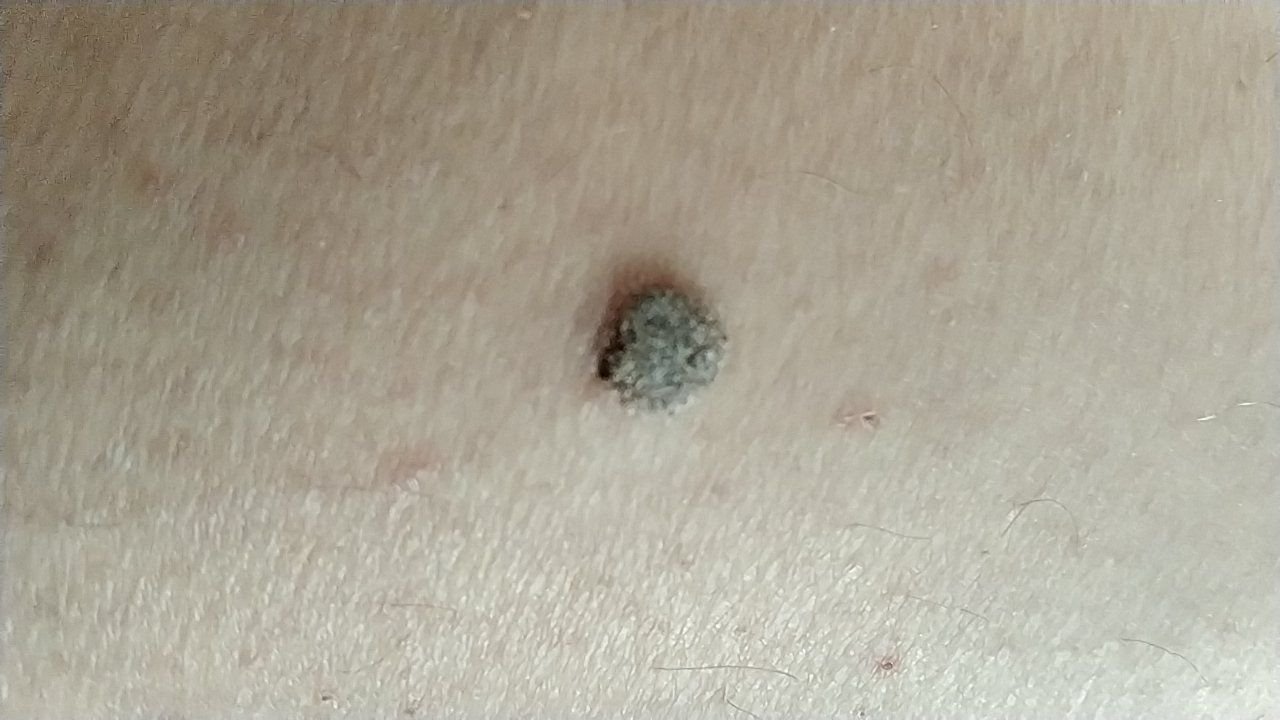
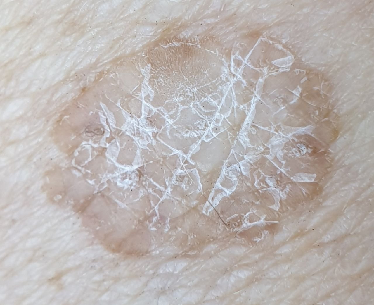
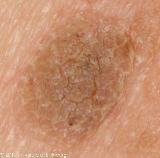
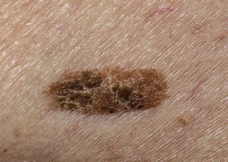
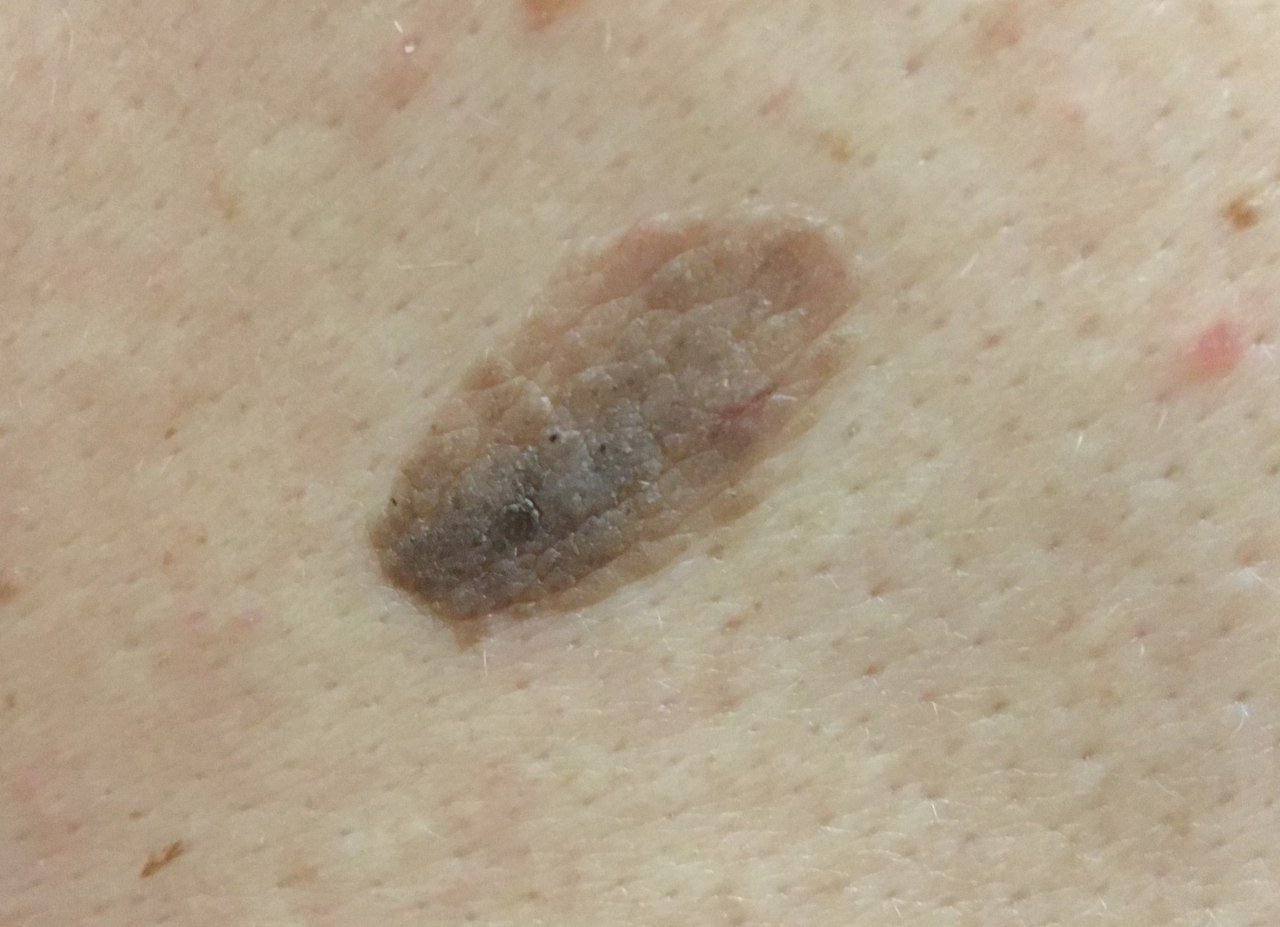
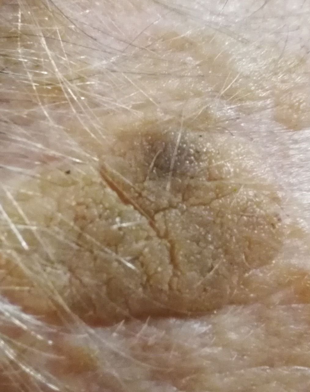
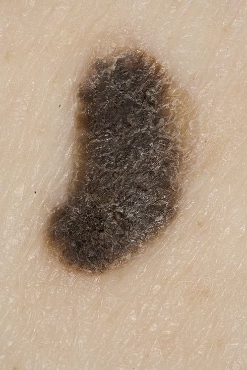
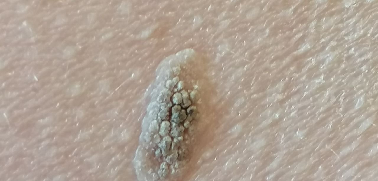
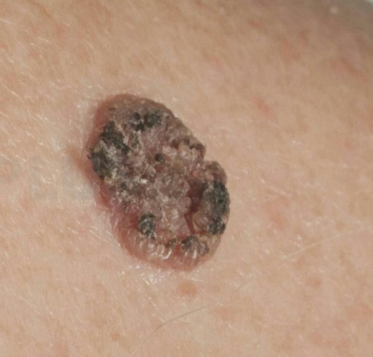
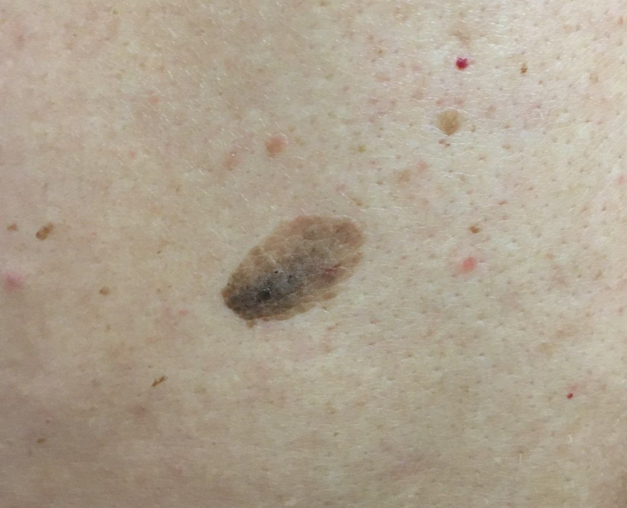
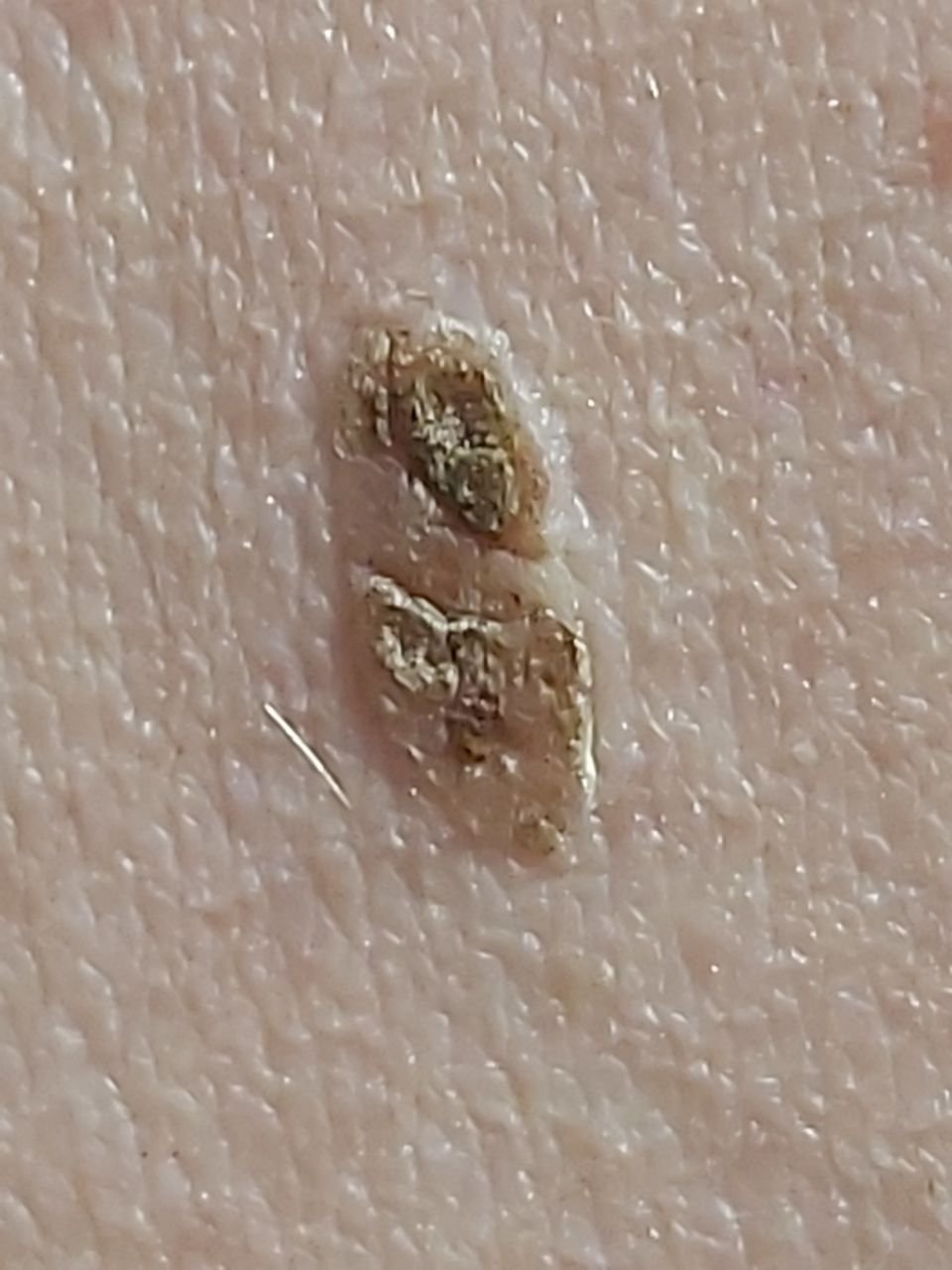
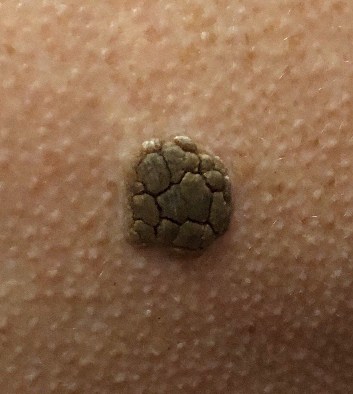
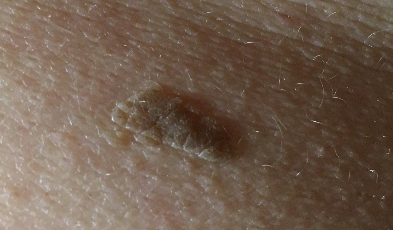
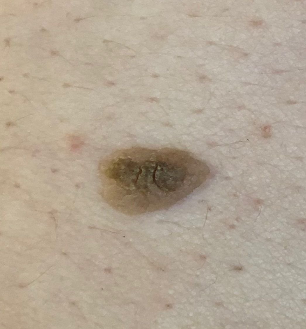
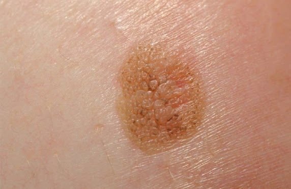
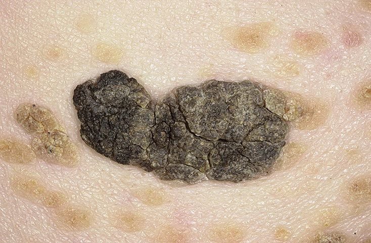
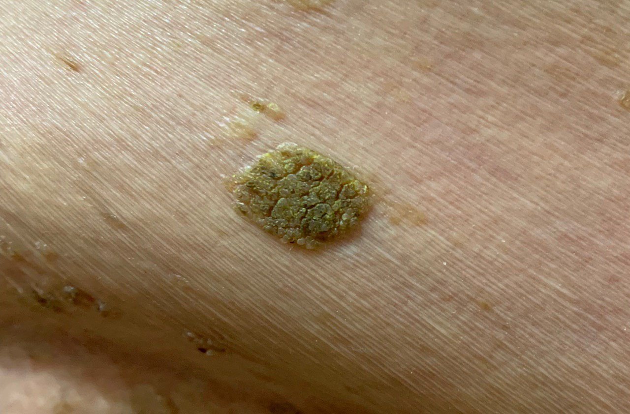
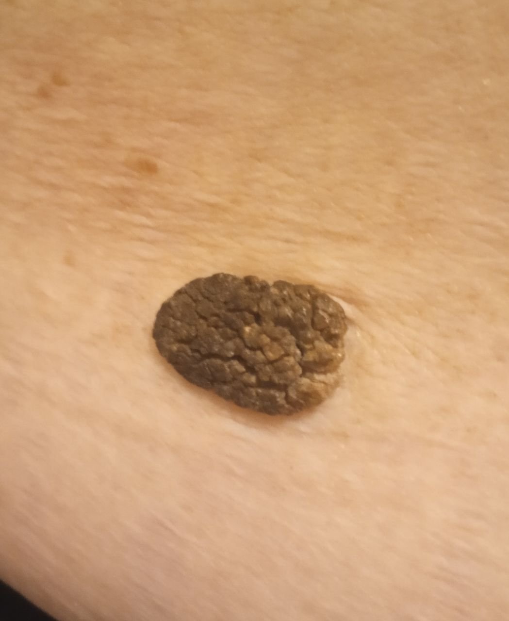
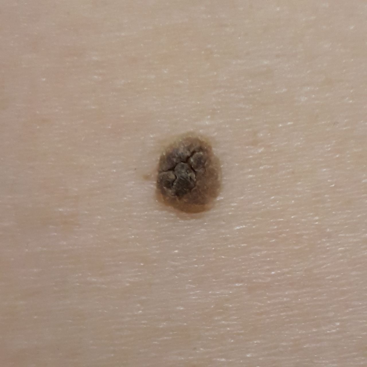
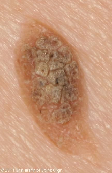
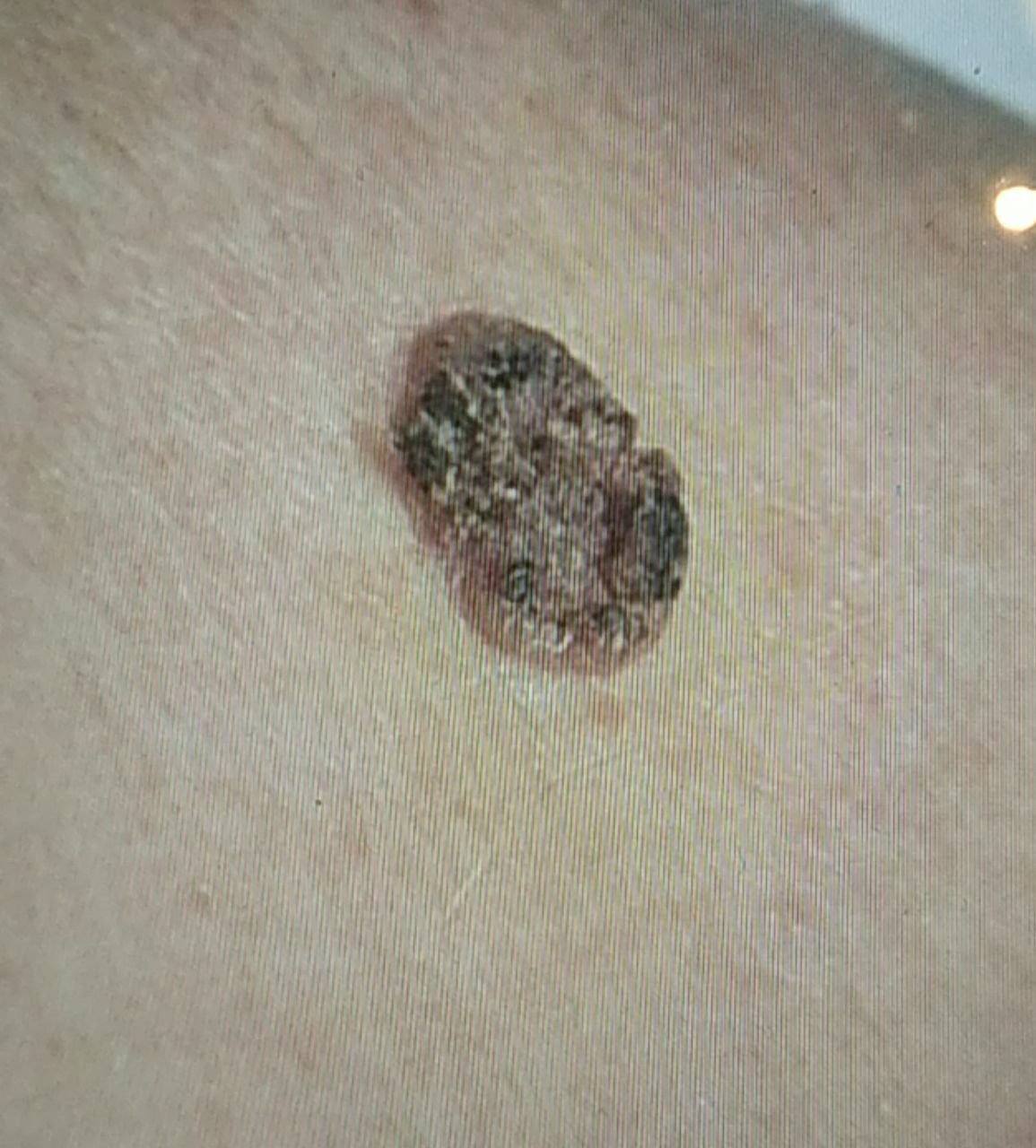
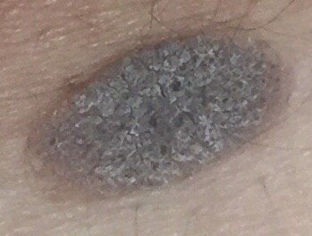
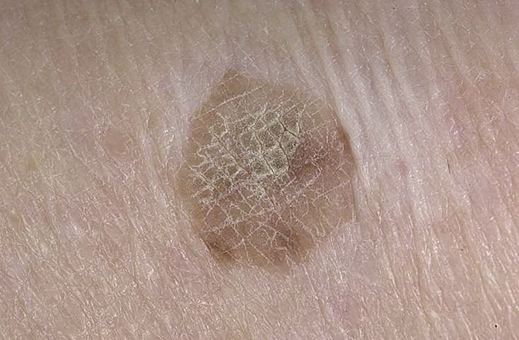
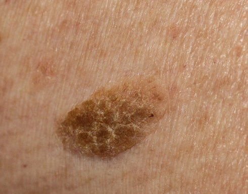
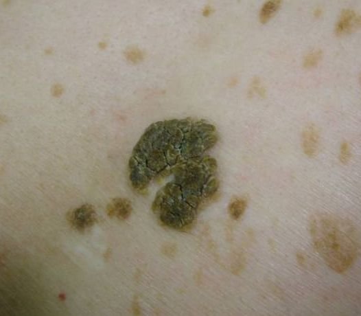
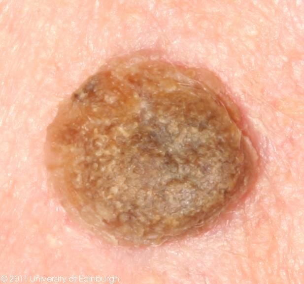
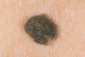
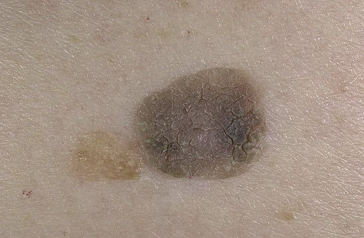
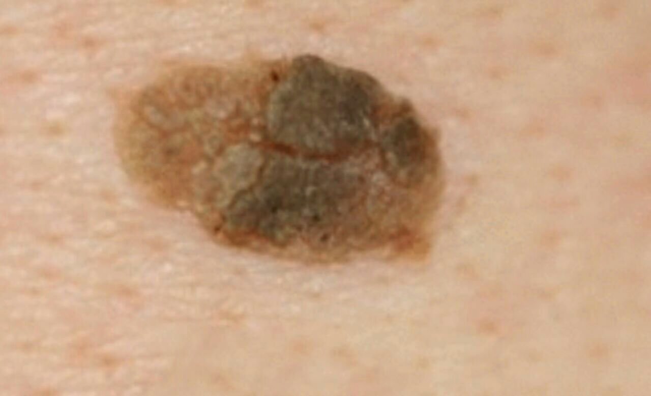
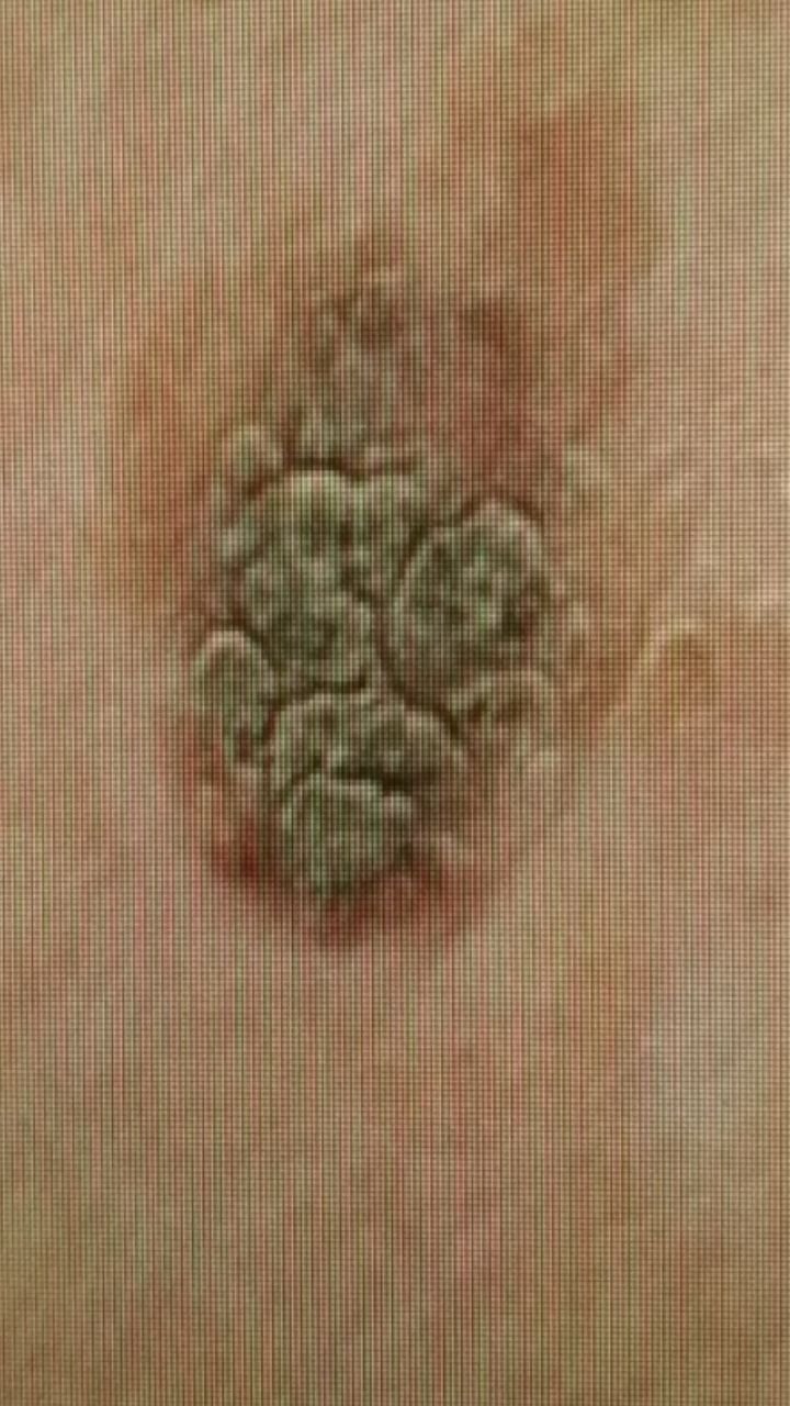
During a visual examination, seborrheic keratosis appears as a flattened, hemispherical, or short-stalked lesion that rises above the skin. These lesions are often symmetrical, typically oval or round, although larger ones can have irregular shapes. The surface texture of seborrheic keratosis differs from that of normal skin: it is rough, dry, and may show signs of peeling. In some cases, especially for larger lesions over 10 mm, the surface may have a warty texture, resembling large, uneven dry papillae. In certain instances, the keratinization is so pronounced that parts of the keratosis may fall off on their own or with minimal physical impact.
The boundaries of seborrheic keratosis are usually clear and even. However, in large keratomas, the edges can be uneven. The color of seborrheic keratosis varies from nude or gray to gray-brown. In some cases, shades of pink or pink-red may appear. Surrounding the lesion, the unchanged skin might show slight redness.
Hair growth in the area of seborrheic keratosis is typically not affected. However, in some cases, hair may grow in the lesion if it is congenital, or in the areas of keratomas with hypopigmentation.
The size of seborrheic keratosis varies, ranging from 5 mm to 20 mm in diameter. Lesions larger than 15 mm are rare. The height of the lesion above the skin surface usually does not exceed 5-7 mm.
On palpation, seborrheic keratosis feels dense and dry, with some areas potentially showing peeling. There are usually no subjective sensations, although mild itching may occur, especially in long-standing lesions.
Seborrheic keratosis is most commonly located on the trunk, upper limbs, and face. In older individuals, these lesions may also appear on other parts of the body.
Dermatoscopic Description
Dermatoscopy of seborrheic keratosis reveals the following characteristic features:
- Comedone-like Openings: Clear gray-brown keratotic inclusions that are well-formed within the lesion.
- Milium-like Cysts: Small, spherical structures with a dull white or yellowish tint that are visible in some cases of seborrheic keratosis.
- Fissures and Ridges: Furrows and raised folds across the surface, which together give the lesion its characteristic texture.
- Cerebriform (Brain-like) Pattern: Fissures and ridges arranged in a pattern resembling the convolutions of the brain.
- Blood Vessels: The presence of blood vessels in the form of hairpin shapes can be seen in dermatoscopic images.
- Background Pigmentation: The background ranges from pale gray to brown and is often fairly uniform. On its own this is not a diagnostic feature, since it occurs in other pigmented lesions as well.
- Pseudo-Network: A uniform pigment network with uneven, sharply breaking edges, particularly visible on the face.
- Dot Pigmentation: Small dots of gray-black color may be seen within the lesion.
Differential Diagnosis
It is important to differentiate seborrheic keratosis from other pigmented skin neoplasms, such as:
- Actinic keratosis
- Papillomatous nevus
- Dermatofibroma
- Bowen’s disease
- Keratoacanthoma
- Basal cell carcinoma
- Squamous cell carcinoma
- Melanoma
Risks
Seborrheic keratosis is generally a harmless condition, with a low risk of malignancy. Seborrheic keratoses are benign; if a lesion changes in appearance or behavior, it should be evaluated to rule out another diagnosis. Seborrheic keratosis is benign, and malignant transformation is very rare; if a lesion changes, it should be evaluated to rule out another diagnosis.
Multiple seborrheic keratoses are typically a benign age-related finding and do not by themselves indicate increased risk of other skin malignancies. This can complicate the timely detection and differential diagnosis of malignant lesions.
Tactics
A lesion you have not had looked at is a different matter from one a clinician has already identified. Monitoring follows assessment, not the other way round: a keratotic lesion that has not been examined should be shown to a dermatologist, because other things — including pigmented basal cell carcinoma, squamous cell carcinoma and melanoma — can look like a seborrheic keratosis, and a photograph cannot settle which it is.
Once a lesion has been assessed as a seborrheic keratosis and is stable, keeping an eye on it between reviews is reasonable. Go back sooner if it changes shape or colour, starts bleeding, crusts or ulcerates, becomes itchy or painful, grows quickly, or no longer resembles your other keratoses.
The healthcare provider will assess whether further monitoring or removal of the lesion is necessary. Seborrheic keratoses that are constantly exposed to chronic trauma due to clothing, jewelry, or occupational activities should be considered for removal. Removal is also indicated if the patient experiences cosmetic discomfort or psychological distress.
For dynamic monitoring, it is helpful to take photos of the seborrheic keratosis to track any changes in appearance over time. A skin neoplasm map is a valuable tool for ongoing observation, helping to identify new or altered lesions.
Treatment
A typical seborrheic keratosis does not have to be treated. Where a lesion is removed — because it is repeatedly traumatised, because it is symptomatic, or for cosmetic reasons — the method is chosen according to the lesion’s morphology, its site, the symptoms it causes, and how certain the diagnosis is. Cryotherapy, curettage or shave removal, laser and electrosurgery are all used; surgical excision with a classic or radiofrequency scalpel is not required for a typical lesion.
Histological examination is not mandatory for every lesion that is removed. It is appropriate whenever the diagnosis is uncertain or the lesion is atypical — and in that case the lesion should be excised so that tissue can be examined, rather than destroyed by a method that leaves nothing to examine.
Some lesions recur after treatment and need more than one session.
Prevention
Seborrheic keratoses are benign, so prevention is not a matter of averting malignancy, and their appearance cannot reliably be prevented. What is worth doing is general skin care, alongside noticing any lesion that looks atypical or is changing:
- Avoiding excessive ultraviolet radiation, including the use of tanning beds and prolonged sun exposure.
- Using sunscreen and protective clothing during periods of intense sunlight exposure.
- Minimizing chronic skin trauma that can cause irritation and damage to keratoses.
- Adhering to safety protocols when handling skin-damaging substances.
- Maintaining good personal hygiene and monitoring skin changes.
Looking the lesions over from time to time, and seeking advice when one of them changes, is what matters — not because a seborrheic keratosis is expected to turn malignant, but because a lesion that changes may prove to be a different diagnosis.